. 2023 Oct 24;2(4):e141.
doi: 10.1002/imt2.141. eCollection 2023 Nov.

Melatonin supplementation protects against traumatic colon injury by regulating SERPINA3N protein expression

Affiliations

Melatonin supplementation protects against traumatic colon injury by regulating SERPINA3N protein expression

Bo Cao et al. Imeta. .

Abstract

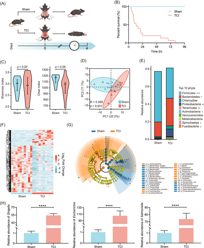
Traumatic colon injury (TCI) is a typical injury with high mortality. Prolongation of the intervention time window is a potentially useful approach to improving the outcomes of TCI casualties. This study aimed to identify the pathological mechanisms of TCI and to develop effective strategies to extend the survival time. A semicircular incision was made to prepare a TCI model using C57BL/6 mice. An overview of microbiota dysregulation was achieved by metagenome sequencing. Protein expression reprogramming in the intestinal epithelium was investigated using proteomics profiling. The mice that were subjected to TCI died within a short period of time when not treated. Gut symbiosis showed abrupt turbulence, and specific pathogenic bacteria rapidly proliferated. The protein expression in the intestinal epithelium was also reprogrammed. Among the differentially expressed proteins, SERPINA3N was overexpressed after TCI modeling. Deletion of Serpina3n prolonged the posttraumatic survival time of mice with TCI by improving gut homeostasis in vivo. To promote the translational application of this research, the effects of melatonin (MLT), an oral inhibitor of the SERPINA3N protein, were further investigated. MLT effectively downregulated SERPINA3N expression and mitigated TCI-induced death by suppressing the NF-κB signaling pathway. Our findings prove that preventive administration of MLT serves as an effective regimen to prolong the posttraumatic survival time by restoring gut homeostasis perturbed by TCI. It may become a novel strategy for improving the prognosis of patients suffering from TCI.

Keywords: SERPINA3N; gut homeostasis; melatonin; microbiota dysbiosis; posttraumatic survival time; traumatic colon injury.


Conflict of interest statement

The authors declare no conflict of interest.

Figures

Figure 1
Figure 1
The abundance of pathogenic bacteria is abruptly increased after TCI modeling. (A) Flow diagram used to establish sham and TCI mouse models. TCI was induced in mice by the incision of the colon that was 3 cm from the cecum end. Sham mice were only subjected to laparotomy without colon incision. (B) Survival curve showing the survival differences between sham and TCI groups (n = 15 per group). (C) Violin plots comparing alpha diversities of gut microbiota in the sham and TCI groups. (D) Scatter plot comparing the beta diversity of gut microbiota as in (C). (E) Stacked histogram showing the relative abundances of top 10 phyla of gut microbiota as in (C). (F) Heatmap showing the bacterial genera with different abundances as in (C) (p < 0.05). (G) Cladogram showing the differential bacteria as in (C). The circles from the inside to the outside indicate the taxonomic ranks from phylum to genus. The blue and orange points indicate the bacteria with important functions in the corresponding groups. The yellow points indicate the species without significant differences in relative abundances. (H) qRT‐PCR analysis to determine the relative abundances of Shigella, Escherichia, and Salmonella in intestinal contents of sham and TCI mice (n = 15 per group). *p < 0.05, **p < 0.01, ***p < 0.001, ****p < 0.0001. qRT‐PCR, quantitative real‐time polymerase chain reaction; TCI, traumatic colon injury.
Figure 2
Figure 2
TCI reprograms the protein expression profile of the intestinal epithelium following TCI challenge. (A) Volcano plot showing the differential proteins in intestinal epithelium (n = 10 per group, log2FC < −1 or >1, p < 0.05). (B–D) Bubble plots showing the top enriched 10 items of BP, CC, and MF categories by GO analysis. (E) Heatmap showing differential proteins reported to be associated with inflammation, infection defense, and barrier integrity. (F) IHC examination used to detect the SERPINA3N expression in the intestinal epithelium of sham and TCI groups. The histogram is displayed below the IHC pictures. Scale bar: 100 μm. ****p < 0.0001. BP, biological process; CC, cellular component; GO, Gene Ontology; IHC, immunohistochemistry; MF, molecular functional; TCI, traumatic colon injury.
Figure 3
Figure 3
Knockout of SERPINA3N protein prolongs the survival time and gut homeostasis of TCI mice. (A) Flow diagram used to establish the Serpina3n KO mice that were subjected to sham or TCI operation and WT mice that were subjected to the TCI operation (n = 15 per group). (B) Survival curve comparing the survival time of mice as in (A). (C) ELISA assay used to detect the concentrations of IL‐6 and TNF‐α in the plasma of WT or Serpina3n KO mice that were subjected to sham or TCI operation (n = 15, 15, 11, and 13, respectively). (D) ELISA assay used to detect the concentrations of IL‐6 and TNF‐α in the intestines of mice as in (C). (E) IHC examination used to determine ZO‐1 and occludin expression in the intestinal epithelium of mice as in (C). The histogram is displayed on the right of the pictures. Scale bar: 100 μm. (F) qRT‐PCR analysis used to determine the relative abundances of Shigella, Escherichia, and Salmonella in the intestinal contents of mice as in (C). ***p < 0.001, ****p < 0.0001. ELISA, enzyme‐linked immunosorbent assay; IHC, immunohistochemistry; IL‐6, interleukin‐6; KO, knockout; n.s, not significant; qRT‐PCR, quantitative real‐time polymerase chain reaction; TCI, traumatic colon injury; TNF‐α, tumor necrosis factor‐α; WT, wild type.
Figure 4
Figure 4
Administration of MLT extends the survival time and gut homeostasis of TCI mice by downregulation of SERPINA3N expression. (A) Flow diagram used to generate the mice in the sham or TCI groups that were administered with vehicle or MLT as indicated (n = 15 per group). (B) Survival curve comparing the survival time of mice as in (A). (C) ELISA assay used to detect the concentrations of IL‐6 and TNF‐α in the plasma of mice in the sham or TCI group that were administered with vehicle or MLT (n = 15, 15, 10, and 13, respectively). (D) ELISA assay used to detect the concentrations of IL‐6 and TNF‐α in the intestines of mice as in (C). (E) IHC examination used to determine ZO‐1 and occludin expression in the intestinal epithelium of mice as in (C). The histogram is displayed on the right of the pictures. Scale bar: 100 μm. (F) qRT‐PCR analysis used to determine the relative abundances of Shigella, Escherichia, and Salmonella in intestinal contents of mice as in (C). **p < 0.01, ***p < 0.001, ****p < 0.0001. ELISA, enzyme‐linked immunosorbent assay; IHC, immunohistochemistry; IL‐6, interleukin‐6; MLT, melatonin; n.s, not significant; qRT‐PCR, quantitative real‐time polymerase chain reaction; TCI, traumatic colon injury; TNF‐α, tumor necrosis factor‐α; WT, wild type.
Figure 5
Figure 5
MLT suppresses SERPINA3N expression by inhibiting the NF‐κB signaling pathway. (A) Histogram showing the signaling pathways that ranked top 10 according to KEGG analysis based on proteomics profiling. (B) WB analysis used to determine the p‐NF‐κB, NF‐κB, and SERPINA3N expression in Caco‐2 cells treated with 0.01, 0.1, and 1 μM MLT for 24 h. (C) WB analysis used to determine the p‐NF‐κB, NF‐κB, and SERPINA3N expression in Caco‐2 cells treated with vehicle, MLT, 100 μM JSH‐23, or MLT plus 100 μM JSH‐23. (D and E) Transwell assay used to determine cell migration capabilities of cells as in (C). The histogram is displayed on the right. Scale bar: 100 μm. (F) Survival curve comparing the survival time of TCI mice treated with vehicle, MLT, 3 mg/kg JSH‐23, and MLT plus 3 mg/kg JSH‐23 (n = 15 per group). (G) IHC examination used to determine SERPINA3N, p‐NF‐κB, and NF‐κB expression in the intestinal epithelium of mice that were subjected to the same treatment as in (F). Scale bar: 100 μm. (H) Histogram showing IHC scores as in (G). ***p < 0.001, ****p < 0.0001. KEGG, Kyoto Encyclopedia of Genes and Genomes; IHC, immunohistochemistry; MLT, melatonin; n.s not significant; TCI, traumatic colon injury; WB, western blot.
Figure 6
Figure 6
Schematic illustration of the mechanisms by which MLT administration prolongs the posttraumatic survival time of TCI mice. TCI leads to significant disruption of gut homeostasis within a short period of time, indicated by inflammatory responses, intestinal barrier hyperpermeability, and microbiota dysregulation. MLT administration can restore gut homeostasis by suppressing SERPINA3N expression in the intestinal epithelium, subsequently prolonging the survival time of TCI mice. MLT, melatonin; TCI, traumatic colon injury.

Similar articles

Cited by

References

    1. Belmont Philip J. Jr., McCriskin Brendan J., Sieg Ryan N., Burks Robert, and Schoenfeld Andrew J.. 2012. “Combat Wounds in Iraq and Afghanistan From 2005 To 2009.” Journal of Trauma and Acute Care Surgery 73: 3–12. 10.1097/TA.0b013e318250bfb4 - DOI - PubMed
    1. Owens, Brett D. , Kragh John F. Jr., Wenke Joseph C., Macaitis Joseph, Wade Charles E., and Holcomb John B.. 2008. “Combat Wounds in Operation Iraqi Freedom and Operation Enduring Freedom.” Journal of Trauma 64: 295–299. 10.1097/TA.0b013e318163b875 - DOI - PubMed
    1. Arafat, Shawqi , Alsabek Mhd Belal, Ahmad Mousa, Hamo Iman, and Munder Eskander. 2017. “Penetrating Abdominal Injuries During the Syrian War: Patterns and Factors Affecting Mortality Rates.” Injury 48: 1054–1057. 10.1016/j.injury.2017.02.005 - DOI - PubMed
    1. Fouda, E. , Emile S., Elfeki H., Youssef M., Ghanem A., Fikry A. A., Elshobaky A., et al. 2016. “Indications for and Outcome of Primary Repair Compared with Faecal Diversion in the Management of Traumatic Colon Injury.” Colorectal Disease 18: O283–O291. 10.1111/codi.13421 - DOI - PubMed
    1. Smith, Iain M. , Naumann David N., Marsden Max E. R., Ballard Mark, and Bowley Douglas M.. 2015. “Scanning and War: Utility of FAST and CT in the Assessment of Battlefield Abdominal Trauma.” Annals of Surgery 262: 389–396. 10.1097/sla.0000000000001002 - DOI - PubMed

LinkOut - more resources