

Periosteum for Root Coverage in an Isolated Gingival Recession as an Autogenous Graft: A Case Report
- PMID: 38868280
- PMCID: PMC11168571
- DOI: 10.7759/cureus.60207
Periosteum for Root Coverage in an Isolated Gingival Recession as an Autogenous Graft: A Case Report
Abstract
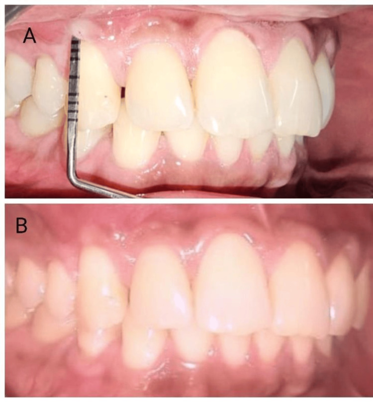
In periodontal care, where patient results are crucial in guiding the development of surgical techniques, gingival recession management is a critical issue. The periosteum eversion technique (PET) emerges as a modern strategy that leverages the intrinsic regenerative capabilities of the periosteum to attain root coverage. A detailed case study showcases the effectiveness of PET in managing a Miller Class I gingival recession alongside an adjunctive platelet-rich fibrin (PRF) procedure. This approach entailed the deliberate elevation and eversion of the periosteal flap to encompass the recession area, securing it meticulously through suturing. Across a six-month observation period, this method exhibited successful root coverage, augmentation of keratinized tissue, and enhanced patient comfort, as reported, with no significant complications observed. These outcomes provide support for the incorporation of PET into standard periodontal protocols, underscoring its capacity to reshape the treatment landscape for gingival recession.
Keywords: autograft; isolated gingival recession; leukocyte platelet rich fibrin (l-prf); periosteum; root coverage.
Copyright © 2024, S et al.
Conflict of interest statement
The authors have declared that no competing interests exist.
Figures


References
-
- The etiology and prevalence of gingival recession. Kassab MM, Cohen RE. J Am Dent Assoc. 2003;134:220–225. - PubMed
-
- Gingival recession and root coverage up to date, a literature review. Mostafa D, Fatima N. Dent Rev. 2022;2:100008.
-
- Miller classification of marginal tissue recession revisited after 35 years. Miller PD. https://pubmed.ncbi.nlm.nih.gov/30188152/ Compend Contin Educ Dent. 2018;39:514–520. - PubMed
-
- Etiopathogenesis of gum recession: the current aspects [Article in Russian] Postnikov MA, Vinnik AV, Rakhimov RR, Kostionova-Ovod IA, Vinnik SV. Aspirantskiy Vestnik Povolzhiya. 2022;22:27–32.
-
- Consensus report. Mucogingival therapy. Ann Periodontol. 1996;1:702–706. - PubMed
Publication types
LinkOut - more resources
Full Text Sources