Review
. 2024 May 15;22(3):191-202.
doi: 10.18502/ijrm.v22i3.16163. eCollection 2023 Mar.

The categorization of opaque pathologies outside of contrast media in hysterosalpingography which facilitate interpretation: A pictorial review

Affiliations
Review

The categorization of opaque pathologies outside of contrast media in hysterosalpingography which facilitate interpretation: A pictorial review

Fereshteh Hosseini et al. Int J Reprod Biomed. .

Abstract

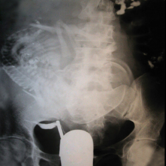
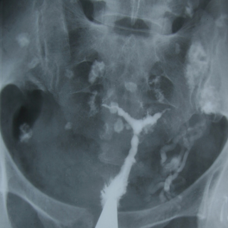
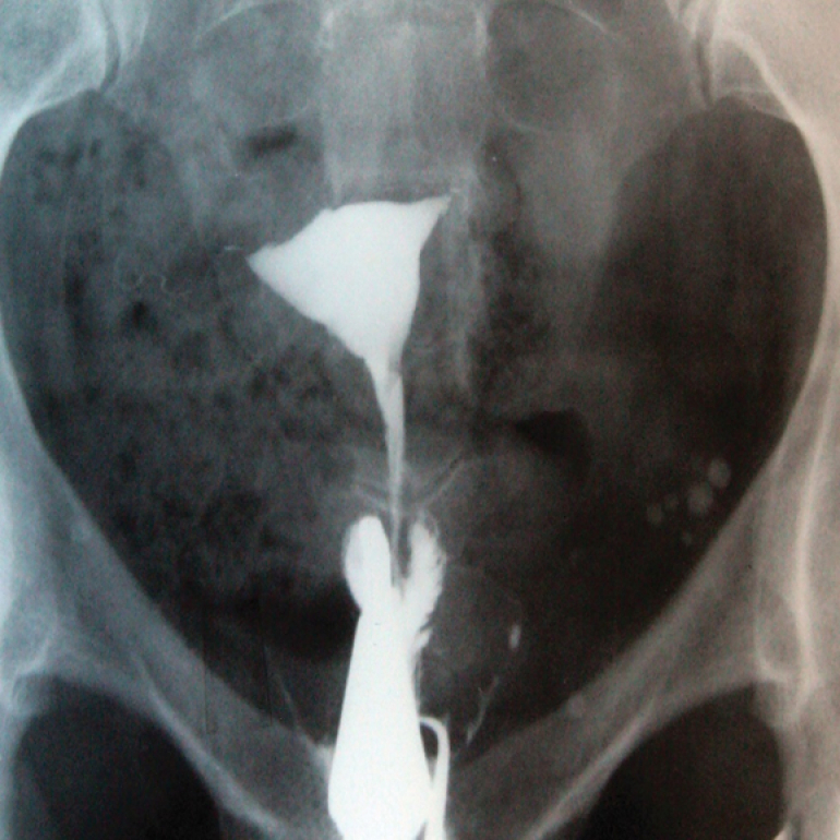
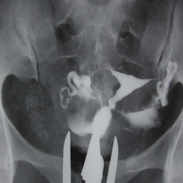
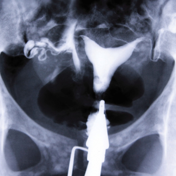
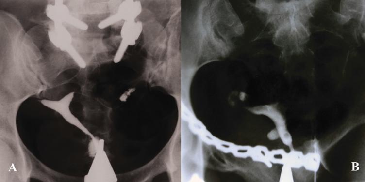
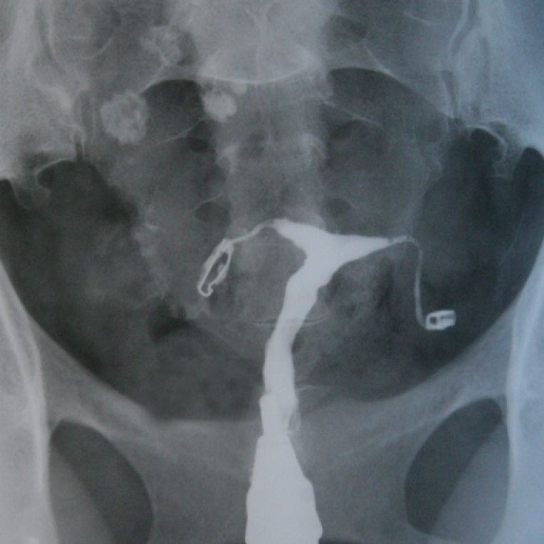
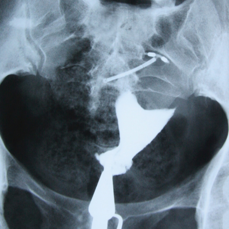
Hysterosalpingography (HSG) is a practical and reliable imaging method to evaluate the cervical canal, uterine isthmus, uterine cavity, and fallopian tubes. Using HSG, opaque pathologies outside of contrast media can be detected as well as pathologies of uterus and fallopian tubes. We aim to present and categorize some uncommon and interesting abnormal findings that are located outside of the contrast areas in HSG. This is a pictorial review that depicts various types of HSG images that include opaque pathologies outside of the contrast areas. Images have been extracted from valuable archives collected over 50 yr by professor Shahrzad. A plain pelvic film contains soft tissues of the pelvis, bony structures, artifacts, or foreign bodies. Categorization might easily help the radiologist to interpret the HSG cliché. Opaque pathologies outside of contrast area in HSG can be categorized into 2 groups: "Pelvic Tissue Related" and "Foreign Bodies". Pelvic tissue abnormalities might have a gynecologic or non-gynecologic source. Foreign bodies can be located in the pelvis or outside of the body. HSG is a reliable and inexpensive procedure. Familiarity with the pathologies of pelvic tissues and the accurate interpretation of HSG images are important.

Keywords: Abnormalities; Fallopian tubes.; Uterine; Hysterosalpingography; Opaque.


Conflict of interest statement

The authors declare that there is no conflict of interest.

Figures

Figure 1
Figure 1
A) HSG of a 35-yr-old woman with primary infertility. The uterine cavity and fallopian tubes have a normal view. A calcified area is seen on the left side of the uterine cavity. B) The fibroma was approved by a 2-D ultrasound examination.
Figure 2
Figure 2
HSG of a 30-yr-old woman with secondary infertility, the uterine cavity is normal. A mild hydrosalpinx is evident at the end of the left tube. The right tube is displaced due to a soft tissue mass. Multiple dense areas with teeth structure appearance are seen inside the mass that represents a large dermoid cyst.
Figure 3
Figure 3
A) After injecting contrast materials, only the cervix was opaque in HSG. The uterus and fallopian tubes were calcified due to advanced TB infection. Calcified lymph nodes are also evident. Genital TB was approved by supplementary investigations. B) Normal uterine cavity. Obstruction in the middle portion of the isthmic segment of both fallopian tubes is seen. No peritoneal spillage was detected. The round calcified area on the left side of the pelvis denotes a calcified ovary caused by genital tuberculosis.
Figure 4
Figure 4
A) HSG of a 27-yr-old patient with primary infertility. A large soft tissue mass causing the elongation of the right wall and dislocation of the uterus to the left. B) An ultrasound image of patient “A”, which confirms the presence of a large fibroid. C) Dislocation of the left fallopian tube due to a soft tissue mass in HSG of a 37-yr-old patient. D) Ultrasound image of patient “C”, that shows an ovarian endometrioma cyst.
Figure 5
Figure 5
HSG of a patient referred with possibility of abdominal mass. (Taken 50 yr ago). An image of a fetus with overlapping cranial sutures is seen on the right portion of the abdomen and pelvis. The uterine cavity is visible on the right side of the pelvis. This finding suggests pregnancy on the left uterine cavity of the uterus didelphys.
Figure 6
Figure 6
A 40-yr-old patient with secondary infertility. Uterine cavity with irregular borders and trifoliate appearance due to adhesions caused by genital tuberculosis. The calcified lymph nodes secondary to TB and intravasation are observed.
Figure 7
Figure 7
Normal uterine cavity. The concentration of tiny phleboliths on the left side of the pelvis and outside of the uterine cavity. Phleboliths are frequently seen in pelvic x-ray and have no clinical value.
Figure 8
Figure 8
A shapeless calcified mass is seen on the right side of the pelvis in HSG. A calcified intestinal parasite was suspected based on the mass features and the patient's history. The patient was referred to an infectious disease specialist. After the medical laboratory test including a stool exam, the presence of the parasite was confirmed, and the patient was treated.
Figure 9
Figure 9
HSG of a 38-yr-old woman with secondary infertility. An arcuate uterus and normal fallopian tubes in HSG. Osteopoikilosis was diagnosed based on the small area of sclerotic bony lesions in the pelvic bones.
Figure 10
Figure 10
Platinum prosthesis. HSG of 2 different patients with primary infertility and history of surgery. A) A pin related to a previous orthopedic vertebral surgery. B) A chain associated with a pelvic surgery.
Figure 11
Figure 11
HSG of a 31-yr-old patient with primary infertility and a history of genital tuberculosis. 2 clips located in the middle of the fallopian tubes observed in HSG had been used to prevent the transmission of genitalia tuberculosis to the uterine cavity. Calcified lymph nodes secondary to TB are seen.
Figure 12
Figure 12
A dislocated IUD outside the uterine cavity in HSG. The patient has mentioned the history of putting in an IUD, but it was not detected during the examination by the gynecologist. After HSG, the patient underwent surgery, and the IUD was removed. (Patient thought that the IUD was already removed from her uterus).
Figure 13
Figure 13
HSG of a 28-yr-old woman with primary infertility. A short fundal septum is evident. Bilateral tubal obstruction is seen. Medical powder (Bismuth) can be seen as scattered and fine granules or clumps through the bowel as observed in HSG. Tiny, dense, and diffuse light spots outside the uterus are caused by the opaque effect of Bismuth.
Figure 14
Figure 14
HSG of a patient with primary infertility. Cervix, uterine cavity, and fallopian tubes have normal view. Metal foreign body is seen on the left side of the uterine cavity in HSG. The patient had swallowed a needle. In complementary imaging assessment, no needle was detected after 3 days. It shows that the needle did not penetrate the soft tissue and has been excreted from the intestines.
Figure 15
Figure 15
HSG of a patient with secondary infertility. Normal uterine cavity. The left fallopian tube failed to opacify but the right tube is filled and peritoneal spillage is observed. Retained surgical gas with radio-opaque markers above the uterine cavity is seen in HSG. The patient had pelvic surgery a few months ago; and referred for infertility work-up.
Figure 16
Figure 16
Normal uterine cavity and fallopian tubes. The safety pin and zipper attached to the patient's cloth are seen in the upper part of the HSG.

References

    1. Luczynska E, Zbigniew K. Diagnostic imaging in gynecology. Ginekol Pol. 2022;93:63–69. - PubMed
    1. Baușic A, Coroleucă C, Coroleucă C, Comandașu D, Matasariu R, Manu A, et al. Transvaginal ultrasound vs. magnetic resonance imaging (MRI) value in endometriosis diagnosis Diagnostics. 2022;12:1767. - PMC - PubMed
    1. Gündüz R, Ağaçayak E, Okutucu G, Karuserci ÖK, Peker N, Çetinçakmak MG, et al. - PMC - PubMed
    1. Panda SR, Kalpana BJC. The diagnostic value of hysterosalpingography and hysterolaparoscopy for evaluating uterine cavity and tubal patency in infertile patients. Cureus. 2021;13:e12526. - PMC - PubMed
    1. Martí-Bonmatí L. Hysterosalpingography and fertility: A technical relationship. Fertil Steril. 2018;110:642. - PubMed

LinkOut - more resources