. 2023 Dec 31;17(4):299-320.
doi: 10.18502/jad.v17i4.15294. eCollection 2023 Dec.

Anti-Leishmanial Effects of a Novel Biocompatible Non-Invasive Nanofibers Containing Royal Jelly and Propolis against Iranian Strain of Leishmania major (MRHO/IR/75/ER): an In-Vitro Study

Affiliations

Anti-Leishmanial Effects of a Novel Biocompatible Non-Invasive Nanofibers Containing Royal Jelly and Propolis against Iranian Strain of Leishmania major (MRHO/IR/75/ER): an In-Vitro Study

Mohsen Mahmoudi et al. J Arthropod Borne Dis. .

Abstract

Background: Current medications especially the pentavalent antimonial compounds have been used as the first line treatment of cutaneous leishmaniasis (CL), but they have limitations due to serious side effects such as drug resistance, cardio and nephrotoxicity, and high costs. Hence, the demand to find more usable drugs is evident. Synthesis and development of natural, effective, biocompatible, and harmless compounds against Leishmania major is the principal priority of this study.

Methods: By electrospinning method, a new type of nanofiber were synthesized from royal jelly and propolis with different ratios. Nanofibers were characterized by Scanning Electron Microscope (SEM), Transmission Electron Microscopy (TEM), Thermogravimetric Analysis (TGA), Contact angle, and Fourier-transform infrared spectroscopy (FTIR). The Half-maximal inhibitory concentration (IC50), Half-maximal effective concentration (EC50) and the 50% cytotoxic concentration (CC50) for different concentrations of nanofibers were determined using quantitative calorimetric methods. Inductively coupled plasma-optical emission spectrometry (ICP-OES) and flow cytometry were performed as complementary tests.

Results: The results showed that the proposed formulas provide a new achievement that, despite the significant killing activity on L. major, has negligible cytotoxicity on the host cells. Royal jelly nanofibers have significantly shown the best 72 hours results (IC50= 35 μg/ml and EC50=16.4 μg/ml) and the least cytotoxicity.

Conclusion: This study presents a great challenge to introduce a new low-cost treatment method for CL, accelerate wound healing, and reduce scarring with minimal side effects and biocompatible materials. Royal jelly and propolis nanofibers significantly inhibit the growth of L. major in-vitro.

Keywords: In-vitro; Leishmania major; Nanofiber; Propolis; Royal jelly.


Figures

Fig. 1.
Fig. 1.
FESEM images for the studied nanofibers with different proportions of royal jelly, propolis and PVA. (Sample D;/40% Royal jelly and Propolis equally/60% PVA; was selected for study)
Fig. 2.
Fig. 2.
TEM images for the 3 selective studied nanofibers: (A) Royal jelly+ Propolis, (B) Royal jelly (C) Propolis
Fig. 3.
Fig. 3.
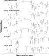
FTIR spectra of different nanofibers compared to PVA, royal jelly, and propolis spectra (spectral range: 350–4000 cm-1
Fig. 4.
Fig. 4.
Contact angle for electrospun samples: (1) Royal jelly Nanofibers (2) Propolis nanofibers (3) Royal jelly – Propolis Nanofibers (4) PVA
Fig. 5.
Fig. 5.
Thermogravimetric (TGA) graphs of different electrospun nanofibers; Royal jelly, Propolis, Royal jelly-propolis combination
Fig. 6.
Fig. 6.
Scanning Elctron microscope Images (SEM) of macrophages on Royal jelly-Propolis nano-fibers for biocompabilty confirmation of nano-fiber. Viable macrophages surrounded by fibers can be seen
Fig. 7.
Fig. 7.
Anti- Leishmainal effct (IC50) of Glucantime® - free nanofibers (A) and nanofibers containing different concentrations of Glucantime® (The composition contains 2%, 4%, 6% Glucantime®) (B) in 24–48–72 h. A p< 0.05 is considered statistically significant
Fig. 7.
Fig. 7.
Anti- Leishmainal effct (IC50) of Glucantime® - free nanofibers (A) and nanofibers containing different concentrations of Glucantime® (The composition contains 2%, 4%, 6% Glucantime®) (B) in 24–48–72 h. A p< 0.05 is considered statistically significant
Fig. 8.
Fig. 8.
Cytotoxicity assay (EC50) of Glucantime® - free nanofibers (A) and nanofibers containing different concentrations of Glucantime® on Leishmania major amastigotes (B) in 24–48–72 h. A P< 0.05 is considered statistically significant
Fig. 9.
Fig. 9.
Invitro cytotoxicity assessment of Glucantime® - free nanofibers (A) and nanofibers containing different concentrations of Glucantime® (The composition contains 2%, 4%, and 6% Glucantime®) (B) on macrophages in 24–48–72hours. A P< 0.05 is considered statistically significant
Fig. 10.
Fig. 10.
Flow cytometry analysis of studied drugs in 24 hours
Fig. 10.
Fig. 10.
Flow cytometry analysis of studied drugs in 24 hours

References

    1. Torres-Guerrero E, Quintanilla-Cedillo MR, Ruiz-Esmenjaud J, Arenas R. (2017) Leishmaniasis: a review.F1000Res. 6: 750. - PMC - PubMed
    1. Izri A, Bendjaballah-Laliam A, Sereno D, Akhoundi M. (2021) Updates on geographical dispersion of Leishmania parasites causing cutaneous affections in Algeria. Pathogens. 10(3): 267. - PMC - PubMed
    1. Haldar AK, Sen P, Roy S. (2011) Use of antimony in the treatment of leishmaniasis: current status and future directions. Mol Biol Int. 2011: 571242. - PMC - PubMed
    1. Frézard F, Demicheli C, Ribeiro RR. (2009) Pentavalent antimonials: new perspectives for old drugs. Molecules. 14(7): 2317–2336. - PMC - PubMed
    1. Nabas Z, Haddadin MS, Haddadin J, Nazer IK. (2014) Chemical composition of royal jelly and effects of synbiotic with two different locally isolated probiotic strains on antioxidant activities. Pol J Food Nutr Sci. 64(3): 171–180.

LinkOut - more resources