. 2024 Sep 3;33(18):1575-1583.
doi: 10.1093/hmg/ddae096.

The maternal protein NLRP5 stabilizes UHRF1 in the cytoplasm: implication for the pathogenesis of multilocus imprinting disturbance

Affiliations

The maternal protein NLRP5 stabilizes UHRF1 in the cytoplasm: implication for the pathogenesis of multilocus imprinting disturbance

Motoko Unoki et al. Hum Mol Genet. .

Abstract

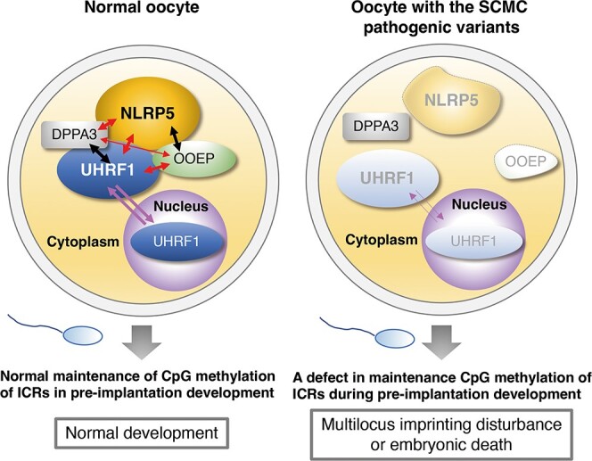
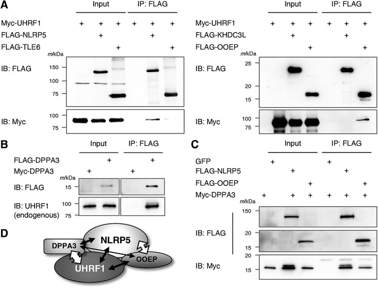
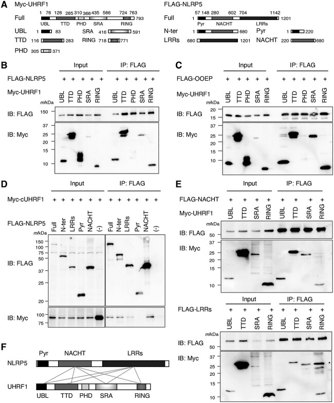
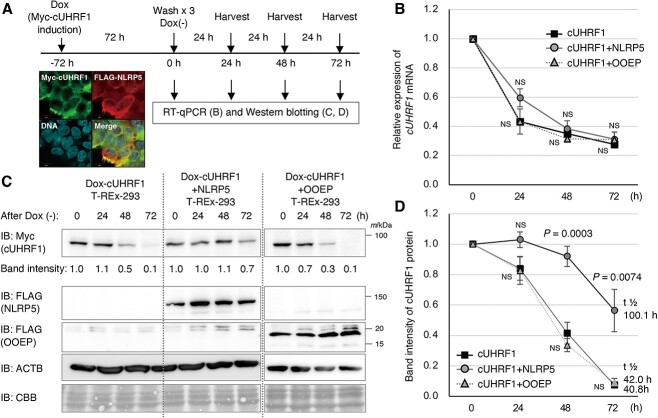
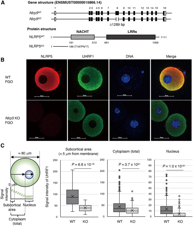
We have recently discovered that the so-called subcortical maternal complex (SCMC) proteins composing of cytoplasmic lattices are destabilized in Uhrf1 knockout murine fully grown oocytes (FGOs). Here we report that human UHRF1 interacts with human NLRP5 and OOEP, which are core components of the SCMC. Moreover, NLRP5 and OOEP interact with DPPA3, which is an essential factor for exporting UHRF1 from the nucleus to the cytoplasm in oocytes. We identify that NLRP5, not OOEP, stabilizes UHRF1 protein in the cytoplasm utilizing specifically engineered cell lines mimicking UHRF1 status in oocytes and preimplantation embryos. Further, UHRF1 is destabilized both in the cytoplasm and nucleus of Nlrp5 knockout murine FGOs. Since pathogenic variants of the SCMC components frequently cause multilocus imprinting disturbance and UHRF1 is essential for maintaining CpG methylation of imprinting control regions during preimplantation development, our results suggest possible pathogenesis behind the disease, which has been a long-standing mystery.

Keywords: MLID; NLRP5; SCMC; UHRF1; multilocus imprinting disturbance.


Figures

Graphical Abstract
Graphical Abstract
Figure 1
Figure 1
Interactions among human UHRF1, NLRP5, OOEP, and DPPA3. (A) Myc-tagged UHRF1 and FLAG-tagged NLRP5, TLE6, KHDC3L, and OOEP were co-expressed in HEK293T cells. Immunoprecipitation was performed using FLAG affinity gel. Indicated antibodies were used for western blotting. (B) FLAG-tagged or Myc-tagged DPPA3 were expressed in HEK293T cells. Immunoprecipitation was performed using FLAG affinity gel. Indicated antibodies were used for western blotting. (C) FLAG-NLRP5 and FLAG-OOEP were co-expressed with Myc-DPPA3 in HEK293T cells. Immunoprecipitation was performed using FLAG affinity gel. Indicated antibodies were used for western blotting. (D) Interactions among UHRF1, NLRP5, OOEP, and DPPA3. White arrows indicate previously reported interactions [15, 18, 19, 26], and black arrows indicate the interactions detected in this study.
Figure 2
Figure 2
Human UHRF1 interacts with NLRP5 and OOEP via its multiple domains. (A) Plasmid constructs for expressing domains of UHRF1 (left) and NLRP5 (right). UBL, ubiquitin-like; TTD, tandem Tudor domain; PHD, plant homeodomain; SRA, SET and RING-associated; RING, really interesting new gene; Pyr, Pyrin-NALPs; LRRs, leucine repeats. (B and C) Myc-tagged UBL, TTD, PHD, SRA, and RING domains of UHRF1 were co-expressed with FLAG-NLRP5 (B) or FLAG-OOEP (C) in HEK293T cells. Immunoprecipitation was performed using FLAG affinity gel. Indicated antibodies were used for western blotting. (D) Myc-cUHRF1 and FLAG-NLRP5 mutants were co-expressed in HEK293T cells. Immunoprecipitation was performed using FLAG affinity gel. Indicated antibodies were used for western blotting. (E) Myc-UHRF1 domains and FLAG-NACHT or FLAG-LRRs were co-expressed in HEK293T cells. Immunoprecipitation was performed using FLAG affinity gel. Indicated antibodies were used for western blotting. *, IgG. (F) Schematic summary of the interactions between NLRP5 and UHRF1. The gray dotted lines indicate the identified interactions by immunoprecipitation in this study.
Figure 3
Figure 3
NLRP5 stabilizes UHRF1 protein in the cytoplasm. (A) Dox-inducible Myc-cytoplasmic UHRF1 (cUHRF1) T-REx-293 (Dox-cUHRF1) and Dox-inducible Myc-cUHRF1 T-REx-293 stably expressing FLAG-NLRP5 (Dox-cUHRF1 + NLRP5) or FLAG-OOEP (Dox-cUHRF1 + OOEP) were treated with Dox for 72 h to induce Myc-cUHRF1 (see Supplementary Material, Fig. S4 and S6). Then cells were washed three times and harvested for RT-qPCR and western blotting. A representative image of subcellular localization of Myc-cUHRF1 and FLAG-NLRP5 is shown (see Supplementary Material, Fig. S6). (B) Relative expression of cUHRF1 mRNA in Dox-cUHRF1 (black rectangle), Dox-cUHRF1 + NLRP5 (gray circle), and Dox-cUHRF1 + OOEP (gray triangle) T-REx-293 cells was examined by RT-qPCR using a primer on UHRF1 and another primer on the vector. ACTB was used for normalization. ΔΔCT was calculated compared with the expression of cUHRF1 at 0 h. Experiments were performed in biological and technical triplicate (n = 9). Data are presented as the mean ± standard deviation (SD). NS, not significant by Student’s t-test. (C) cUHRF1 protein levels in Dox-cUHRF1, Dox-cUHRF1 + NLRP5, and Dox-cUHRF1 + OOEP T-REx-293 cells were examined by western blotting using anti-Myc antibody to detect Myc-cUHRF1. A representative image of the experiment performed in biological and technical triplicate (n = 9) is shown. Anti-ACTB antibody was used for loading control, and anti-FLAG antibody was used to confirm the stable expression of FLAG-NLRP5 and FLAG-OOEP. Band intensity of cUHRF1 at 24, 48, and 72 h was calculated compared with that of cUHRF1 at 0 h. CBB, Coomassie Brilliant Blue. (D) Summary of western blotting results. The black rectangle, gray circle, and gray triangle indicate the relative expression of cUHRF1 protein in Dox-cUHRF1, Dox-cUHRF1 + NLRP5, and Dox-cUHRF1 + OOEP T-REx-293 cells, respectively. The estimated protein half-life (t 1/2) is indicated at right. Data are presented as the mean ± standard deviation (SD). P-values were obtained by Student’s t-test. NS, not significant.
Figure 4
Figure 4
UHRF1 protein was destabilized in Nlrp5 KO murine fully grown oocytes (FGOs). (A) Gene (transcript ID: ENSMUST00000015866.14) and protein structure of NLRP5 in wild-type (WT) and Nlrp5 KO mice. Boxes in gene structure indicate exons, with black indicating coding regions. Gray and black boxes in protein structure indicate the NACHT domain and LRRs, respectively. The knockout mice possess a total of 1289 bp deletion in exon 8, resulting in a truncated protein (T187Pfs*7). (B) Representative image of subcellular localization of NLRP5 and UHRF1 in WT and Nlrp5 KO FGOs. DNA was stained by DAPI. Scale bars: 50 μm. (C) Signal intensities of UHRF1 in WT and Nlrp5 KO FGOs (n = 5 for each) were obtained using the profile function of the Zeiss ZEN 3.6 software. Each box indicates the 25th to 75th percentile, with a bar in the box indicating the median. “x” indicates the mean. P-values were obtained using the Wilcoxon signed-rank test.

Similar articles

Cited by

References

    1. Hirasawa R, Chiba H, Kaneda M. et al. Maternal and zygotic Dnmt1 are necessary and sufficient for the maintenance of DNA methylation imprints during preimplantation development. Genes Dev 2008;22:1607–1616. - PMC - PubMed
    1. Maenohara S, Unoki M, Toh H. et al. Role of UHRF1 in de novo DNA methylation in oocytes and maintenance methylation in preimplantation embryos. PLoS Genet 2017;13:e1007042. - PMC - PubMed
    1. Unoki M, Sasaki H. The UHRF protein family in epigenetics, development, and carcinogenesis. Proc Jpn Acad Ser B Phys Biol Sci 2022;98:401–415. - PMC - PubMed
    1. Docherty LE, Rezwan FI, Poole RL. et al. Mutations in NLRP5 are associated with reproductive wastage and multilocus imprinting disorders in humans. Nat Commun 2015;6:8086. - PMC - PubMed
    1. Kagami M, Hara-Isono K, Matsubara K. et al. ZNF445: a homozygous truncating variant in a patient with temple syndrome and multilocus imprinting disturbance. Clin Epigenetics 2021;13:119. - PMC - PubMed

Publication types

MeSH terms