. 2024 Jul 23;98(7):e0017424.
doi: 10.1128/jvi.00174-24. Epub 2024 Jun 13.

Deficiency in Ever2 does not increase susceptibility of mice to pathogenesis by the mouse papillomavirus, MmuPV1

Affiliations

Deficiency in Ever2 does not increase susceptibility of mice to pathogenesis by the mouse papillomavirus, MmuPV1

Alexandra D Torres et al. J Virol. .

Abstract

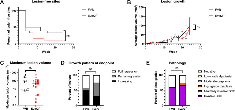
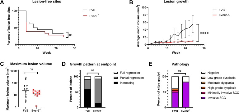
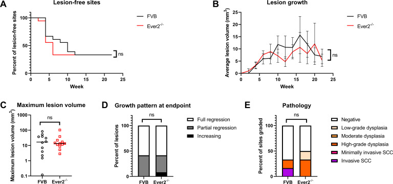
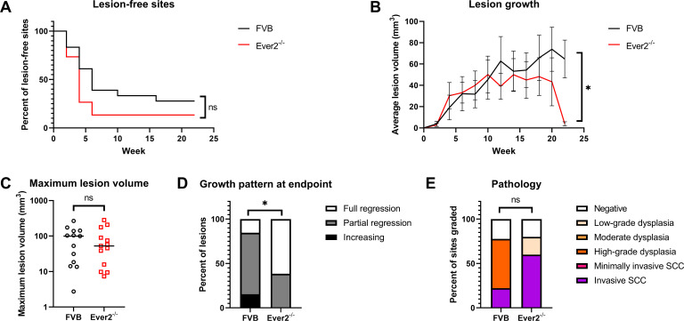
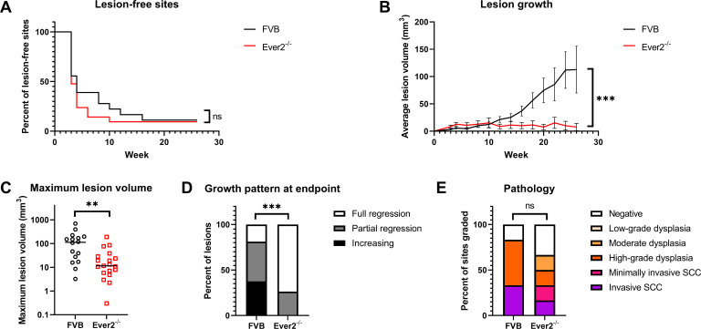
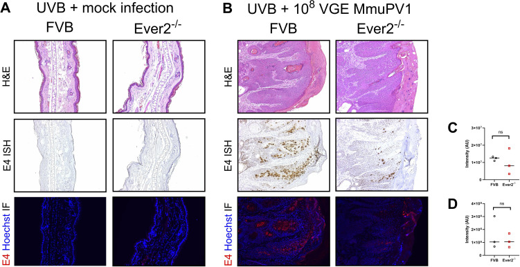
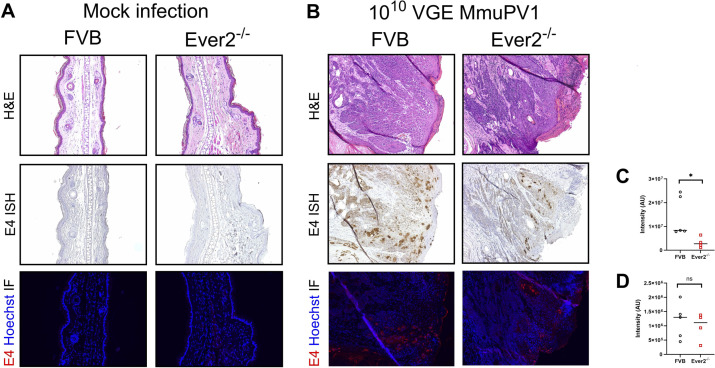
Epidermodysplasia verruciformis (EV) is a rare genetic skin disorder that is characterized by the development of papillomavirus-induced skin lesions that can progress to squamous cell carcinoma (SCC). Certain high-risk, cutaneous β-genus human papillomaviruses (β-HPVs), in particular HPV5 and HPV8, are associated with inducing EV in individuals who have a homozygous mutation in one of three genes tied to this disease: EVER1, EVER2, or CIB1. EVER1 and EVER2 are also known as TMC6 and TMC8, respectively. Little is known about the biochemical activities of EVER gene products or their roles in facilitating EV in conjunction with β-HPV infection. To investigate the potential effect of EVER genes on papillomavirus infection, we pursued in vivo infection studies by infecting Ever2-null mice with mouse papillomavirus (MmuPV1). MmuPV1 shares characteristics with β-HPVs including similar genome organization, shared molecular activities of their early, E6 and E7, oncoproteins, the lack of a viral E5 gene, and the capacity to cause skin lesions that can progress to SCC. MmuPV1 infections were conducted both in the presence and absence of UVB irradiation, which is known to increase the risk of MmuPV1-induced pathogenesis. Infection with MmuPV1 induced skin lesions in both wild-type and Ever2-null mice with and without UVB. Many lesions in both genotypes progressed to malignancy, and the disease severity did not differ between Ever2-null and wild-type mice. However, somewhat surprisingly, lesion growth and viral transcription was decreased, and lesion regression was increased in Ever2-null mice compared with wild-type mice. These studies demonstrate that Ever2-null mice infected with MmuPV1 do not exhibit the same phenotype as human EV patients infected with β-HPVs.IMPORTANCEHumans with homozygous mutations in the EVER2 gene develop epidermodysplasia verruciformis (EV), a disease characterized by predisposition to persistent β-genus human papillomavirus (β-HPV) skin infections, which can progress to skin cancer. To investigate how EVER2 confers protection from papillomaviruses, we infected the skin of homozygous Ever2-null mice with mouse papillomavirus MmuPV1. Like in humans with EV, infected Ever2-null mice developed skin lesions that could progress to cancer. Unlike in humans with EV, lesions in these Ever2-null mice grew more slowly and regressed more frequently than in wild-type mice. MmuPV1 transcription was higher in wild-type mice than in Ever2-null mice, indicating that mouse EVER2 does not confer protection from papillomaviruses. These findings suggest that there are functional differences between MmuPV1 and β-HPVs and/or between mouse and human EVER2.

Keywords: EVER2; MmuPV1; TMC8; epidermodysplasia verruciformis; papillomavirus.


Conflict of interest statement

The authors declare no conflict of interest.

Figures

Fig 1
Fig 1
Lesion characteristics in Ever2-null and wild-type mice infected with 108 VGE MmuPV1 and treated with UVB. Ear and tail sites were wounded to induce scarification and topically treated with MmuPV1 virions, then 24 hours later irradiated with UVB. Onset of overt lesions was monitored, and lesions were measured every other week over a period of 24 weeks postinfection. (A) Onset of lesions did not differ between Ever2-null and wild-type mice (P = 0.090, log-rank test, n = 18 infected sites per genotype). (B) Lesion volume did not differ between Ever2-null and wild-type mice (P = 0.13, type 3 test of interaction effect of Genotype × Week in a quadratic mixed model, n = 12–16 lesions per genotype). (C) Maximum lesion volume did not differ between Ever2-null and wild-type mice (P = 0.77, Wilcoxon rank-sum test, n = 12–16 lesions per genotype). (D) Lesion regression did not differ between Ever2-null and wild-type mice (P = 0.84, Wilcoxon rank-sum test, n = 12–16 lesions per genotype). (E) Disease severity did not differ between Ever2-null and wild-type mice (P = 0.60, Wilcoxon rank-sum test, n = 15–18 infected sites scored per genotype).
Fig 2
Fig 2
Lesion characteristics in Ever2-null and wild-type mice infected with 107 VGE MmuPV1 and treated with UVB. Ear and tail sites were wounded to induce scarification and topically treated with MmuPV1 virions, then 24 hours later irradiated with UVB. Onset of overt lesions was monitored, and lesions were measured every other week over a period of 24 weeks postinfection. (A) Onset of lesions did not differ between Ever2-null and wild-type mice (P = 0.39, log-rank test, n = 18 infected sites per genotype). (B) Lesion volume was lower in Ever2-null mice than in wild-type mice (P < 0.0001, type 3 test of interaction effect of Genotype × Week in a linear mixed model, n = 12–14 lesions per genotype). (C) Maximum lesion volume did not differ between Ever2-null and wild-type mice (P = 0.40, Wilcoxon rank-sum test, n = 12–14 lesions per genotype). (D) Lesion regression did not differ between Ever2-null and wild-type mice (P = 0.059, Wilcoxon rank-sum test, n = 12–14 lesions per genotype). (E) Disease severity did not differ between Ever2-null and wild-type mice (P = 0.075, Wilcoxon rank-sum test, n = 18 infected sites scored per genotype).
Fig 3
Fig 3
Lesion characteristics in Ever2-null and wild-type mice infected with 108 VGE MmuPV1 without irradiation. Ear and tail sites were wounded to induce scarification and topically treated with MmuPV1 virions. Onset of overt lesions was monitored, and lesions were measured every other week over a period of 22 weeks postinfection. (A) Onset of lesions did not differ between Ever2-null and wild-type mice (P = 0.64, log-rank test, n = 18 infected sites per genotype). (B) Lesion volume did not differ between Ever2-null and wild-type mice (P = 0.53, type 3 test of interaction effect of Genotype × Week in a quadratic mixed model, n = 12 lesions per genotype). (C) Maximum lesion volume did not differ between Ever2-null and wild-type mice (P = 0.98, Wilcoxon rank-sum test, n = 12 lesions per genotype). (D) Lesion regression did not differ between Ever2-null and wild-type mice (P > 0.99, Wilcoxon rank-sum test, n = 12 lesions per genotype). (E) Disease severity did not differ between Ever2-null and wild-type mice (P > 0.99, Wilcoxon rank-sum test, n = 6 infected sites scored per genotype).
Fig 4
Fig 4
Lesion characteristics in Ever2-null and wild-type mice infected with 109 VGE MmuPV1 without irradiation. Ear and tail sites were wounded to induce scarification and topically treated with MmuPV1 virions. Onset of overt lesions was monitored, and lesions were measured every other week over a period of 22 weeks postinfection. (A) Onset of lesions did not differ between Ever2-null and wild-type mice (P = 0.13, log-rank test, n = 15–18 infected sites per genotype). (B) Lesions grew more slowly in Ever2-null mice than in wild-type mice (P = 0.018, type 3 test of interaction effect of Genotype × Week2 in a quadratic mixed model, n = 13 lesions per genotype). (C) Maximum lesion volume did not differ between Ever2-null and wild-type mice (P = 0.69, Wilcoxon rank-sum test, n = 13 lesions per genotype). (D) Lesion regression was higher in Ever2-null mice than in wild-type mice (P = 0.021, Wilcoxon rank-sum test, n = 13 lesions per genotype). (E) Disease severity did not differ between Ever2-null and wild-type mice (P = 0.57, Wilcoxon rank-sum test, n = 5–9 infected sites scored per genotype).
Fig 5
Fig 5
Lesion characteristics in Ever2-null and wild-type mice infected with 1010 VGE MmuPV1 without irradiation. Ear and tail sites were wounded to induce scarification and topically treated with MmuPV1 virions. Onset of overt lesions was monitored, and lesions were measured every other week over a period of 26 weeks postinfection. (A) Onset of lesions did not differ between Ever2-null and wild-type mice (P = 0.43, log-rank test, n = 18–21 infected sites per genotype). (B) Lesions grew more slowly in Ever2-null mice than in wild-type mice (P = 0.0003, type 3 test of interaction effect of Genotype × Week2 in a quadratic mixed model, n = 16–19 lesions per genotype). (C) Maximum lesion volume was lower in Ever2-null mice than in wild-type mice (P = 0.0018, Wilcoxon rank-sum test, n = 16–19 lesions per genotype). (D) Lesion regression was higher in Ever2-null mice than in wild-type mice (P = 0.0003, Wilcoxon rank-sum test, n = 16–19 lesions per genotype). (E) Disease severity did not differ between Ever2-null and wild-type mice (P = 0.58, Wilcoxon rank-sum test, n = 6 infected sites scored per genotype).
Fig 6
Fig 6
MmuPV1 biomarkers in lesions from Ever2-null and wild-type mice infected with 108 VGE MmuPV1 and treated with UVB. (A) Normal tissues in mock-infected Ever2-null and wild-type mice irradiated with UVB. Serial sections of tissues stained with H&E (top row), MmuPV1 E4 RNAscope ISH (brown, middle row), and MmuPV1 E4 IF (red, bottom row). (B) Lesional tissue in MmuPV1-infected wild-type mice and Ever2-null mice irradiated with UVB. Depicted examples represent high-grade dysplasia with minimally invasive carcinoma seen at the base of the squamous epithelium in both genotypes. Serial sections stained with H&E (top row), MmuPV1 E4 RNAscope ISH (brown, middle row), and MmuPV1 E4 IF (red, bottom row). (C) E4 transcript staining intensity did not differ between Ever2-null and wild-type mice (P = 0.70, Wilcoxon rank-sum test, n = 3 lesions per genotype). (D) E4 protein staining intensity did not differ between Ever2-null and wild-type mice (P > 0.99, Wilcoxon rank-sum test, n = 3 lesions per genotype).
Fig 7
Fig 7
MmuPV1 biomarkers in lesions from Ever2-null and wild-type mice infected with 1010 VGE MmuPV1 without irradiation. (A) Normal tissues in mock-infected Ever2-null and wild-type mice. Serial sections of tissues stained with H&E (top row), MmuPV1 E4 RNAscope ISH (brown, middle row), and MmuPV1 E4 IF (red, bottom row). (B) Minimally invasive squamous cell carcinoma in the background of high-grade dysplasia in MmuPV1-infected Ever2-null and wild-type mice. Serial sections stained with H&E (top row), MmuPV1 E4 RNAscope ISH (brown, middle row), and MmuPV1 E4 IF (red, bottom row). (C) E4 transcript staining intensity was lower in Ever2-null mice than in wild-type mice (P = 0.016, Wilcoxon rank-sum test, n = 4–5 lesions per genotype). (D) E4 protein staining intensity did not differ between Ever2-null and wild-type mice (P = 0.56, Wilcoxon rank-sum test, n = 4–5 lesions per genotype).

Similar articles

Cited by

References

    1. Jablonska S, Orth G. 1985. Epidermodysplasia verruciformis. Clin Dermatol 3:83–96. doi:10.1016/0738-081x(85)90052-5 - DOI - PubMed
    1. Majewski S, Jabłońska S. 1995. Epidermodysplasia verruciformis as a model of human papillomavirus-induced genetic cancer of the skin. Arch Dermatol 131:1312–1318. - PubMed
    1. Orth G. 2006. Genetics of epidermodysplasia verruciformis: insights into host defense against papillomaviruses. Semin Immunol 18:362–374. doi:10.1016/j.smim.2006.07.008 - DOI - PubMed
    1. McLaughlin-Drubin ME. 2015. Human papillomaviruses and non-melanoma skin cancer. Semin Oncol 42:284–290. doi:10.1053/j.seminoncol.2014.12.032 - DOI - PubMed
    1. Orth G. 1986. Epidermodysplasia verruciformis: a model for understanding the oncogenicity of human papillomaviruses, p 157–174. In Ciba foundation symposium 120 - papillomaviruses. John Wiley & Sons, Ltd. - PubMed

MeSH terms

Substances