. 2025 Jan 1;28(1):79-87.
doi: 10.1227/ons.0000000000001226. Epub 2024 Jun 13.

Feasibility of Endovascular Deep Brain Stimulation of Anterior Nucleus of the Thalamus for Refractory Epilepsy

Affiliations

Feasibility of Endovascular Deep Brain Stimulation of Anterior Nucleus of the Thalamus for Refractory Epilepsy

Varun Kashyap et al. Oper Neurosurg. .

Abstract

Background and objectives: Deep brain stimulation (DBS) has developed into an effective therapy for several disease states including treatment-resistant Parkinson disease and medically intractable essential tremor, as well as segmental, generalized and cervical dystonia, and obsessive-compulsive disorder (OCD). Dystonia and OCD are approved with Humanitarian Device Exemption. In addition, DBS is also approved for the treatment of epilepsy in the anterior nucleus of the thalamus. Although overall considered an effective treatment for Parkinson disease and epilepsy, a number of specific factors determine the treatment success for DBS including careful patient selection, effective postoperative programming of DBS devices and accurate electrode placement. Furthermore, invasiveness of the procedure is a rate limiter for patient adoption. It is desired to explore a less invasive way to deliver DBS therapy.

Methods: Here, we report for the first time the direct comparison of endovascular and parenchymal DBS in a triplicate ovine model using the anterior nucleus of the thalamus as the parenchymal target for refractory epilepsy.

Results: Triplicate ovine studies show comparable sensing resolution and stimulation performance of endovascular DBS with parenchymal DBS.

Conclusion: The results from this feasibility study opens up a new frontier for minimally invasive DBS therapy.


Figures

FIGURE 1.
FIGURE 1.
Ovine NvDBS lead implantation. Figure shows dorsal view of sheep's head, brain, cerebral veins, brain, and cerebral veins visible through transparent external anatomy. Pathway of endovascular catheter visible—(Jugular Vein -> Maxillary Vein -> Transverse Sinus -> confluence of Sinuses -> Straight Sinus -> Vein of Galen -> Venous Angle -> Thalamostriate Vein). Inset shows close-up of lateral ventricles (dorsal view). Deployed electrodes are visible inside transparent thalamostriate vein over ANT. ANT, anterior nucleus of the thalamus; NvDBS, neurovascular deep brain stimulation.
FIGURE 2.
FIGURE 2.
NvDBS device features and navigation to the thalamostriate vein. A, Low metal coverage device for NvDBS. B, Contrast run showing the thalamostriate vein in both the anteroposterior (AP) and lateral views. C, Illustrations of the relevant intracranial venous vasculature with respect to the parenchymal structures with parenchymal DBS leads implanted in the ANT. D, Final placement of parenchymal and NvDBS leads. E, Relative positions of parenchymal and NvDBS leads in AP and lateral views that may correspond to distance-related performance. ANT, anterior nucleus of the thalamus; DBS, deep brain stimulation; HC, hippocampus; NvDBS, neurovascular deep brain stimulation; Pt-Ir, platinum iridium.
FIGURE 3.
FIGURE 3.
Preclinical evoked potentials. A, Representative evoked potentials elicited by ramped parenchymal and NvDBS stimuli at ANT, sensing from HC parenchymal electrodes. B, Growth curves for parenchymal and endovascular stimulation in 3 ovine subjects. C, Representative evoked potentials elicited by ramped parenchymal stimuli at HC, sensing at parenchymal and NvDBS electrodes at ANT, respectively. D, Growth curves for parenchymal and endovascular sensing in 3 ovine subjects. Green markers indicate 5 uVp (where Vp—peak voltage) evoked amplitude (above baseline) in each test condition. ANT, anterior nucleus of the thalamus; DBS, deep brain stimulation; HC, hippocampus; NvDBS, neurovascular deep brain stimulation.
FIGURE 4.
FIGURE 4.
In vitro LFP analysis A, Signal attenuation as a function of radial distance is shown. 80% signal attenuation is observed at 3.5 mm distance from the neural target. The inset shows NvDBS lead placed within a synthetic vessel (syndaver) with a sense electrode being moved for distance variations. B, Signal attenuation is plotted as a function of vertical (z) distance for each electrode pair. The inset shows the NvDBS device with the electrodes annotated. LFP, local field potential; NvDBS, neurovascular deep brain stimulation.
FIGURE 5.
FIGURE 5.
E-field simulation comparison between DBS and NvDBS in multimodal imaging-based detailed anatomical model. Three possible placements and associated e-field for endovascular device, from posterior (optimal, closest to standard DBS lead) to anterior, are shown to target different regions of ANT. Coronal, sagittal view and transverse views are shown. ANT, anterior nucleus of the thalamus; DBS, deep brain stimulation; e-field, electric fields; NvDBS, neurovascular deep brain stimulation.
FIGURE 6.
FIGURE 6.
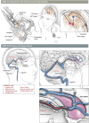
Minimally invasive DBS therapy. Conventional DBS: patient with headset showing drilling of craniotomy, patient with headset showing insertion of electrode to ANT, and patient with bilateral electrodes into ANT, bone caps, subcutaneous lead, and INS units in chest. NvDBS: lateral view of the patient's head showing cerebral veins and brain visible through transparent external anatomy (shows pathway of the catheter). Inset close-up of lateral ventricles (superior view). Deployed electrodes are visible inside transparent thalamostriate vein over ANT. ANT, anterior nucleus of the thalamus; DBS, deep brain stimulation; NvDBS, neurovascular deep brain stimulation.

References

    1. Salanova V, Witt T, Worth R, et al. Long-Term Efficacy and Safety of Thalamic Stimulation for Drug-Resistant Partial Epilepsy. Marshfield Clinic; 2015. - PMC - PubMed
    1. Li MCH, Cook MJ. Deep brain stimulation for drug-resistant epilepsy. Epilepsia. 2018;59(2):273-290. - PubMed
    1. Zhong XL, Yu JT, Zhang Q, Wang ND, Tan L. Deep brain stimulation for epilepsy in clinical practice and in animal models. Brain Res Bull. 2011;85(3-4):81-88. - PubMed
    1. Bouwens van der Vlis TAM, Schijns OEMG, Schaper FLWVJ, et al. Deep brain stimulation of the anterior nucleus of the thalamus for drug-resistant epilepsy. Neurosurg Rev. 2019;42(2):287-296. - PMC - PubMed
    1. Quraishi IH. Is MORE better? Accumulating evidence for ANT DBS in epilepsy. Epilepsy Curr. 2023;23(6):357-359. - PMC - PubMed

Grants and funding

LinkOut - more resources