. 2024 Jun 26;16(25):31997-32016.
doi: 10.1021/acsami.4c04265. Epub 2024 Jun 13.

Surface-Bioengineered Extracellular Vesicles Seeking Molecular Biotargets in Lung Cancer Cells

Affiliations

Surface-Bioengineered Extracellular Vesicles Seeking Molecular Biotargets in Lung Cancer Cells

Agata Kowalczyk et al. ACS Appl Mater Interfaces. .

Abstract

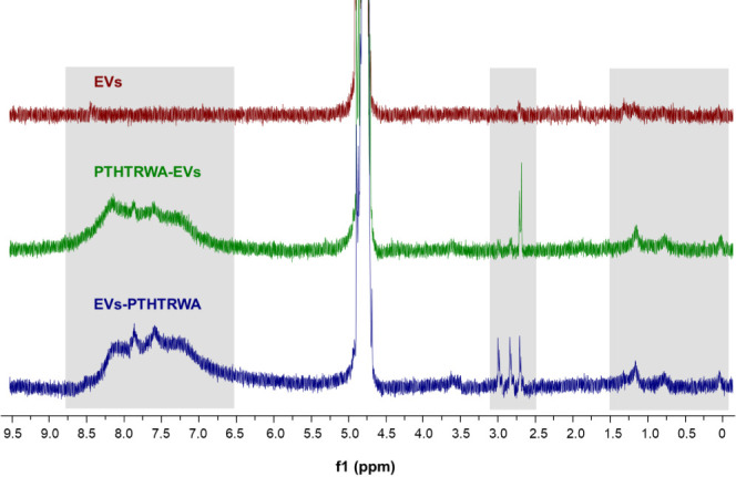
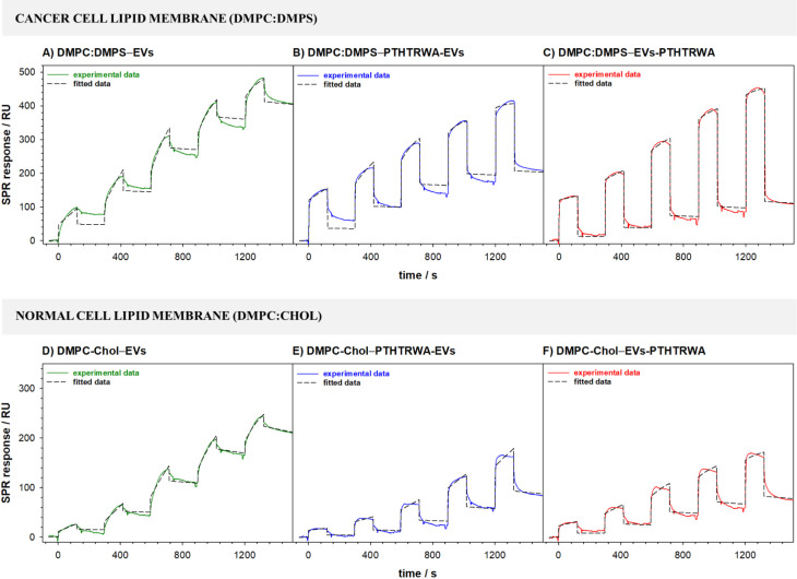
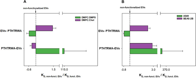
Personalized medicine is a new approach to modern oncology. Here, to facilitate the application of extracellular vesicles (EVs) derived from lung cancer cells as potent advanced therapy medicinal products in lung cancer, the EV membrane was functionalized with a specific ligand for targeting purposes. In this role, the most effective heptapeptide in binding to lung cancer cells (PTHTRWA) was used. The functionalization process of EV surface was performed through the C- or N-terminal end of the heptapeptide. To prove the activity of the EVs functionalized with PTHTRWA, both a model of lipid membrane mimicking normal and cancerous cell membranes as well as human adenocarcinomic alveolar basal epithelial cells (A549) and human normal bronchial epithelial cells (BEAS-2B) have been exposed to these bioconstructs. Magnetic resonance imaging (MRI) showed that the as-bioengineered PTHTRWA-EVs loaded with superparamagnetic iron oxide nanoparticle (SPIO) cargos reach the growing tumor when dosed intravenously in NUDE Balb/c mice bearing A549 cancer. Molecular dynamics (MD) in silico studies elucidated a high affinity of the synthesized peptide to the α5β1 integrin. Preclinical safety assays did not evidence any cytotoxic or genotoxic effects of the PTHTRWA-bioengineered EVs.

Keywords: NUDE mice; extracellular vesicle bioengineering; lung cancer cells; model lipid membrane; preclinical safety.


Conflict of interest statement

The authors declare no competing financial interest.

Figures

Figure 1
Figure 1
Scheme of the bioconjugation process of EVs with heptapeptide.
Figure 2
Figure 2
Mean size, PDI, and ZP of nonfunctionalized EVs or EVs decorated with heptapeptide dispersed in PBS buffer based on DLS studies (n = 5).
Figure 3
Figure 3
1H NMR spectra (600 MHz, D2O) of nonfunctionalized EVs (red), PTHTRWA-EVs (green), and EVs-PTHTRWA (blue). The regions of the spectra in which significant differences were observed were marked with gray frames.
Figure 4
Figure 4
TEM images of nonfunctionalized EVs (A–D), PTHTRWA-EVs (E–H), and EVs-PTHTRWA (I–L).
Figure 5
Figure 5
Possible optimized structures of heptapeptide anchored to the EV surface through its N- and C-terminus ends.
Figure 6
Figure 6
(A) The difference in the position of the υ(CH)as band is between a model membrane of a healthy (DMPC:Chol) and cancer cell (DMPC:DMPS) after interaction with appropriate EVs and the membrane. (B) The ratio of the υ(CH)as band absorbance for the membrane after the interaction with nonfunctionalized and functionalized EVs (EVs, PTHTRWA-EVs, and EVs-PTHTRWA) and the model system of cell membranes of a healthy or cancer cell.
Figure 7
Figure 7
Deconvoluted spectra of the C=O bond for the membrane DMPC:DMPS, what represents the cancer cell membrane (A), and membrane after interaction with nonfunctionalized (B) and appropriately functionalized EVs (C and D).
Figure 8
Figure 8
Deconvoluted spectra of the C=O bond for the membrane DMPC:Chol, which represents the normal cell membrane (A), and membrane after interaction with nonfunctionalized (B) and appropriately functionalized EVs (C and D).
Figure 9
Figure 9
Sensorgrams recorded during the interactions of nonfunctionalized and appropriately functionalized EVs with cancer (A–C) and normal (D–F) cell lipid membranes. Experimental conditions: 0.01 M PBST-Gibco (pH 7.4), CEVs: 0.16–1.60 pM (1.0 × 10⁸–1.0 × 10⁹ particles·mL–1).
Figure 10
Figure 10
Ratio of KD values for nonfunctionalized EVs and EVs functionalized with heptapeptide for the interaction with model membranes (A) and A549 and BEAS-2B cells (B).
Figure 11
Figure 11
Scheme of the interaction of PTHTRWA-EVs with model cancer and a normal cell lipid membrane.
Figure 12
Figure 12
Sensorgrams recorded during the interactions of nonfunctionalized and appropriately functionalized EVs with human adenocarcinomic alveolar basal epithelial (A–C) and human normal bronchial epithelial (D–F) cells. Experimental conditions: 0.01 M PBST- Gibco (pH 7.4), CEVs: 0.16–1.60 pM (1.0 × 10⁸–1.0 × 10⁹ particles·mL–1).
Figure 13
Figure 13
Uptake of EVs and PTHTRWA-EVs by A549 and BEAS-2B cells. Cells were exposed to DiOC18(3)-labeled EVs or PTHTRWA-EVs (both shown in green) for 2 h, fixed, and counterstained with wheat germ agglutinin-Alexa Fluor 647 (magenta) to visualize the cell membrane. EVs and PTHTRWA-EVs vesicles are internalized by A549 and Beas-2B cells (arrows). Controls (background staining control) were incubated with EV-free PBS that was treated like the EV solutions during the DiOC18(3) staining procedure. No residual dye was observed in the control-treated cells. Scalebar is 20 μm.
Figure 14
Figure 14
Binding mode of PTHTRWA to α5β1 integrin headpiece resulting from MD simulation. (A) Hydrogen bond surface of heptapeptide with α5β1. The hydrogen donor is presented using a pink color, while the hydrogen acceptor is presented as a lime surface. (B) The hydrophobic and hydrophilic amino acid residues surrounding the heptapeptide. Surface hydrophobicity is depicted using brown color—the hydrophobic and blue color—the lipophilic regions. (C) 2D view of all α5β1 residue interacting with the heptapeptide (residues involved in hydrogen bonds indicated as green and cyan circles; in hydrophobic interactions indicated as pink circles and electrostatic interactions indicated as orange circles).
Figure 15
Figure 15
Predicted binding mode of N-terminated heptapeptide (6-AHA-PTHTRWA) and C-terminated heptapeptide (PTHTRWA-3-APA) to the α5β1 integrin headpiece resulting from MD simulation. (A) Superposition of compounds: PTHTRWA (C atoms shown in green), 6-AHA-PTHTRWA (C atoms shown in turquoise and orange), and PTHTRWA-3-APA (C atoms shown in pink and orange). (B) Hydrogen bond surface of PTHTRWA-3-APA with α5β1. (C) Hydrogen bond surface of 6-AHA-PTHTRWA with α5β1. The hydrogen donor is presented using the pink color, while the hydrogen acceptor is presented as a lime surface. (D) Molecular interactions between PTHTRWA-3-APA and α5β1. (E) Molecular interactions between 6-AHA-PTHTRWA and α5β1.
Figure 16
Figure 16
Representative T2-images of NUDE Balb/c mice bearing human lung A549 cancer. Mice were imaged before (left/top panel) and 1 h (right/top panel), 18 h (left/down panel), and 24 h (right/down panel) after intravenous injection (0.2 mg·mL–1) of SPIO-loaded PTHTRWA-EVs into the tail vain. In the tumor, numerous dark areas were appeared at 18 and 24 h postinjection. This was associated with decreased relaxation times (T2) in postinjected mice. MRI was performed using the Turbo RARE sequence (TR 4500 ms, TE 30 ms, FA 180.0 deg, TA 19 m 12 s, FOV 3.20 cm, MTX 256) in the axial plane. There is a clear blackening of the tumor areas accompanied by decreased signal intensities on T2-weighted images observed at the 18 and 24 h postinjection.
Figure 17
Figure 17
Effect of extracellular vesicles derived from lung cancer cells on lung cancerous and noncancerous cell growth and function assessed by electric cell–substrate impedance sensing. A549 cells (A, B), BEAS-2B cells (C, D). EVs – pristine extracellular vesicles; PTHTRWA-EVs – bioengineered extracellular vesicles. Cell index was normalized to the baseline control. EVs and PTHTRWA-EVs are expressed as particles·mL–1 unit.

References

    1. Song Y.; Kim Y.; Ha S.; Sheller-Miller S.; Yoo J.; Choi C.; Park C. H. The Emerging Role of Exosomes as Novel Therapeutics: Biology, Technologies, Clinical Applications, and the Next. Am. J. Reprod. Immunol. 2021, 85 (2), e1332910.1111/aji.13329. - DOI - PMC - PubMed
    1. Colombo M.; Moita C.; Van Niel G.; Kowal J.; Vigneron J.; Benaroch P.; Manel N.; Moita L. F.; Théry C.; Raposo G. Analysis of ESCRT Functions in Exosome Biogenesis, Composition and Secretion Highlights the Heterogeneity of Extracellular Vesicles. J. Cell Sci. 2013, 126 (24), 5553–5565. 10.1242/jcs.128868. - DOI - PubMed
    1. Wang Z. -G.; He Z. -Y.; Liang S.; Yang Q.; Cheng P.; Chen A. -M. Comprehensive Proteomic Analysis of Exosomes Derived from Human Bone Marrow, Adipose Tissue, and Umbilical Cord Mesenchymal Stem Cells. Stem Cell Res. Ther. 2020, 11 (1), 511.10.1186/s13287-020-02032-8. - DOI - PMC - PubMed
    1. Chen H.; Wang L.; Zeng X.; Schwarz H.; Nanda H. S.; Peng X.; Zhou Y. Exosomes, a New Star for Targeted Delivery. Front. Cell Dev. Biol. 2021, 9, 751079.10.3389/fcell.2021.751079. - DOI - PMC - PubMed
    1. Qiu H.; Shi S.; Wang S.; Peng H.; Ding S. J.; Wang L. Proteomic Profiling Exosomes from Vascular Smooth Muscle Cell. Proteomics: Clin. Appl. 2018, 12 (5), e170009710.1002/prca.201700097. - DOI - PMC - PubMed