. 2024 Jan-Dec:38:3946320241254083.
doi: 10.1177/03946320241254083.

Corilagin relieves atherosclerosis via the toll-like receptor 4 signaling pathway in vascular smooth muscle cells

Affiliations

Corilagin relieves atherosclerosis via the toll-like receptor 4 signaling pathway in vascular smooth muscle cells

Yujie Wang et al. Int J Immunopathol Pharmacol. 2024 Jan-Dec.

Abstract

Introduction: Corilagin possesses a diverse range of pharmacologic bioactivities. However, the specific protective effects and mechanisms of action of corilagin in the context of atherosclerosis remain unclear. In this study, we investigated the impact of corilagin on the toll-like receptor (TLR)4 signaling pathway in a mouse vascular smooth muscle cell line (MOVAS) stimulated by oxidized low-density lipoprotein (ox-LDL). Additionally, we examined the effects of corilagin in Sprague-Dawley rats experiencing atherosclerosis.

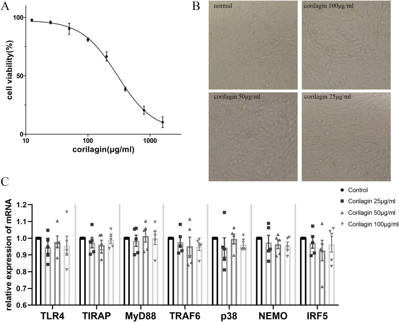
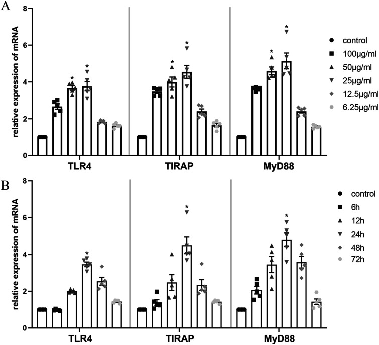
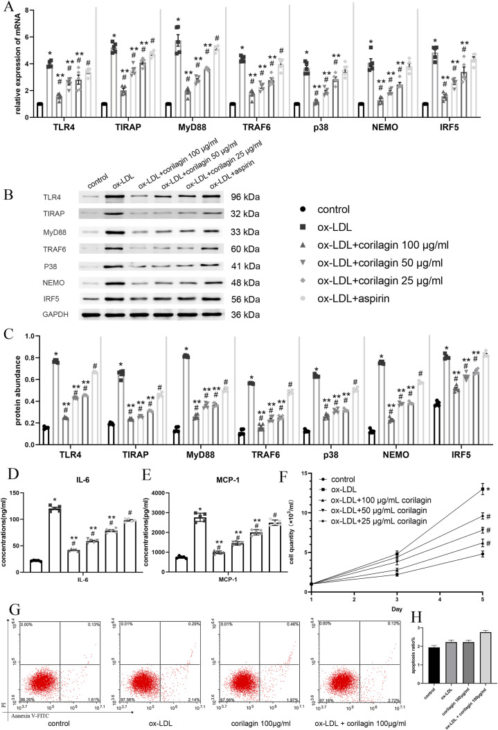
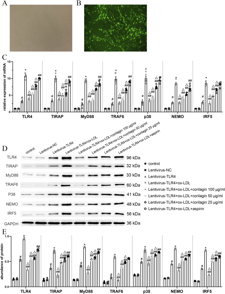
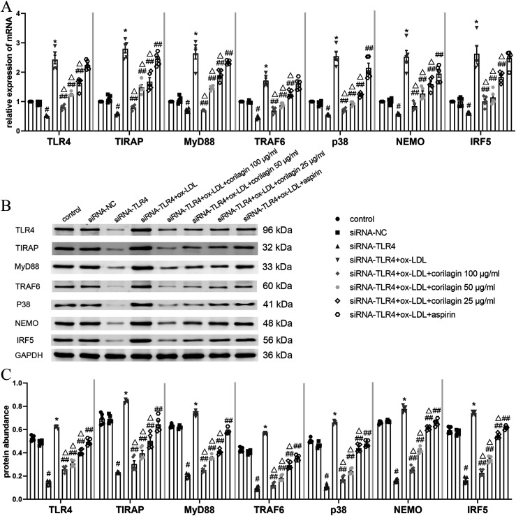
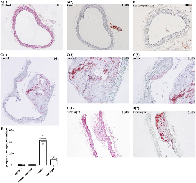
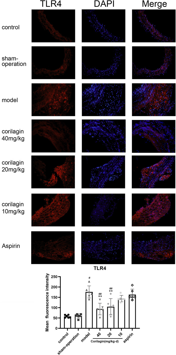
Methods: The cytotoxicity of corilagin was assessed using the CCK8 assay. MOVAS cells, pre-incubated with ox-LDL, underwent treatment with varying concentrations of corilagin. TLR4 expression was modulated by either downregulation through small interfering (si)RNA or upregulation via lentivirus transfection. Molecular expression within the TLR4 signaling pathway was analyzed using real-time polymerase chain reaction (PCR) and Western blotting. The proliferation capacity of MOVAS cells was determined through cell counting. In a rat model, atherosclerosis was induced in femoral arteries using an improved guidewire injury method, and TLR4 expression in plaque areas was assessed using immunofluorescence. Pathological changes were examined through hematoxylin and eosin staining, as well as Oil-Red-O staining.

Results: Corilagin demonstrated inhibitory effects on the TLR4 signaling pathway in MOVAS cells pre-stimulated with ox-LDL, consequently impeding the proliferative impact of ox-LDL. The modulation of TLR4 expression, either through downregulation or upregulation, similarly influenced the expression of downstream molecules. In an in vivo context, corilagin exhibited the ability to suppress TLR4 and MyD88 expression in the plaque lesion areas of rat femoral arteries, thereby alleviating the formation of atherosclerotic plaques.

Conclusion: Corilagin can inhibit the TLR4 signaling pathway in VSMCs, possibly by downregulating TLR4 expression and, consequently, relieving atherosclerosis.

Keywords: atherosclerosis; corilagin; ox-LDL; toll-like receptor 4; vascular smooth muscle cell.


Conflict of interest statement

Declaration of conflicting interestsThe author(s) declared no potential conflicts of interest with respect to the research, authorship, and/or publication of this article.

Figures

Figure 1.
Figure 1.
Cytotoxicity of corilagin (a) determined using the CCK8 assay for MOVAS cell viability after treatment with various concentrations of corilagin for 24 h. The equation for the dose–effect curve was Y = (94.54/(1 + 10^ (log X − 2.475))) + 3.035 (IC50 = 298.7 μg/mL. (b) Morphology of MOVAS cells after corilagin treatment for 24 h. (c) mRNA expression was measured by qRT-PCR. Data are presented as mean ± SEM. No significant difference was found between groups, as determined by one-way ANOVA and subsequent Student–Newman–Keuls q-test (n = 5).
Figure 2.
Figure 2.
Effect of ox-LDL on the TLR4 signaling pathway in MOVAS cells. (a) mRNA expression of TLR4, TIRAP and MyD88 was measured by qRT-PCR. *p < .05 compared with other concentrations of ox-LDL groups or control group as determined by one-way ANOVA and subsequent Student–Newman–Keuls q-test (n = 5). (b) *p < .05 compared with either time point or control groups determined by one-way ANOVA and subsequent Student–Newman–Keuls q-test (n = 5).
Figure 3.
Figure 3.
Effect of corilagin on the TLR4 signaling pathway in MOVAS cells stimulated by ox-LDL. (a) mRNA expression of TLR4, TIRAP, MyD88, TRAF6, p38, NEMO and IRF5 was measured by qRT-PCR. *p < .05 compared with the control group, #p < .05 compared with the ox-LDL group, **p < .05 compared with the aspirin group as determined by one-way ANOVA and subsequent Student–Newman–Keuls q-test (n = 5). (B. C) Protein abundance was measured by western blotting. *p < .05 compared with the control group, #p < .05 compared with the ox-LDL group, **p < .05 compared with the aspirin group, as determined by one-way ANOVA and subsequent Student–Newman–Keuls q-test (n = 5). (D, E) Abundance of IL-6 and MCP-1 in cell culture supernatant was measured by ELISAs. *p < .05 compared with the control group, #p < .05 compared with the ox-LDL group, **p < .05 compared with the aspirin group as determined by one-way ANOVA and subsequent Student–Newman–Keuls q-test (n = 5). (F) Effect of corilagin on the MOVAS cells proliferation. *p < .05 compared with the control group, #p < .05 compared with the ox-LDL group determined by one-way ANOVA and subsequent Student–Newman–Keuls q-test (n = 5). (G, H) apoptosis ratio of MOVAS were detected by Annexin V staining. No significant difference was found among four groups determined by one-way ANOVA and subsequent Student–Newman–Keuls q-test (n = 5).
Figure 4.
Figure 4.
Effect of corilagin on the TLR4 signaling pathway after upregulation of TL4 expression in MOVAS cells stimulated by ox-LDL. (A. B) 72 hours after transfection of the lentiviral vector, >85% of MOVAS cells expressed green fluorescent protein as observed with a fluorescence microscope. (C) mRNA expression was measured by qRT-PCR. #p < .05 compared with the control group, *p < .05 compared with the lentivirus-TLR4 group, ##p < .05 compared with the model group (lentivirus-TLR4 + ox-LDL), △p < .05 compared with the aspirin group, as determined by one-way ANOVA and Student–Newman–Keuls q-test (n = 5). (D. E) Protein abundance was measured by western blotting. #p < .05 compared with the control group, *p < .05 compared with the lentivirus-TLR4 group, ##p < .05 compared with the model group, △p < .05 compared with the aspirin group, as determined by one-way ANOVA and subsequent Student–Newman–Keuls q-test (n = 5).
Figure 5.
Figure 5.
Effect of corilagin on the TLR4 signaling pathway after downregulation of TLR4 expression in MOVAS cells stimulated by ox-LDL. (A) mRNA expression of molecules in the TLR4 signaling pathway was measured by qRT-PCR. #p < .05 compared with the control group, *p < .05 compared with the siRNA-TLR4 group, ##p < .05 compared with the model group (siRNA-TLR4 + ox-LDL), △p < .05 compared with the aspirin group, as determined by one-way ANOVA and subsequent Student–Newman–Keuls q-test (n = 5). (B. C) Protein abundance was measured by western blotting. #p < .05 compared with the control group, *p < .05 compared with the siRNA-TLR4 group, ##p < .05 compared with the model group, △p < .05 compared with the aspirin group, as determined by one-way ANOVA and subsequent Student–Newman–Keuls q-test (n = 5). siRNA-TLR4: TLR4 was knocked down in MOVAS cells by siRNA. siRNA-NC: siRNA-negative control group.
Figure 6.
Figure 6.
(A1-2) HE and O-R-O staining of one femoral artery in the control group viewed at 100x magnification; (B) O-R-O staining of one femoral artery in the sham-operation group viewed at 100x magnification; (C1-3) O-R-O staining of one femoral artery in the model group. C1 was viewed at 40x magnification and C2 and C3 were viewed at 200x magnification. (D1-2) HE and O-R-O staining of one femoral artery in the corilagin group viewed at 200 times magnification.
Figure 7.
Figure 7.
Effect of corilagin on TLR4 in atheromatous plaque in the femoral arteries of rats. #p < .05 compared with the control group, **p < .05 compared with the model group, ##p < .05 compared with the aspirin group, as determined by one-way ANOVA and subsequent Student–Newman–Keuls q-test (n = 6).

Similar articles

Cited by

References

    1. Hiramoto JS, Teraa M, de Borst GJ, et al. (2018) Interventions for lower extremity peripheral artery disease. Nat Rev Cardiol 15(6): 332–350. DOI: 10.1038/s41569-018-0005-0. - DOI - PubMed
    1. Lusis AJ. (2000) Atherosclerosis. Nature 407(6801): 233–241. DOI: 10.1038/35025203. - DOI - PMC - PubMed
    1. Reinecke H, Unrath M, Freisinger E, et al. (2015) Peripheral arterial disease and critical limb ischaemia: still poor outcomes and lack of guideline adherence. Eur Heart J 36(15): 932–938. DOI: 10.1093/eurheartj/ehv006. - DOI - PubMed
    1. Owens GK, Kumar MS, Wamhoff BR. (2004) Molecular regulation of vascular smooth muscle cell differentiation in development and disease. Physiol Rev 84(3): 767–801. DOI: 10.1152/physrev.00041.2003. - DOI - PubMed
    1. Alexander MR, Owens GK. (2012) Epigenetic control of smooth muscle cell differentiation and phenotypic switching in vascular development and disease. Annu Rev Physiol 74: 13–40. DOI: 10.1146/annurev-physiol-012110-142315. - DOI - PubMed

MeSH terms