. 2024 Jun 18;121(25):e2317285121.
doi: 10.1073/pnas.2317285121. Epub 2024 Jun 13.

A highly reproducible and efficient method for retinal organoid differentiation from human pluripotent stem cells

Affiliations

A highly reproducible and efficient method for retinal organoid differentiation from human pluripotent stem cells

Jade Harkin et al. Proc Natl Acad Sci U S A. .

Abstract

Human pluripotent stem cell (hPSC)-derived retinal organoids are three-dimensional cellular aggregates that differentiate and self-organize to closely mimic the spatial and temporal patterning of the developing human retina. Retinal organoid models serve as reliable tools for studying human retinogenesis, yet limitations in the efficiency and reproducibility of current retinal organoid differentiation protocols have reduced the use of these models for more high-throughput applications such as disease modeling and drug screening. To address these shortcomings, the current study aimed to standardize prior differentiation protocols to yield a highly reproducible and efficient method for generating retinal organoids. Results demonstrated that through regulation of organoid size and shape using quick reaggregation methods, retinal organoids were highly reproducible compared to more traditional methods. Additionally, the timed activation of BMP signaling within developing cells generated pure populations of retinal organoids at 100% efficiency from multiple widely used cell lines, with the default forebrain fate resulting from the inhibition of BMP signaling. Furthermore, given the ability to direct retinal or forebrain fates at complete purity, mRNA-seq analyses were then utilized to identify some of the earliest transcriptional changes that occur during the specification of these two lineages from a common progenitor. These improved methods also yielded retinal organoids with expedited differentiation timelines when compared to traditional methods. Taken together, the results of this study demonstrate the development of a highly reproducible and minimally variable method for generating retinal organoids suitable for analyzing the earliest stages of human retinal cell fate specification.

Keywords: organoid; retina; stem cell.


Conflict of interest statement

Competing interests statement:J.H. and J.S.M. have filed a patent related to the methodology described in this paper.

Figures
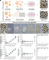
Fig. 1.
Fig. 1.
Reproducibility of early-stage cellular aggregates. (A and B) Schematic of traditional and standardized methods of differentiation. The schematic shows representative images of cellular aggregates differentiated until Day 6 using the traditional and standardized methods. (C) Representative images of cellular aggregates differentiated until Day 3 using the traditional method. (DI) Representative images of cellular aggregates at different densities (250, 500, 1,000, 2,000, 4,000, and 8,000 cells per well) differentiated until Day 3 using the standardized method. (JO) Compared to the traditional method, the standardized method is highly reproducible generating cellular aggregates that are more consistent in both their size and circularity at both Day 3 and Day 6. (Scale bars equal 500 μm (AC).) The scale bar in (C) applies to (DI). n = 3 biological replicates for each cell line.
Fig. 2.
Fig. 2.
Establishing the appropriate cell density for efficient retinal organoid formation. (A) Schematic demonstrating the process of establishing standardized cellular aggregates of defined cell densities for the subsequent differentiation of retinal organoids. (B) Quantification of retinal organoid differentiation efficiency based upon the size of original cell aggregates at Day 16, as determined by the expression of a SIX6:GFP reporter (n = 4 biological replicates). (CT) Representative images at Day 16 by brightfield microscopy, fluorescence microscopy for SIX6:GFP expression, as well as merged images for each starting cell density. The yellow arrowhead indicates an aggregate that flattened out and failed to differentiate into retinal lineage. White arrowheads indicate aggregates that remained three-dimensional but did not differentiate into retinal lineage. The scale bar equals 1,000 μm.
Fig. 3.
Fig. 3.
Highly efficient differentiation of retinal organoids via standardized methods. (AL) Brightfield and fluorescent images of organoids at Day 25 differentiated using the SIX6-GFP reporter cell line by either the traditional method or the standardized method. (A, B, and M) Organoids differentiated using the traditional method without BMP4 treatment generated retinal organoids at an efficiency of 38.67% ± 6.12% expressing the SIX6:GFP reporter. (C, D, and M) Organoids differentiated using the traditional method with BMP4 supplementation improved the efficiency of retinal organoid differentiation to 84.33% ± 2.91% of organoids expressing SIX6-GFP. (E, F, and M) Organoids differentiated using the standardized method without BMP4 treatment generated approximately 51.67% ± 20.93% (SEM) of organoids that express the SIX6:GFP reporter. (G, H, and M) Organoids differentiated using the standardized method with BMP4 treatment improved the efficiency of retinal organoid production to 100% based upon SIX6-GFP expression. (IM) Inhibition of BMP4 signaling with LDN-193189, using both the traditional and standardized differentiation methods, produced organoids that lacked expression SIX6-GFP, indicating an inability to differentiate toward a retinal lineage. (N and O) Quantification of the size and circularity of traditional vs. standardized methods of retinal organoid differentiation at Day 25. Error bars represent SEM (*P < 0.05, ***P < 0.001, and ****P < 0.0001). (Scale bars equal 500 μm (AL) and n = 3 biological replicates (MO).)
Fig. 4.
Fig. 4.
Reproducibility of highly efficient retinal organoid differentiation with standardized methods across hPSC lines. (AF) Organoids differentiated using the traditional method across multiple hPSC lines until Day 25. (G–L) Organoids differentiated using the standardized method across the same hPSC lines until Day 25. (AL) VSX2 is stained in green and DAPI is stained in blue. (MR) Quantification of retinal organoid differentiation efficiency demonstrating that the traditional method of differentiation generated variable yields of VSX2-positive retinal organoids depending on the cell line used, while organoids differentiated using the standardized method produced 100% VSX2-positive organoids consistently across all hPSC lines. Error bars represent SEM. (*P < 0.05, **P < 0.01, ***P < 0.001, and ****P < 0.0001). The scale bar equals 1,000 μm (AL) and n = 3 biological replicates (MR).
Fig. 5.
Fig. 5.
Standardized methods of retinal organoid differentiation allow for the analysis of transcriptional changes associated with the earliest stages of retinogenesis. (A) Schematic of organoid sample collection and RNA-seq analyses of early-stage cellular aggregates. (BG) Volcano plots showing differentially expressed genes (DEGs) and associated pathway enrichment analyses that were significantly up-regulated (blue) or down-regulated between (aquamarine). (B and C) Day 8 after treatment with BMP4 and Day 6 untreated, (D and E) Day 8 after treatment with LDN193-189 and Day 6 untreated, and (F and G) Day 8 after treatment with BMP4 and Day 8 after treatment with LDN-193189. DEGs, *Padj < 0.05, **Padj < 0.01. (Scale bars, 500 μm in (A) for Day 6 and 100 μm in (A) for Day 8 and Day 25.)
Fig. 6.
Fig. 6.
Expedited retinal ganglion cell and photoreceptor differentiation. Retinal organoids at 30 d, 60 d, and 150 d of total differentiation, derived using both the traditional and standardized methods. (A and B) Brightfield images of retinal organoids at Day 30 of differentiation that were derived using a BRN3b:GFP reporter cell line, (CF) whole sections and high magnification images of retinal organoids expressing the retinal progenitor marker VSX2 (magenta) and the retinal ganglion cell (RGC) marker BRN3b (green), identified with a BRN3b:GFP reporter. (G) qRT-PCR analyses of retinal organoids differentiated using both the traditional and standardized methods from Day 30 until Day 80 showing mRNA expression of BRN3b normalized to the Day 30 traditional method as the control. (H and I) Brightfield images of retinal organoids taken after 60 d of differentiation, (JM) whole sections and high magnification images of retinal organoids expressing the cone and rod photoreceptor marker CRX (green) and the retinal ganglion cell (RGC) marker BRN3b:tdTomato (red). (N) qRT-PCR analyses of retinal organoids differentiated using both the traditional and standardized methods from Day 30 until Day 150 showing mRNA expression of CRX normalized to the Day 30 traditional method as the control. (OR) Low-magnification and high-magnification brightfield images of retinal organoids at Day 150 showing photoreceptor outer segments. (SX) Whole sections and high magnification images of retinal organoids at 150 d showing the expression of the cone and rod photoreceptor marker CRX (red), the rod photoreceptor markers NRL (purple) and Rhodopsin (blue), as well as the cone photoreceptor marker ARR3 (green). DAPI (blue) for all images. Error bars represent SEM. (Scale bars equal 500 μm (A, B, H, I, O, and P) or 100 μm (C and D) or 200 μm (J, K, S, and T) or 50 μm (E, F, L, M, Q, and R) and 20 μm (UX). n = 3 to 6 biological replicates (G and N).

References

    1. Gonzalez-Cordero A., et al. , Recapitulation of human retinal development from human pluripotent stem cells generates transplantable populations of cone photoreceptors. Stem Cell Rep. 9, 820–837 (2017). - PMC - PubMed
    1. Hallam D., et al. , Human-induced pluripotent stem cells generate light responsive retinal organoids with variable and nutrient-dependent efficiency. Stem Cells 36, 1535–1551 (2018). - PMC - PubMed
    1. Fligor C. M., et al. , Three-dimensional retinal organoids facilitate the investigation of retinal ganglion cell development, organization and neurite outgrowth from human pluripotent stem cells. Sci. Rep. 8, 14520 (2018). - PMC - PubMed
    1. Nakano T., et al. , Self-formation of optic cups and storable stratified neural retina from human ESCs. Cell Stem Cell 10, 771–785 (2012). - PubMed
    1. Meyer J. S., et al. , Optic vesicle-like structures derived from human pluripotent stem cells facilitate a customized approach to retinal disease treatment. Stem Cells 29, 1206–1218 (2011). - PMC - PubMed

Substances

LinkOut - more resources