. 2024 Jun 18;121(25):e2409269121.
doi: 10.1073/pnas.2409269121. Epub 2024 Jun 13.

SIRT7 promotes lung cancer progression by destabilizing the tumor suppressor ARF

Affiliations

SIRT7 promotes lung cancer progression by destabilizing the tumor suppressor ARF

Poonam Kumari et al. Proc Natl Acad Sci U S A. .

Abstract

Sirtuin 7 (SIRT7) is a member of the mammalian family of nicotinamide adenine dinucleotide (NAD+)-dependent histone/protein deacetylases, known as sirtuins. It acts as a potent oncogene in numerous malignancies, but the molecular mechanisms employed by SIRT7 to sustain lung cancer progression remain largely uncharacterized. We demonstrate that SIRT7 exerts oncogenic functions in lung cancer cells by destabilizing the tumor suppressor alternative reading frame (ARF). SIRT7 directly interacts with ARF and prevents binding of ARF to nucleophosmin, thereby promoting proteasomal-dependent degradation of ARF. We show that SIRT7-mediated degradation of ARF increases expression of protumorigenic genes and stimulates proliferation of non-small-cell lung cancer (NSCLC) cells both in vitro and in vivo in a mouse xenograft model. Bioinformatics analysis of transcriptome data from human lung adenocarcinomas revealed a correlation between SIRT7 expression and increased activity of genes normally repressed by ARF. We propose that disruption of SIRT7-ARF signaling stabilizes ARF and thus attenuates cancer cell proliferation, offering a strategy to mitigate NSCLC progression.

Keywords: ARF; SIRT7; Sirtuins; lung cancer; nucleophosmin.


Conflict of interest statement

Competing interests statement:The authors declare no competing interest.

Figures
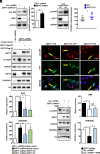
Fig. 1.
Fig. 1.
SIRT7 reduces ARF protein levels independently of its catalytic activity. (A) Western blot analysis of p14ARF levels in scrambled (Scr. shRNA) and SIRT7 Knockdown (KD; SIRT7 shRNA#1) H1299 lung cancer cell lines. Quantification of ARF relative expression ± SD is shown on the Right (n = 6). (B) Western blot analysis of p19ARF levels in age-matched WT and SIRT7 KO lungs. Quantification of p19ARF expression is shown in the histogram on the right (n = 10 WT and 12 KO). (C) Western blot analysis of p14ARF expression in SIRT7 KD (SIRT7 shRNA) H1299 cells, transiently transfected with wild-type (WT) or catalytic inactive mutant (HY) SIRT7. Vectors were titrated to obtain physiological levels of SIRT7. H3K36Ac, a target of SIRT7 catalytic activity was used as a control. Quantification of ARF and H3K36Ac relative expression ± SD normalized on GAPDH and total histone 3 (H3), respectively, is shown in the histograms below (n = 7). (D) Immunofluorescence (IF) staining for p14ARF (red) and yellow fluorescent protein (YFP) in H1299 cells transiently transfected with empty vector (Empty YFP), YFP-tagged WT, and catalytic inactive mutant (HY) SIRT7. Cell nuclei were counterstained with 4′,6-Diamidino-2-Phenylindole (DAPI; n = 3; Scale bar: 20 µm). (E) Western blot analysis of p14ARF levels in SIRT7 KD (SIRT7 shRNA) and scrambled (Scr. shRNA) H1299 treated with NAM (5 mM) for 24 h as indicated. GAPDH and total histone 3 (H3) were used as loading controls. Quantifications of relative p14ARF levels and H3K36Ac (normalized on GAPDH and total H3, respectively) ± SD are shown in the histograms on the Right (n = 3).
Fig. 2.
Fig. 2.
SIRT7 promotes ubiquitination and proteasomal-dependent degradation of ARF. (A) Western blot analysis of p14ARF levels in control (scrambled; Scr. shRNA) and SIRT7 KD (SIRT7 shRNA) H1299 cells at indicated time points after treatment with CHX (50 µg/mL). Quantifications of p14ARF levels are given in the graph below (n = 4; two-way Anova statistic test). (B) Coupled immunoprecipitation (IP; anti-p14ARF antibody) and Western blot analysis (anti-ubiquitin antibody) of control (scrambled; Scr. shRNA) and SIRT7 KD (SIRT7 shRNA) H1299 cells. A representative blot out of three independent experiments is shown (Upper). The membrane was reprobed with anti-p14ARF antibody (Lower). (C) Western blot analysis of p14ARF levels in scrambled and SIRT7 KD cells transiently transfected with Flag-tagged WT and catalytic inactive (HY) SIRT7 as indicated, 5 h after treatment with 10 µM MG-132. DMSO was used as vehicle. Quantification of ARF levels ± SD is shown in the histograms below (n = 5).
Fig. 3.
Fig. 3.
SIRT7 directly interacts with ARF. (A) Coupled IP (anti-Flag antibody) and Western blot analysis (anti-Flag and anti-YFP antibody) of 293T HEK cells transfected with YFP-tagged WT and HY mutant SIRT7 in combination with Flag-tagged p14ARF as indicated. Nonimmune immunoglobulin (IgG) was used as negative control. A representative experiment out of three biological replicates is shown. (B) Coupled IP (anti-p14ARF antibody) and Western blot analysis (anti-p14 and SIRT7 antibodies) of scrambled and SIRT7 KD H1299 cells. Nonimmune IgG was used as a negative control. (C) IF staining for SIRT7 (green) and p14ARF (red) of H1299 lung cancer cells. Nuclei were counterstained with DAPI. (Scale bar: 20 µm); n = 3. (D) Coupled IP (anti-V5 antibody) and Western blot analysis (anti-V5 and SIRT7 antibodies) of stable scrambled and NPM KD H1299 lung cancer cell lines transfected with V5-tagged p14ARF as indicated. No difference in binding of exogenous p14ARF with endogenous SIRT7 was observed upon inhibition of NPM expression (Upper). The inputs of the IP demonstrating efficient depletion of NPM are shown in the Lower. A representative image of three independent experiments is shown. (E) GST-pull down assay of bacterial purified GST-tagged p14ARF and His-tagged SIRT7 demonstrating direct interaction between proteins. A representative image of three independent experiments is shown.
Fig. 4.
Fig. 4.
SIRT7 destabilizes ARF by preventing its interaction with NPM. (A) Coupled IP (anti-NPM antibody) and Western blot analysis (anti-NPM and p14ARF antibodies) of scrambled and SIRT7 KD H1299 lung cancer cells (Upper). The inputs of the IP are shown in the lower histogram. The relative levels of coimmunoprecipitated ARF normalized on the immunoprecipitated NPM ± SD are quantified in the histograms below (n = 6). (B) Coupled IP (anti-ARF antibody) and Western blot analysis (anti-ARF and NPM antibodies) of purified Flag-tagged ARF and NPM incubated in the presence or absence of Flag-tagged SIRT7. A quantification of relative coimmunoprecipitated NPM is shown in the lower histogram (n = 4). (C) Western blot analysis of ARF protein levels in stable H1299 cells expressing SIRT7- and NPM-targeting shRNA alone (KD) or in combination (double KD; dKD) at different time points after treatment with translational inhibitor CHX (50 µg/mL) as indicated. Quantifications of p14ARF levels are given in the graph below (n = 4; two-way Anova statistic test). (D) Molecular model of human ARF (residues 1 to 58) suggests that Phe23 and Leu49 (in red) are positioned at the surface of the protein. (E) Coupled IP (anti-ARF antibody) and Western blot analysis (anti-Flag antibody) of purified Flag-tagged WT, point mutant ARF (substitution of Phe23 and Leu49 into Alanine; 2A) and Flag-tagged NPM. Quantification of relative coimmunoprecipitated NPM normalized to immunoprecipitated ARF ± SD is shown in the histogram on the Right (n = 3). (F) Coupled IP (anti-SIRT7 antibody) and Western blot analysis (anti-Flag antibody) of purified Flag-tagged WT, 2A point mutant ARF and Flag-tagged SIRT7. Quantification of relative coimmunoprecipitated ARF normalized to immunoprecipitated SIRT7 ± SD is shown in the histogram on the Right (n = 4).
Fig. 5.
Fig. 5.
Depletion of SIRT7 represses expression of genes involved in lung cancer progression in an ARF-dependent manner. (A) Venn diagram of significantly up-regulated genes [Log2 fold of change (FC) ≥ 0.58; FDR < 0.05] in ARF KD and SIRT7–V5 overexpressing H1299 cells as assessed by RNA-sequencing. A total of 159 genes were up-regulated in both datasets including Nectin2, XRCC1, SUPT5H, and SIPA1L3. (B) Western blot analysis of p14ARF levels in stable scrambled, SIRT7 KD and SIRT7/p14ARF dKD H1299 cells. (C) RT-qPCR analysis of mRNA expression of indicated genes in cells as in B. GAPDH was used as loading control (n = 7 for Nectin2, SUPT5H, and SIPA1L3 and n = 9 for XRCC1). (D) RT-qPCR analysis of mRNA expression in H226 and H322 ARF-depleted lung cancer cell lines. GAPDH was used as a loading control (n = 4). (E) RT-qPCR analysis of mRNA expression of CCNE1 in stable H1299 cells as in B. GAPDH was used as loading control (n = 7). (F) Chromatin IP analysis of p14ARF (Left) and acetylated H2B at lysine 20 (H2BK20; Right) enrichment at the promoter of CCNE1 gene in SIRT7 KD and SIRT7/ARF dKD cells (n = 3).
Fig. 6.
Fig. 6.
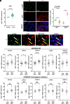
SIRT7 expression levels inversely correlate with expression of ARF-regulated genes in human lung tumors. (A) Scatter plot of Log2 mRNA expression of SIRT7 in healthy tissues and lung adenocarcinoma samples obtained from the lung adenocarcinoma TCGA study (healthy n = 59; tumor n = 517). (B) Representative IF staining of SIRT7 and ARF in healthy human lungs and lung cancers. The histogram on the Right shows a quantification of ARF and SIRT7 intensity staining from 4 healthy tissues and 4 lung tumors. (Scale bar: 50 µm.) (C) IF staining of SIRT7 and ARF in human lung tumor. Note that individual lung cancer cells displaying high levels of SIRT7 exhibit low levels of ARF in the nucleoli (yellow arrow), and vice versa (white arrow). (Scale bar: 10 µm.) (D) Scatter plots of Log2 mRNA expression of indicated genes in lung adenocarcinoma patients with low or high mRNA levels of SIRT7 harboring WT CDKN2A locus or CDKN2A homozygous deletion (WT CDKN2A locus: low SIRT7 n = 65; high SIRT7 n = 53; CDKN2A homozygous deletion low SIRT7 n = 13; high SIRT7 n = 33).
Fig. 7.
Fig. 7.
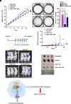
SIRT7 depletion inhibits lung cancer cells growth in vivo and in vitro in an ARF-dependent manner. (A) Growth curves of scrambled, SIRT7 KD, ARF KD, and SIRT7–ARF dKD cells. SIRT7 depletion significantly reduces cell proliferation, which is reverted by concomitant inhibition of ARF (n = 3; Two-way ANOVA). (B) Soft-agar colony formation of scrambled, SIRT7 KD, ARF KD, and SIRT7/ARF dKD H1299 cells. The average number of colonies of 3 independent experiments ± SD is shown in the histogram. (C) Schematic representation of the subcutaneous mouse xenograft model used in this study. 1 × 10⁶ stable scrambled, SIRT7 KD, ARF KD, and SIRT7/ARF dKD cells were injected subcutaneously in 4 to 6-wk-old BALB/c nude mice, and tumor size was measured every 4 d starting from day 14 after injection. (D) Fluorescence-based imaging of tumor volumes in the mouse experiment described in C. (E and F) Fluorescence-based imaging (E) and macroscopic images of excised tumors (F) 34 d after tumor cells injection as described in C and D. (G) Scheme depicting the putative mechanism of SIRT7-mediated ARF regulation in lung cancer cells.

References

    1. Kim W. Y., Sharpless N. E., The regulation of INK4/ARF in cancer and aging. Cell 127, 265–275 (2006). - PubMed
    1. Kamijo T., et al. , Tumor suppression at the mouse INK4a locus mediated by the alternative reading frame product p19ARF. Cell 91, 649–659 (1997). - PubMed
    1. Serrano M., et al. , Role of the INK4a locus in tumor suppression and cell mortality. Cell 85, 27–37 (1996). - PubMed
    1. Sherr C. J., Divorcing ARF and p53: An unsettled case. Nat. Rev. Cancer 6, 663–673 (2006). - PubMed
    1. Seo J., Seong D., Lee S. R., Oh D. B., Song J., Post-translational regulation of ARF: Perspective in cancer. Biomolecules 10, 1143 (2020). - PMC - PubMed