. 2024 Jun 18;121(25):e2315481121.
doi: 10.1073/pnas.2315481121. Epub 2024 Jun 13.

The multifunction Coxiella effector Vice stimulates macropinocytosis and interferes with the ESCRT machinery

Affiliations

The multifunction Coxiella effector Vice stimulates macropinocytosis and interferes with the ESCRT machinery

Arthur Bienvenu et al. Proc Natl Acad Sci U S A. .

Abstract

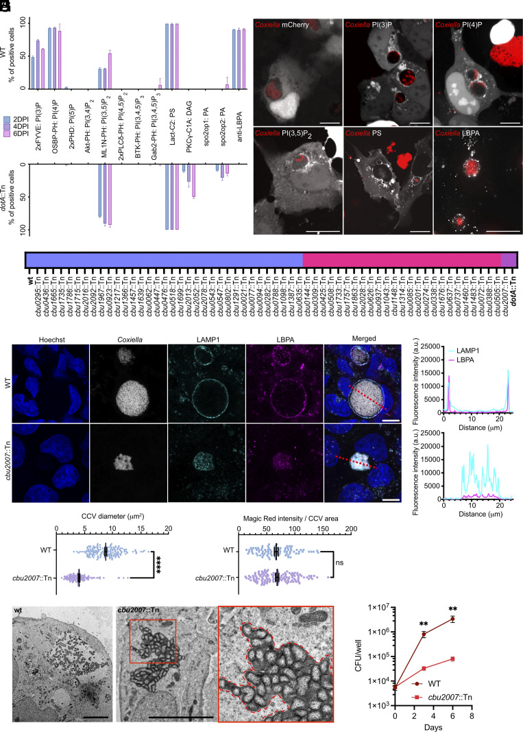
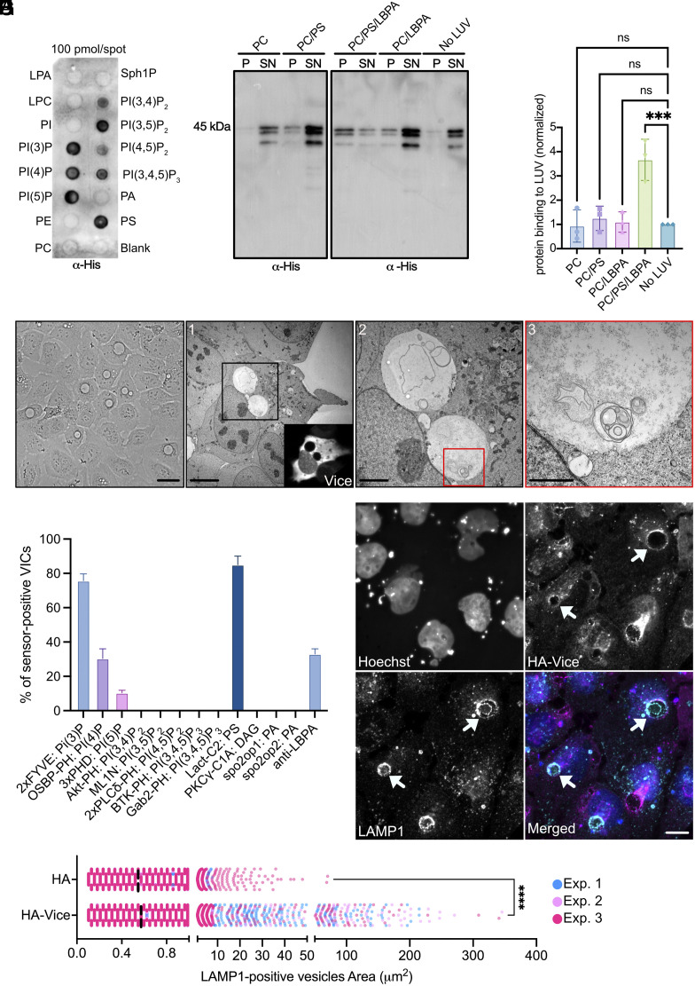
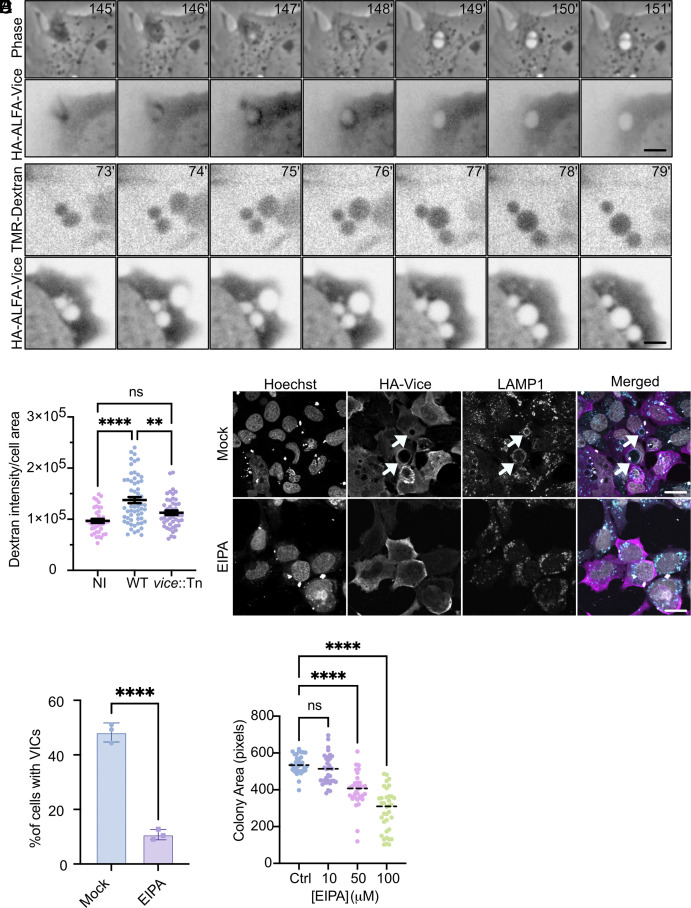
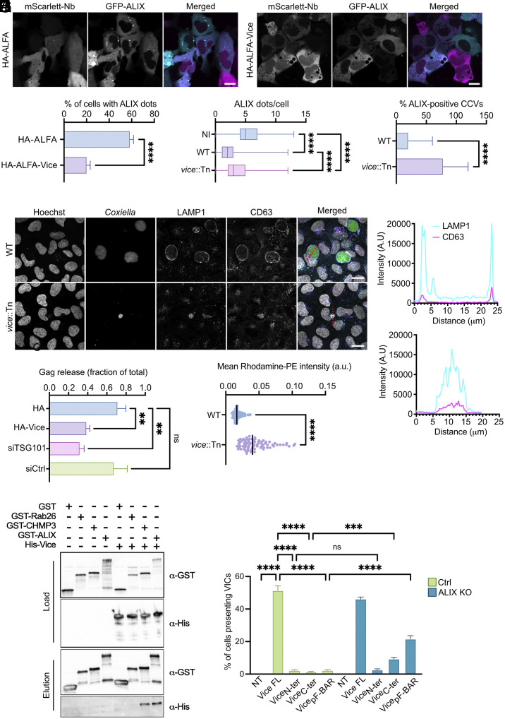
Intracellular bacterial pathogens divert multiple cellular pathways to establish their niche and persist inside their host. Coxiella burnetii, the causative agent of Q fever, secretes bacterial effector proteins via its Type 4 secretion system to generate a Coxiella-containing vacuole (CCV). Manipulation of lipid and protein trafficking by these effectors is essential for bacterial replication and virulence. Here, we have characterized the lipid composition of CCVs and found that the effector Vice interacts with phosphoinositides and membranes enriched in phosphatidylserine and lysobisphosphatidic acid. Remarkably, eukaryotic cells ectopically expressing Vice present compartments that resemble early CCVs in both morphology and composition. We found that the biogenesis of these compartments relies on the double function of Vice. The effector protein initially localizes at the plasma membrane of eukaryotic cells where it triggers the internalization of large vacuoles by macropinocytosis. Then, Vice stabilizes these compartments by perturbing the ESCRT machinery. Collectively, our results reveal that Vice is an essential C. burnetii effector protein capable of hijacking two major cellular pathways to shape the bacterial replicative niche.

Keywords: Coxiella burnetii; ESCRT; effector protein; lipids; macropinocytosis.


Conflict of interest statement

Competing interests statement:The authors declare no competing interest.

Figures

Fig. 1.
Fig. 1.
Lipid profiling of CCVs identifies CBU2007 as an effector required for LBPA localization. (A) U2OS cells were infected with Coxiella dotA::Tn (Bottom) or WT Coxiella (Upper) for 2, 4, and 6 d and transfected with plasmids encoding a mCherry/RFP-tagged catalog of lipid-binding sensors or labeled with an anti-LBPA antibody. The percentage of cells presenting CCVs positive for the indicated lipids was visually scored. Values are means ± SD of duplicate experiments where 100 cells were scored for each condition, DPI: days postinfection. (B) Representative images of U2OS cells treated as described in A (C. burnetii is pseudocolored in red, lipid-binding sensors or LBPA labeling are pseudocolored in white). (C) U2OS cells were infected for 4 d either with a library of mutants carrying transposon insertions in validated or predicted effector-coding genes, with wt C. burnetii as positive control, or with the dotA::Tn mutant as a negative control, fixed and labeled with an anti-LBPA antibody. The presence of LBPA at the CCV was visually scored and ranked as compared to wt C. burnetii infected cells: Unaltered LBPA labeling of CCVs (blue), partial loss of LBPA labeling (pink) and complete loss of LBPA labeling (purple). (D) U2OS cells infected for 4 d either with wt C. burnetii (Upper, gray) or the cbu2007::Tn mutant (Bottom, gray) were labeled with Hoechst (blue), anti-LAMP1 (cyan), and anti-LBPA (magenta) antibodies. (E) The fluorescence intensity of LAMP1 (cyan) and LBPA (magenta) was measured along the red dash lines from (D). (F) The area of CCVs harboring either wt C. burnetii (WT) or the cbu2007::Tn mutant strain (cbu2007::Tn) was measured using LAMP1 labeling from U2OS cells infected for 4 d. Values are means ± SD of three independent experiments (****P < 0.0002; unpaired t test). (G) Proteolysis of cathepsin B-Magic Red was measured for individual CCVs at 4 dpi. Values are means ± SD of three independent experiments (ns: nonsignificant; unpaired t test). (H) Representative electron micrographs of U2OS cells infected with wt C. burnetii (Left) or the cbu2007::Tn mutant strain (Right). The red Inset shows an enlarged view of the CCV generated by the cbu2007::Tn mutant strain, the dashed red line represents the CCV contour. (I) CFU counts from U2OS cells challenged with wt C. burnetii or the C. burnetii cbu2007::Tn mutant strain for 3 and 6 d. Values are means ± SD of four independent experiments (**P < 0.01; multiple unpaired t test). (Scale bars: 10 µm.)
Fig. 2.
Fig. 2.
CBU2007/Vice is a lipid-binding effector involved in CCV biogenesis and LBPA enrichment. (A) Representative protein/lipid overlay assay performed with 6xHIS-CBU2007. (B) Representative western blot of LUVs cosedimentation assay using 6xHIS-CBU2007. CBU2007 in the pellet (P; bound to LUVs) or the supernatant (SN; unbound to LUVs) was detected using an anti-Histidine antibody. (C) CBU2007/LUVs binding was determined by measuring CBU2007 signal detected in B and using the following formula: (Pintensity/(Pintensity+ SNintensity))*100. Values are means ± SD of three independent experiments (***P < 0.0001; ns: nonsignificant. One-way ANOVA, Dunnett’s multiple comparisons test). (D) Representative brightfield image of U2OS cells transfected for 24 h with plasmids encoding HA-Vice, (scale bar: 20 µm.) (E) Correlative Light-Electron Microscopy (CLEM) of U2OS cells cotransfected with plasmids encoding HA-ALFA-Vice and NbALFA-mScarlet. Cells were imaged using confocal microscopy to identify the cell of interest (Vice inset) followed by fixation for electron microscopy [(1) scale bar: 5 µm; (2) scale bar: 2 µm; (3) scale bar: 1 µm).] (F) The lipid composition of VICs was assessed by visually scoring the presence of the indicated lipid sensors in U2OS cells transfected with plasmids encoding HA-Vice and the indicated lipid sensors or HA-Vice alone followed by anti-LBPA labeling. Values are means ± SD of three independent experiments where 100 VICs were scored for each condition. (G) U2OS cells transfected with a plasmid encoding HA-Vice (magenta) and labeled with anti-LAMP1 antibodies (cyan) indicate that VICs are positive for LAMP1 (white arrows). Hoechst was used to label nuclei (white) in all conditions. (Scale bars: 10 µm.) (H) The median size of LAMP1-positive compartments in U2OS cells expressing either HA-tag alone or HA-Vice was measured using CellProfiler. Each spot represents a LAMP1-positive compartment from three independent experiments (blue, pink, red; ****P < 0.0001, unpaired t test).
Fig. 3.
Fig. 3.
Vice stimulates macropinocytosis. (A) Representative images from Movie S1 illustrating U2OS cells cotransfected with plasmids encoding HA-ALFA-Vice and NbALFA-mScarlet and imaged after 8 h in phase contrast (Upper) and 555 nm emission (Lower) channels. (Scale bars: 4 µm.) (B) Representative images from Movie S6 illustrating U2OS cells cotransfected with plasmids encoding HA-ALFA-Vice and NbALFA-mNeonGreen for 8 h prior to TMR-Dextran addition. Images were acquired every minute overnight in 555 nm emission (Upper) and 488 nm emission (Lower) channels. (Scale bars: 4 µm.) (C) Stimulation of macropinocytosis was assessed by measuring the fluorescence intensity of TMR-Dextran from noninfected (NI), wt-infected (WT), or vice::Tn mutant-infected (vice::Tn) U2OS cells. Values are means ± SD of three independent experiments (****P < 0.0001, **P < 0.0022, ns: nonsignificant. One-way ANOVA, Dunnett’s multiple comparisons test). (D) U2OS cells transfected with a plasmid encoding HA-Vice were incubated overnight with either methanol (Mock, Upper) or EIPA (Lower). VIC formation was assessed by using Hoechst to label nuclei (white), anti-HA (magenta), and anti-LAMP1 antibodies (cyan). White arrows point at VICs; (scale bars: 20 µm.) (E) The percentage of cells described in D containing VICs was visually scored. Values are means ± SD of three independent experiments (****P < 0.0001, unpaired t test). (F) U2OS infected with wt C. burnetii were incubated with the indicated concentration of EIPA for 48 h. The median area of C. burnetii colonies was measured from 25 images per condition (corresponding to more than 3,000 colonies per condition). Values are means of two independent experiments (****P < 0.0001, ns: nonsignificant. One-way ANOVA, Dunnett’s multiple comparisons test).
Fig. 4.
Fig. 4.
Vice inhibits the activity of the ESCRT machinery. (A) U2OS cells were cotransfected with plasmids encoding GFP-ALIX (cyan), NbALFA-mScarlet (magenta), and either HA-ALFA (Left) or HA-ALFA-Vice (Right). (B) The percentage of cells presenting GFP-ALIX dots in cells treated as in A, was visually scored. (C) U2OS cells infected for 4 d with either WT Coxiella or vice::Tn C. burnetii mutant were transfected for 24 h with a plasmid encoding GFP-ALIX and the number of GFP-ALIX dots in noninfected (NI) and infected cells was visually scored. (D) GFP-ALIX localization at CCVs was visually scored in both infection conditions described in C. Values are means ± SD of three independent experiments (****P < 0.0001, unpaired t test for B and D, One-way ANOVA, Dunnett’s multiple comparisons test for C). (E) U2OS cells infected for 4 d with either WT Coxiella or vice::Tn C. burnetii mutant (green) were labeled with Hoechst (white), anti-LAMP1 (cyan), and anti-CD63 (magenta). (F) Representative distribution of LAMP1 (cyan) and CD63 (magenta) fluorescence intensity was measured along the corresponding red dash lines illustrated in F. (Scale bars: 20 µm.) (G) HEK293T were cotransfected with plasmids encoding either HA or HA-Vice and GAG, or transfected with plasmids encoding GAG, 24 h after transfection with either TSG101-targeting siRNAs (siTSG101) or scrambled siRNA (siCTRL). Virus-like particles (VLP) release was assessed by western blot using anti-p24 antibodies and the relative percentage of VLPs was measured by quantifying the p24 signal from triplicates experiments using the following formula: (VLPsp24_intensity/(VLPsp24_intensity + Cellsp24_intensity)). Values are means ± SD of three independent experiments (**P < 0.001, ns: nonsignificant, One-way ANOVA, Dunnett’s multiple comparisons test). (H) The CCV-associated fluorescence intensity of Rhodamine-PE was measured from maximum intensity projection of confocal images of U2OS cells infected either with WT C. burnetii (WT) or the vice::Tn mutant strain (vice::Tn). Values are mean of three independent experiments where 100 CCVs were measured for each condition (****P < 0.0001, unpaired t test). (I) Protein copurification on agarose-glutathione resin from lysates of LEMO21 bacteria transformed with pGEX4T1 (GST), pGEX4T1-RAB26 (GST-RAB26), pGEX4T1-CHMP3B (GST-CHMP3), or pGEX4T1-ALIX (GST-ALIX) in combination with pTWIST-Vice (His-Vice). Total cell lysates (Load) and purified proteins (Elution) were labeled with anti-GST and anti-His antibodies. (J) Occurrence of VICs was visually scored from control (Ctrl) or ALIX-KO (ALIX KO) U2OS cells transiently expressing the indicated HA-tagged Vice truncations or the HA tag alone as control. Values are means ± SD of three independent experiments (****P < 0.0001, ***P < 0.0022, ns: nonsignificant, one-way ANOVA, Dunnett’s multiple comparisons test).

Similar articles

Cited by

References

    1. Martinez E., Siadous F. A., Bonazzi M., Tiny architects: Biogenesis of intracellular replicative niches by bacterial pathogens. Fems Microbiol. Rev. 42, 425–447 (2018). - PubMed
    1. van Schaik E. J., Chen C., Mertens K., Weber M. M., Samuel J. E., Molecular pathogenesis of the obligate intracellular bacterium Coxiella burnetii. Nat. Rev. Microbiol. 11, 561–573 (2013). - PMC - PubMed
    1. Larson C. L., et al. , Right on Q: Genetics begin to unravel Coxiella burnetii host cell interactions. Fut. Microbiol. 11, 919–939 (2016). - PMC - PubMed
    1. Martinez E., et al. , Coxiella burnetii effector CvpB modulates phosphoinositide metabolism for optimal vacuole development. Proc. Natl. Acad. Sci. U.S.A. 113, E3260–E3269 (2016). - PMC - PubMed
    1. van Schaik E. J., Case E. D., Martinez E., Bonazzi M., Samuel J. E., The SCID mouse model for identifying virulence determinants in Coxiella burnetii. Front. Cell Infect. Microbiol. 7, 25 (2017). - PMC - PubMed

Substances

LinkOut - more resources