Hierarchical structure and chemical composition of complementary segments of the fruiting bodies of Fomes fomentarius fungi fine-tune the compressive properties
- PMID: 38870218
- PMCID: PMC11175439
- DOI: 10.1371/journal.pone.0304614
Hierarchical structure and chemical composition of complementary segments of the fruiting bodies of Fomes fomentarius fungi fine-tune the compressive properties
Abstract
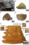
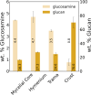
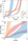
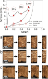
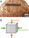
Humanity is often fascinated by structures and materials developed by Nature. While structural materials such as wood have been widely studied, the structural and mechanical properties of fungi are still largely unknown. One of the structurally interesting fungi is the polypore Fomes fomentarius. The present study deals with the investigation of the light but robust fruiting body of F. fomentarius. The four segments of the fruiting body (crust, trama, hymenium, and mycelial core) were examined. The comprehensive analysis included structural, chemical, and mechanical characterization with particular attention to cell wall composition, such as chitin/chitosan and glucan content, degree of deacetylation, and distribution of trace elements. The hymenium exhibited the best mechanical properties even though having the highest porosity. Our results suggest that this outstanding strength is due to the high proportion of skeletal hyphae and the highest chitin/chitosan content in the cell wall, next to its honeycomb structure. In addition, an increased calcium content was found in the hymenium and crust, and the presence of calcium oxalate crystals was confirmed by SEM-EDX. Interestingly, layers with different densities as well as layers of varying calcium and potassium depletion were found in the crust. Our results show the importance of considering the different structural and compositional characteristics of the segments when developing fungal-inspired materials and products. Moreover, the porous yet robust structure of hymenium is a promising blueprint for the development of advanced smart materials.
Copyright: © 2024 Klemm et al. This is an open access article distributed under the terms of the Creative Commons Attribution License, which permits unrestricted use, distribution, and reproduction in any medium, provided the original author and source are credited.
Conflict of interest statement
The authors have declared that no competing interests exist.
Figures




Similar articles
-
An approach to change the basic polymer composition of the milled Fomes fomentarius fruiting bodies.Fungal Biol Biotechnol. 2021 Apr 15;8(1):5. doi: 10.1186/s40694-021-00112-9. Fungal Biol Biotechnol. 2021. PMID: 33858513 Free PMC article.
-
Chemical Composition and Ultraviolet Absorption Activity of an Aqueous Alkali Extract from the Fruiting Bodies of the Tinder Conk Mushroom, Fomes fomentarius (Agaricomycetes).Int J Med Mushrooms. 2021;23(4):23-37. doi: 10.1615/IntJMedMushrooms.2021038160. Int J Med Mushrooms. 2021. PMID: 33822505
-
Decomposition of Fomes fomentatius fruiting bodies - transition of healthy living fungus into a decayed bacteria-rich habitat is primarily driven by Arthropoda.FEMS Microbiol Ecol. 2024 Apr 10;100(5):fiae044. doi: 10.1093/femsec/fiae044. FEMS Microbiol Ecol. 2024. PMID: 38640440 Free PMC article.
-
Medicinal Potential of the Insoluble Extracted Fibers Isolated from the Fomes fomentarius (Agaricomycetes) Fruiting Bodies: A Review.Int J Med Mushrooms. 2023;25(3):21-35. doi: 10.1615/IntJMedMushrooms.2022047222. Int J Med Mushrooms. 2023. PMID: 37017659 Review.
-
Tasting the fungal cell wall.Cell Microbiol. 2010 Jul;12(7):863-72. doi: 10.1111/j.1462-5822.2010.01474.x. Epub 2010 May 6. Cell Microbiol. 2010. PMID: 20482553 Review.
References
-
- Hawksworth DL, Lücking R. Fungal Diversity Revisited: 2.2 to 3.8 Million Species. Microbiol Spectr [Internet]. 2017. Jul;5(4). Available from: https://pubmed.ncbi.nlm.nih.gov/28752818/ doi: 10.1128/microbiolspec.FUNK-0052-2016 - DOI - PMC - PubMed
-
- Fungi Boddy L., Ecosystems, and Global Change. In 2016. p. 361–400.
-
- GBIF Backbone Taxonomy. [cited 2023 Sep 28]; Available from: https://www.gbif.org/dataset/d7dddbf4-2cf0-4f39-9b2a-bb099caae36c
-
- Hein W, Trommer F, EXARC Experimental Archaeology Collection Manager. Brennt wie Zunder! Steinzeitliche Feuererzeugung im Experiment. In: Scheer A, editor. Eiszeitwerkstatt, experimentelle Archäologie. Blaubeuren: Urgeschichtliches Museum Blaubeuren; 1995. p. 73–7.
-
- Seeberger F. Steinzeit selbst erleben!: Waffen, Schmuck und Instrumente—nachgebaut und ausprobiert. Stuttgart WL, Blaubeuren UM, Buchau FB, editors. Stuttgart: wbg Theiss in Wissenschaftliche Buchgesellschaft; 2003. 78 p.
MeSH terms
Substances
LinkOut - more resources
Full Text Sources

