. 2024 Jun 14;384(6701):1196-1202.
doi: 10.1126/science.adk9428. Epub 2024 Jun 13.

In vivo editing of lung stem cells for durable gene correction in mice

Affiliations

In vivo editing of lung stem cells for durable gene correction in mice

Yehui Sun et al. Science. .

Abstract

In vivo genome correction holds promise for generating durable disease cures; yet, effective stem cell editing remains challenging. In this work, we demonstrate that optimized lung-targeting lipid nanoparticles (LNPs) enable high levels of genome editing in stem cells, yielding durable responses. Intravenously administered gene-editing LNPs in activatable tdTomato mice achieved >70% lung stem cell editing, sustaining tdTomato expression in >80% of lung epithelial cells for 660 days. Addressing cystic fibrosis (CF), NG-ABE8e messenger RNA (mRNA)-sgR553X LNPs mediated >95% cystic fibrosis transmembrane conductance regulator (CFTR) DNA correction, restored CFTR function in primary patient-derived bronchial epithelial cells equivalent to Trikafta for F508del, corrected intestinal organoids and corrected R553X nonsense mutations in 50% of lung stem cells in CF mice. These findings introduce LNP-enabled tissue stem cell editing for disease-modifying genome correction.


Figures

Fig. 1.
Fig. 1.. Direct in vivo gene editing was achieved in mouse lungs and persisted for >1.8 years.
(A) Schematic representation of LNP-mediated gene editor delivery into lung cells after systemic administration. (B) Diagram showing the experimental procedure used to evaluate the efficiency of lung cell editing in Ai14 tdTom reporter mice, in which Cre recombinase can excise the loxP flanked stop cassette, thereby enabling fluorescent tdTom protein expression. Mice were injected with LNP-Cre at 2 mg/kg total RNA (20:1, total lipid to RNA weight ratio) with two sequential doses, 48 hours apart. Mice treated with phosphate-buffered saline (PBS) were used as negative control. (C) Ex vivo fluorescence imaging analyses of mouse lungs 2, 7, 21, 42, 60, 120, 180, 360, and 660 days after the last injection. (D and E) Quantitative analysis of ex vivo lung images was shown as average radiance (D) and as total flux (E). (F to K) Time-course flow cytometry analyses showing the percentage of tdTom-positive (tdTOM+) cells reported among lung cells (F), endothelial cells (G), immune cells (H), epithelial cells (I), NGFR+ stem cells (J), and KRT5+ stem cells (K). Data are means ± SEMs (n = 3 biologically independent replicates) for treated groups.
Fig. 2.
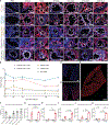
Fig. 2.. Lung SORT LNPs mediated efficient delivery into diverse lung cell types with enhanced delivery to VtnR-expressing cells.
(A) Representative immunofluorescence images of lung sections from LNP-Cre–treated Ai14 reporter mice (2, 7, 60, 120, 270, and 360 days after treatment) to assess LNP-mediated editing in mature lung epithelial cells (HOPX for AT1 cells, ABCA3 for AT2 cells, MUC5AC for goblet cells, tubulin for ciliated cells, and SCGB1A1 for club cells). PBS-treated mice served as negative controls. Scale bars, 30 μm. Markers are white, tdTom are red, and nuclei are blue. (B) Quantification of LNP-Cre–mediated editing in mature lung epithelium based on immunofluorescence images. Results were obtained from five to six random segments per whole slide. Data are presented as means ± SEMs. (C and D) Representative whole-slide immunofluorescence images from PBS-treated (C) and LNP-Cre–treated (D) mice. 4′,6-diamidino-2-phenylindole (DAPI) is blue, and tdTom is red. Scale bars, 1 mm. (E) Representative tissuecyte image of a LNP-Cre–treated mouse’s lung whole left lobe (3D rendering provided in movie S1). Scale bar, 1 mm. (F to K) VtnR (CD51+CD61+) abundance (F) and quantification of tdTom positivity in VtnR+ (CD51+CD61+) fraction and VtnR (CD51CD61) fraction in lung endothelial cells (G), immune cells (H), epithelial cells (I), NGFR+ cells (J), and KRT5+ stem cells (K). Data are means ± SEMs (n = 3 biologically independent replicates). Unpaired t test. P values of <0.05 were considered statistically significant. ns, not significant.
Fig. 3
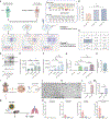
Fig. 3. Efficient adenine base editing was achieved in lung basal cells in patient-derived HBE cells and CF mouse model.
(A) Workflow for differentiation of HBEs from a healthy donor and a CF person with CFTRR553X/F508del into airway epithelium and base editing strategy to correct CF R553X mutation. Untreated CF HBE cells were used as negative control, and HBEs from a healthy donor with wild-type (WT) CFTR gene were used for comparison. (B) LNP-ABE–mediated 60% of allelic base editing in both undifferentiated P2 (n = 1) and fully differentiated P3 culture (n = 4). (C) The frequency of desired product (the box highlighted in blue) and bystander editing was evaluated using NGS sequencing. (D) Efficacy of LNP-ABE with or without Trikafta in CFTR protein restoration in CFTRR553X/F508del HBE culture measured by capillary Western blotting. (E and F) Band B (E) and band C (F) intensities from (D) were normalized to vinculin (20 μg/mL) as an internal standard. Data are means ± SEMs of n =3 independent replicates. One-way analysis of variance (ANOVA); P values of <0.05 were considered statistically significant. (G) Quantitative data for average current calculated from AUC representing CFTR activity further confirmed CFTR restoration. Data are means ± SEMs of n = 4. One-way ANOVA; P values of <0.05 were considered statistically significant. (H) CFTR function in HBE culture from non-CF individuals who are wild-type for the CFTR gene (n = 4 independent replicates). (I) Workflow of R553X correction in intestinal organoids using LNP-ABEs and the mechanism of FIS assay. Intestinal stem cells were isolated from R553X homozygous mice to generate intestinal organoids as an ex vivo model to evaluate CFTR function restoration after LNP-ABE treatment. Forskolin-induced CFTR activation can facilitate ion-water transportation leading to organoid swelling. (J to L) No swelling was observed from untreated group (J), whereas the LNP-ABE–treated group (K) exhibited 80% of intestinal organoid swelling. Scale bars, 1000 μm. (M) Approximately 50% base editing was confirmed using DNA sequencing. Data are presented as means ± SEMs of n = 8 independent replicates in (L) and (M). Unpaired t test; P values of <0.05 were considered statistically significant. (N) Workflow for assessing LNP-ABE–mediated base editing in mouse lung basal cells after a single administration. CF heterozygous R553X mice were injected intravenously with LNP-ABE (1.5 mg/kg total RNA, ABE mRNA:sgR553X = 2:1, weight ratio). Mice were euthanized 10 days after the injection. Whole lung tissue, trachea, and isolated lung NGFR+ basal stem cells populations were used for DNA extraction, PCR amplification, and NGS sequencing. (O) Base editing efficiency of all adenines within the target protospacer in three lung populations (n =4 independent replicates). Data are presented as means ± SEMs.

Comment in

  • Gene editing flows to the lungs.
    Bulcaen M, Carlon MS. Bulcaen M, et al. Science. 2024 Jun 14;384(6701):1175-1176. doi: 10.1126/science.adq0059. Epub 2024 Jun 13. Science. 2024. PMID: 38870313
  • A rush of CRISPR to the lungs.
    Villanueva MT. Villanueva MT. Nat Rev Drug Discov. 2024 Aug;23(8):580. doi: 10.1038/d41573-024-00117-0. Nat Rev Drug Discov. 2024. PMID: 38987628 No abstract available.

References

    1. Wang JY, Doudna JA, Science 379, eadd8643 (2023). - PubMed
    1. Hodges CA, Conlon RA, Genes Dis. 6, 97–108 (2018). - PMC - PubMed
    1. Hajj KA, Whitehead KA, Nat. Rev. Mater 2, 17056 (2017).
    1. Wei T. et al. , ACS Nano 14, 9243–9262 (2020). - PMC - PubMed
    1. Raguram A, Banskota S, Liu DR, Cell 185, 2806–2827 (2022). - PMC - PubMed

Publication types

Substances