. 2024 Jun 14;384(6701):eadj4301.
doi: 10.1126/science.adj4301. Epub 2024 Jun 14.

Metabolic inflexibility promotes mitochondrial health during liver regeneration

Affiliations

Metabolic inflexibility promotes mitochondrial health during liver regeneration

Xun Wang et al. Science. .

Abstract

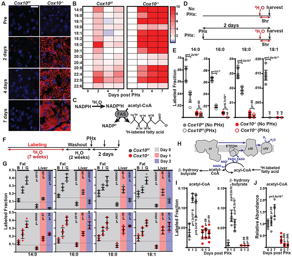
Mitochondria are critical for proper organ function and mechanisms to promote mitochondrial health during regeneration would benefit tissue homeostasis. We report that during liver regeneration, proliferation is suppressed in electron transport chain (ETC)-dysfunctional hepatocytes due to an inability to generate acetyl-CoA from peripheral fatty acids through mitochondrial β-oxidation. Alternative modes for acetyl-CoA production from pyruvate or acetate are suppressed in the setting of ETC dysfunction. This metabolic inflexibility forces a dependence on ETC-functional mitochondria and restoring acetyl-CoA production from pyruvate is sufficient to allow ETC-dysfunctional hepatocytes to proliferate. We propose that metabolic inflexibility within hepatocytes can be advantageous by limiting the expansion of ETC-dysfunctional cells.


Figures

Figure 1.
Figure 1.. Zone-specific mitochondrial metabolomics in murine livers.
A, Schematic of Cre-expression patterns from Albumin-Cre and Zone-specific Cre driver alleles. CV: central vein. B, Representative immunofluorescent images of hepatic HA-GFP-OMP25 expression in the indicated Cre-driver mice. HA (green), DAPI (blue). Scale bar, 100 μm. C, Schematic of rapid liver mitochondrial isolation. 70% PHx was performed 7 days after a single tamoxifen injection (Day 0). Samples were collected at Day 0 and Day 2 post PHx, and HA-labelled mitochondria were rapidly pulled down using anti-HA beads. D, Heatmap (z-score values) for 173 detected mitochondrial metabolites from Alb-Cre; LSL-HA-eGFP-OMP25 liver mitochondria at Day 0 (black) and Day 2 (red) post PHx. E, Principal components analysis of the metabolomes from total hepatocyte mitochondria at Day 0 and 2 post PHx. The percentages of total variance of principal components 1 and 2 (PC1 and PC2) are indicated on the x and y axes. F, Relative abundance of the indicated metabolites from total hepatocyte mitochondria. n=5 per group. nd, not detected. Same color scheme as E. G, Principal components analysis of the mitochondrial metabolomes from Zone 1, 2, or 3 hepatocyte mitochondria at Days 0 and 2 post PHx. The percentages of total variance of principal components 1 and 2 (PC1 and PC2) are indicated on the x and y axes. H, Relative abundance of the indicated metabolites from zone 1, 2, or 3 hepatocyte mitochondria at Day 0 and Day 2 post PHx. n=5 mice per group. nd, not detected. Same color scheme as G. Statistical significance was assessed using Welch’s t-test (F,H), with corrections for multiple comparisons. Dot and whisker plots indicate median values and interquartile ranges. Ovals indicate 95% confidence intervals. The number of biological replicates in each group and false discovery rates (q value) are indicated in the figure or above.
Figure 2.
Figure 2.. The mitochondrial ETC is required for liver regeneration.
A, Schematic of experiment of low-dose AAV tracing protocol. 70% PHx was performed 14 days after AAV-Cre (0.125x10¹⁰ genome copies (GC) per mouse) administration. Samples were harvested 14 days later, and assessed for DENDRA2 expression. B, Representative immunofluorescent images of indicated livers at Day 0 and Day 14 post PHx. Scale bar, 100 μm. C, Quantitation of DENDRA2+ cells (as a percentage of HNF4α+ cells) in livers of the indicated mice. p values reflect comparisons with the control (mDendra2f/f) group for each timepoint. D, Schematic of experiment of high-dose AAV protocol. 70% PHx (Day 0) was performed 28 days after AAV-Cre and AAV-GFP (5x10¹⁰ GC per mouse) injection. Livers were harvested and analyzed at various time points post PHx. E, Representative BrdU immunohistochemistry and H&E images of indicated mice at the indicated timepoints post PHx. Scale bar, 50 μm. F, Quantitation of BrdU+ cell numbers (normalized to liver area) of indicated mice at various timepoints post PHx. G, Liver/body weight ratio of indicated mice at various time points post PHx. Same color scheme as F. Statistical significance was assessed using two-way ANOVA with adjustments for multiple comparisons (C,F,G). Dot and whisker plots indicate median values and interquartile ranges. The number of biological replicates in each group and p values are indicated in the figure.
Figure 3.
Figure 3.. Transdifferentiation of biliary epithelial cells is stimulated after mitochondrial complex IV inhibition in hepatocytes.
A, Survival fraction of Cox10f/f and Cox10−/− mice post PHx. B, Representative H&E liver images at timepoints post PHx. Scale bar, 50 μm. C, Representative immunofluorescent liver images at 2 days post PHx. Scale bar, 100 μm. D, Protocol for cholangiocyte lineage tracing: AAV-sgControl or AAV-sgCox10 were administered to tamoxifen-treated CK19-CreERT2; Rosa26-tdTomato; Rosa26-Cas9 mice. A 70% PHx was performed 14 days later, and livers were analyzed 4 weeks post PHx. E, Western blot analysis of liver lysates 14 days after AAV injection. ACTB levels are shown as a loading control. Molecular weight markers are indicated in kilodaltons. F, Representative immunofluorescent images of livers at timepoints post PHx. Scale bar, 100 μm. G, Representative H&E and BrdU immunohistochemistry of livers at 2 days post PHx. Scale bar, 50 μm. H, Survival fraction of Alb-Cre; Cox10f/f and Alb-Cre; Cox10+/+ mice post PHx. I, UMAP visualization from snRNA sequencing in Cox10f/f and Cox10−/− livers at 4 weeks post PHx. J, UMAP plots showing expression of Gls2, Igfbp2, and Glul in hepatocyte clusters. K, Dot plot showing relative expression of the indicated genes in hepatocyte clusters. Dot size indicates the percentage of cells expressing each gene, and color indicates average mRNA level. L, Violin plots for expression of the indicated genes in hepatocyte clusters. M, UMAP visualization of hepatocyte and cholangiocyte clusters from Cox10−/− liver. N, Violin plots for expression of the indicated genes in hepatocyte and cholangiocyte clusters of Cox10−/− liver. O, Top 5 upregulated pathways in GSEA of stage 2 vs. stage 1 TLPCs of Cox10−/− liver. Statistical significance was assessed using a log-rank test (A and H). The number of biological replicates in each group and p values are indicated in the figure.
Figure 4.
Figure 4.. Hepatic oxidation of peripheral fatty acids is stimulated during liver regeneration.
A, Representative immunofluorescent images of liver Nile Red staining at the indicated timepoints post PHx. Nile Red (red), DAPI (blue). Scale bar, 20 μm. B, Heatmap (z-score values) of the indicated fatty acid species abundances in livers at various time points post PHx. C, Schematic of expected labeling of fatty acids with 2H2O administration. FAS, Fatty acid synthase. D, Experimental protocol for short-term 2H2O labeling: At 5 hours before liver collection, a single dose of 2H2O was administered to mice. E, Total labeled fraction of indicated liver fatty acid species by short-term (5 hours) 2H2O tracing (from D) in the indicated genotypes. F, Schematic of long-term (7 weeks) 2H2O labeling experiment to evaluate fate of peripheral fatty acids. G, Total labeled fraction of indicated fatty acid species by long-term 2H2O tracing in liver and different adipose tissues at the indicated time points post PHx. p values reflect comparison with the labeling in fat pads for that species and genotype on Day 0. B, brown fat. I, inguinal fat pad. G, gonadal fat pad. H, Schematic of mitochondrial fatty acid oxidation to produce acetyl-CoA and β-hydroxybutyrate. I, Total labeled fraction of liver acetyl-CoA and β-hydroxybutyrate from long-term 2H2O tracing (from F) at the indicated time points post PHx. p values reflect comparison with the Day 0 group for each genotype. J, Relative abundance of acetyl-CoA in livers at the indicated time points post PHx. p values reflect comparison with the day 0 group for each genotype. The same color scheme is used throughout. Statistical significance was assessed using two-way ANOVA (E, I, J), or Kruskal-Wallis (G) tests with adjustments for multiple comparisons. Dot and whisker plots indicate median values and interquartile ranges. n=6 mice in each group were used in B, E, G, I, J. p values are indicated in the figure.
Figure 5.
Figure 5.. Peripheral fatty acid mobilization promotes hepatocyte proliferation.
A, Schematic of free fatty acid (FFA) mobilization from adipose tissue to liver to produce acetyl-CoA via mitochondrial fatty acid oxidation. ATGL, HSL inhibitors are indicated (green). B, Western blots for HADHA levels in livers from sgRNA-treated mice. ACTB levels are shown as a loading control; molecular weight markers are in kilodaltons. C, Liver H&E and BrdU immunohistochemistry at 2 days post PHx. Scale bar, 50 μm. D, BrdU+ cell numbers (per liver area) and liver/body weight ratios. p values reflect comparison with sgControl group for each timepoint. E, (Top) Protocol for lipolysis inhibitor administration: Vehicle or inhibitors were administered to C57BL/6J mice every 8 hours during PHx. (Bottom) Serum FFA concentrations at the indicated timepoints. F, Liver H&E and BrdU immunohistochemistry of treated C57BL/6J mice at 2 days post PHX. Scale bar, 50 μm. G, BrdU+ cell numbers (per liver area) and liver/body weight ratio in treated C57BL/6J mice. H, Relative abundance of liver acetyl-CoA in treated C57BL/6J mice at 2 days post PHx. Same color scheme as E. I, Protocol for [U-13C]glucose tracing post PHx. J, Labeled fraction of plasma glucose m+6 in treated C57BL/6J during [U-13C]glucose infusions. K, Fractional enrichment of the indicated isotopologues in livers of treated C57BL/6J mice following [U-13C]glucose infusion at 2 days post PHx. L, Liver acetyl-CoA m+2/pyruvate m+3 ratio in treated C57BL/6J mice following [U-13C]glucose infusion at 2 days post PHx. Same color scheme as J. Statistical significance was assessed using Brown-Forsythe ANOVA (BrdU in D), or two-way ANOVA (D, G, E and J), or Student’s t-test for (BrdU in G, H,K,L) with adjustments for multiple comparisons. Dot and whisker plots indicate median values and interquartile ranges. The number of biological replicates in each group and p values are indicated in the figure.
Figure 6.
Figure 6.. Fatty acid accumulation inhibits acetyl-CoA production.
A, Schematic of [U-13C]glucose tracing in Cox10f/f and Cox10−/− mice during liver regeneration. B, Fractional enrichment of the indicated isotopologues following [U-13C] glucose infusion at 2 days post PHx. C, Schematic of [U-13C]acetate tracing in Cox10f/f and Cox10−/− mice during liver regeneration. D, Fractional enrichment of the indicated isotopologues following [U-13C]acetate infusion at 2 days post PHx. E, Western blot analysis in Cox10f/f and Cox10−/− liver protein lysates at the indicated time points post PHx. ACTB levels are shown as a loading control. F, Western blot analysis in liver protein lysates from animals treated with vehicle or ATGL and HSL inhibitors. ACTB levels are shown as a loading control. G, Representative BrdU immunohistochemistry images of Cox10−/− mice treated with vehicle or lipolysis inhibitors at 2 days post PHx. Scale bar, 50 μm. H, BrdU+ cell numbers (normalized to liver area) in the indicated mice. I, Proposed model: In the setting of ETC dysfunction, fatty acid (FA) buildup in hepatocytes inhibits acetyl-CoA production via the induction of PDK4 and suppression of ACSS2 expression. Statistical significance was assessed using Student’s t-test (B,D) or Welch’s t-test (H). Dot and whisker plots indicate median values and interquartile ranges. The number of biological replicates in each groups and p values are indicated in the figure. Molecular weight markers are indicated in kilodaltons.
Figure 7.
Figure 7.. PDK4 inhibition promotes expansion of hepatocytes with ETC dysfunction.
A, Schematic of DCA treatment in Cox10−/− mice (top), and PDK4-PDH pathway inhibition by DCA (bottom). B, Western blots from livers of treated Cox10−/− mice at 2 days post PHx. ACTB levels are shown as a loading control. C, Fractional enrichment of the indicated liver isotopologues in treated Cox10−/− mice (following [U-13C]glucose infusion at 2 days post PHx). D, Relative abundance of liver acetyl-CoA from treated Cox10−/− mice at 2 days post PHx. Same color scheme as A. E, Liver BrdU immunohistochemistry images at 2 days post PHx. Scale bar, 50 μm. F, BrdU+ cell numbers (per liver area) of Cox10−/− livers at 2 days post PHx. Same color scheme as A. G, (Top) Schematic of simultaneous deletion of Cox10 and Pdk family members. (Bottom) Liver BrdU immunohistochemistry from Cas9 mice administered the indicated AAVs. Scale bar, 50 μm. H, Western blots from liver of Cas9 mice administered the indicated AAVs. ACTB is shown as a loading control. I, Liver BrdU immunohistochemistry from mice administered the indicated AAVs. Scale bar, 50 μm. J, BrdU+ cell numbers (per liver area) at 2 days post PHx. n=4 mice per group. K, Schematic for DCA treatment in animals with mosaic livers induced by low titer AAV. L, Liver immunofluorescent images from DCA-treated mice at the indicate timepoints post PHx. Scale bar, 100 μm. M, Quantitation of DENDRA2+ cells (as a percentage of HNF4α+ cells) in livers at Day 0 and 14 post PHx. Statistical significance was assessed using Student’s t-test (C), Welch’s t-test (C,F), or Mann-Whitney (D), Brown-Forsythe (J), or two-way ANOVA (M) tests with adjustments for multiple comparisons. Dot and whisker plots indicate median values and interquartile ranges. The number of biological replicates in each groups and p values are indicated above or in the figure. Molecular weight markers are indicated in kilodaltons.

Similar articles

Cited by

References

    1. Koliaki C. et al., Adaptation of hepatic mitochondrial function in humans with non-alcoholic fatty liver is lost in steatohepatitis. Cell Metab 21, 739–746 (2015). - PubMed
    1. Perez-Carreras M. et al., Defective hepatic mitochondrial respiratory chain in patients with nonalcoholic steatohepatitis. Hepatology 38, 999–1007 (2003). - PubMed
    1. Cortez-Pinto H. et al., Alterations in liver ATP homeostasis in human nonalcoholic steatohepatitis: a pilot study. JAMA 282, 1659–1664 (1999). - PubMed
    1. Fromenty B, Roden M, Mitochondrial alterations in fatty liver diseases. J Hepatol 78, 415–429 (2023). - PubMed
    1. Lesner NP et al., Differential requirements for mitochondrial electron transport chain components in the adult murine liver. Elife 11, (2022). - PMC - PubMed

Publication types