. 2024 Jun 12;32(6):913-924.e7.
doi: 10.1016/j.chom.2024.04.012.

Bacterial esterases reverse lipopolysaccharide ubiquitylation to block host immunity

Affiliations

Bacterial esterases reverse lipopolysaccharide ubiquitylation to block host immunity

Magdalena Szczesna et al. Cell Host Microbe. .

Abstract

Aspects of how Burkholderia escape the host's intrinsic immune response to replicate in the cell cytosol remain enigmatic. Here, we show that Burkholderia has evolved two mechanisms to block the activity of Ring finger protein 213 (RNF213)-mediated non-canonical ubiquitylation of bacterial lipopolysaccharide (LPS), thereby preventing the initiation of antibacterial autophagy. First, Burkholderia's polysaccharide capsule blocks RNF213 association with bacteria and second, the Burkholderia deubiquitylase (DUB), TssM, directly reverses the activity of RNF213 through a previously unrecognized esterase activity. Structural analysis provides insight into the molecular basis of TssM esterase activity, allowing it to be uncoupled from its isopeptidase function. Furthermore, a putative TssM homolog also displays esterase activity and removes ubiquitin from LPS, establishing this as a virulence mechanism. Of note, we also find that additional immune-evasion mechanisms exist, revealing that overcoming this arm of the host's immune response is critical to the pathogen.

Keywords: Autophagy; Burkholderia; RNF213; TssM; Ubiquitin esterase; bacterial effector; cell-autonomous immunity; non-canonical ubiquitylation.


Conflict of interest statement

Declaration of interests The authors declare no competing interests.

Figures

Figure 1.
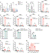
Figure 1.. Burkholderia effector TssM counteracts the activity of RNF213.
(A) Representative confocal microscopy images of RNF213-knockout (RNF213KO) mouse embryonic fibroblasts (MEFs) stably expressing GFP-RNF213, infected with B. thailandensis E264 (bottom panel) or E264 pH4-GroS-RFP (top panel) and fixed at 6 h post-invasion (p.i.). Samples were labelled with an anti-ubiquitin (Ub) antibody when indicated. Scale bar - 10 μm. (B) Immunofluorescence-based quantification of marker-positive B. thailandensis E264 and E555 strains in RNF213KO MEFs stably expressing GFP-RNF213. Ub localization was quantified following infection of WT MEFs and immunolabelling. (C) Immunoblots of indicated Salmonella (Sal) and B. thailandensis (Bt) strains isolated from infected MEFs at 4 h or 24 h p.i., respectively or non-infected (NI) cells as a control. Cell lysates and isolated intracellular bacteria were immunoblotted with the indicated antibodies. DnaK and GroEL were used as loading controls for Salmonella and Burkholderia, respectively. (D) Immunoblot analysis of Δrfc Salmonella strain isolated from infected HEK293ET cells that were transiently transfected with plasmids encoding the indicated N-terminal GFP-tagged effector from B. pseudomallei or B. thailandensis. Cell lysates and isolated intracellular bacteria were immunoblotted with the indicated antibodies. (E) Immunofluorescence-based quantification of the percentage of Ub-coated bacteria over time in WT MEFs infected with indicated B. thailandensis strains. (F) Immunoblot analysis of indicated B. thailandensis strains isolated from infected WT or RNF213KO MEFs at 24 h p.i. (G) Immunoblot analysis of HEK293ET cells transiently transfected with plasmids encoding the indicated GFP-tagged effector and infected with the Bt E264::tssMpknock strain. Mock transfected cells (mock TF), refers to cells that did not receive DNA. Values show mean of three biological repeats ± SEM (B,E). Other data are representative of at least three biological repeats. Statistical significance was assessed by two-way ANOVA with Sidak’s multiple comparisons test (E); **** < 0.0001. See also Figure S1.
Figure 2.
Figure 2.. TssM blocks ubiquitin accumulation and autophagy receptor recruitment.
Colony forming unit assays were used to determine the fold replication at 6 h p.i. of (A) the indicated Burkholderia strains in MEFs or RAW264.7 macrophages, and (B) of Salmonella in HEK293ET cells expressing the indicated GFP-tagged protein. Quantification of the percentage of (C) K63-linked or (D) M1-linked ubiquitin-positive bacteria. Quantification of the percentage of indicated B. thailandensis strain colocalising with (E) GFP-NDP52, (F) GFP-p62, (G) GFP-OPTN, (H) GFP-RNF213, (I) Ubiquitin, (J) GFP-NDP52, (K) GFP-LC3B, and (L) GFP-WIPI2B, as determined by microscopy at 6 h p.i. (M) Percentage of marker-positive B. thailandensis among ubiquitin-coated bacteria in WT MEFs. Data represent the mean ± SEM of at least three independent biological repeats. Statistical significance was assessed by two-way ANOVA with Tukey’s multiple comparisons test (C-G,K,L) or one-way ANOVA (A,B, H-J, M); * P < 0.05; ** P < 0.01; **** < 0.0001. See also Figure S1.
Figure 3.
Figure 3.. TssM is a ubiquitin esterase that hydrolyses ubiquitylated LPS.
(A) Emerald 300 stain of LPS from indicated B. thailandensis strains grown in LB. (B) Immunoblot analysis of B. thailandensis strains isolated from infected MEFs, 24 h p.i. (C) Bacteria isolated from MEFs infected with the E555::ΔwbiI,ΔtssM strain were lysed and incubated with 100, 200 or 400 mM NaOH for 30 min prior to immunoblot analysis. CT: non-treated control (D) Representative FP data monitoring cleavage of Rho-S(Ub)G (Ser-Ub), Rho-T(Ub)G (Thr-Ub), and Tamra-K(Ub)G (Lys-Ub) substrates following addition of the DUB (indicated by an asterisk). (E) Catalytic efficiencies (mean ± SEM of three repeats) for all enzyme-substrate combinations, with the exception of JOSD1 which had no detectable isopeptidase activity. (F) Bacteria isolated from MEFs infected with the E555::ΔwbiI,ΔtssM strain were incubated with recombinant His-GST-tagged TssMBpΔN191 (rTssMBp) +/− iodoacetamide (IA) for 30 min prior to immunoblot analysis. Data representative of three biological repeats (A-D and F). See also Figure S2.
Figure 4.
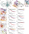
Figure 4.. Structural basis of TssMBp esterase activity.
(A) Crystal structure of TssMBpΔN191 (teal) bound to Ub (grey). (B) Close-up of the TssM USP domain coloured by box regions 1–6, with Ub (grey) shown in the S1 site. Representative 2|Fo|-|Fc| electron density is shown at 1s. (C) TssM catalytic triad C292, H426, and D441, as well as the oxyanion hole N286 are shown. Hydrogen bonds are shown as dashed lines. Effects of mutation on TssM activity are shown for (D) Ser-Ub and (E) Lys-Ub substrates. (F) The extended TssM Cys-loop (red) in comparison to a typical USP Cys-loop (grey). Activity data for TssM truncations ΔL287 and ΔD288 are shown for (G) Ser-Ub and (H) Lys-Ub substrates. (I) Coordination of the Ub C-terminus and R42 by TssM E362 and E469. Activity data of E362R and E469R mutants are shown for (J) Ser-Ub and (K) Lys-Ub substrates. (L) Coordination of the Ub I44 hydrophobic patch by TssM S1 site residues Y402, F459, and V466 is shown. The effects of mutations on cleavage of (M) Ser-Ub and (N) Lys-Ub are shown. In all panels, DUB addition is indicated by an asterisk. WT and control data in panels D and G, E and H, J and M, and K and N are identical and reproduced for clarity as data were collected in the same experiment. All FP data shown represent mean ± SEM of three biological repeats. See also Figure S3, S4 and Table S1.
Figure 5.
Figure 5.. Esterase-specific activity of the TssMBp V466R mutant.
(A) Rate constants and correlating catalytic efficiency (kcat/KM) derived for esterase-specific TssMBpΔN191 V466R are shown as mean ± SEM of three biological repeats. (B) Chemical and enzymatic treatment of K63-linked di-ubiquitin and ester-linked ubiquitylated maltoheptaose. HOIL-1 was used to assemble ubiquitylated maltoheptaose, and samples of the reaction were treated with reducing agent, base, the nonspecific vOTU (marked with an asterisk), or the indicated TssM variants. (C) Immunoblot analysis of indicated B. thailandensis strains isolated from infected WT MEFs at 24 h p.i. Data representative of three biological repeats (B,C). See also Figure S4.
Figure 6.
Figure 6.. Esterase activity in other bacterial peptidases.
(A) Representative FP data monitoring cleavage of Ser-Ub, Thr-Ub, and Lys-Ub substrates following addition of the DUB (indicated by an asterisk). (B) Immunoblot analysis of Δrfc Salmonella isolated at 6 h p.i. from infected HEK293ET cells transiently expressing the indicated GFP-tagged bacterial cysteine hydrolases. The panel includes SseL and SpvD from Salmonella,, ChlaDUB1 and ChlaDUB2 from Chlamydia as well as LotA from Legionella, ShiCE from Shigella and RickCE from Rickettsia. (C) Bt E555::ΔtssM,ΔwbiI mutant bacteria isolated from infected MEF cells were treated with 0.5 μM of the indicated purified bacterial DUB. (D) Immunoblot analysis of Δrfc Salmonella isolated from infected cells expressing the indicated GFP-tagged putative C19 peptidases of Burkholderia (B sp.), Parachlamydia acanthamoebae (Pa), Simkania negevensis (Sn), Waddlia chondrophila (Wc) and Chromobacterium sinusclupearum (Cs). (E) Representative FP data monitoring cleavage of Ser-Ub, Thr-Ub, and Lys-Ub substrates with 0.5 μM TssMCs. Data were collected in triplicate and (F) rate constants and (G) catalytic efficiency of the TssMCs protein towards each substrate are shown as mean ± SEM. (H) Immunoblot analysis of E555::ΔtssM,ΔwbiI B. thailandensis isolated from infected MEFs and incubated with control (CT), recombinant His-GST-tagged TssMBpΔN191 (rTssMBp) or TssMCs +/− iodoacetamide (IA) for 30 min. (I) Structural alignment of the active site of TssM (grey) and the AlphaFold model of TssMCs (coloured) showing similarity between the two proteins surrounding the active site. Catalytic residues for TssM are labelled, with aligned residues in TssMCs labelled in italics. rTssMBp refers to recombinant His-GST-tagged TssMBpΔN191 (C,H). Data representative of three biological repeats (A-F,H) or mean ± SEM of three biological repeats (G). See also Figure S5 and S6.

Update of

References

    1. Huang S, Meng Q, Maminska A, and MacMicking JD (2019). Cell-autonomous immunity by IFN-induced GBPs in animals and plants. Curr Opin Immunol 60, 71. 10.1016/J.COI.2019.04.017. - DOI - PMC - PubMed
    1. Randow F, MacMicking JD, and James LC (2013). Cellular Self-Defense: How Cell-Autonomous Immunity Protects Against Pathogens. Science 340, 701–706. 10.1126/SCIENCE.1233028. - DOI - PMC - PubMed
    1. Thery F, Martina L, Asselman C, Zhang Y, Vessely M, Repo H, Sedeyn K, Moschonas GD, Bredow C, Teo QW, et al. (2021). Ring finger protein 213 assembles into a sensor for ISGylated proteins with antimicrobial activity. Nat Commun 12. 5772. 10.1038/S41467-021-26061-W. - DOI - PMC - PubMed
    1. Walsh SC, Reitano JR, Dickinson MS, Kutsch M, Hernandez D, Barnes AB, Schott BH, Wang L, Ko DC, Kim SY, et al. (2022). The bacterial effector GarD shields Chlamydia trachomatis inclusions from RNF213-mediated ubiquitylation and destruction. Cell Host Microbe 30, 1671–1684.e9. 10.1016/j.chom.2022.08.008. - DOI - PMC - PubMed
    1. Otten EG, Werner E, Crespillo-Casado A, Boyle KB, Dharamdasani V, Pathe C, Santhanam B, and Randow F (2021). Ubiquitylation of lipopolysaccharide by RNF213 during bacterial infection. Nature 594, 111–116. 10.1038/s41586-021-03566-4. - DOI - PMC - PubMed

LinkOut - more resources