. 2024 Jun 20;84(12):2337-2352.e9.
doi: 10.1016/j.molcel.2024.05.018. Epub 2024 Jun 12.

K29-linked free polyubiquitin chains affect ribosome biogenesis and direct ribosomal proteins to the intranuclear quality control compartment

Affiliations

K29-linked free polyubiquitin chains affect ribosome biogenesis and direct ribosomal proteins to the intranuclear quality control compartment

Harsha Garadi Suresh et al. Mol Cell. .

Abstract

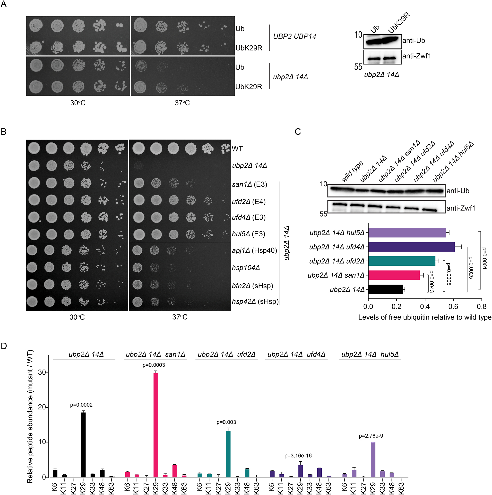
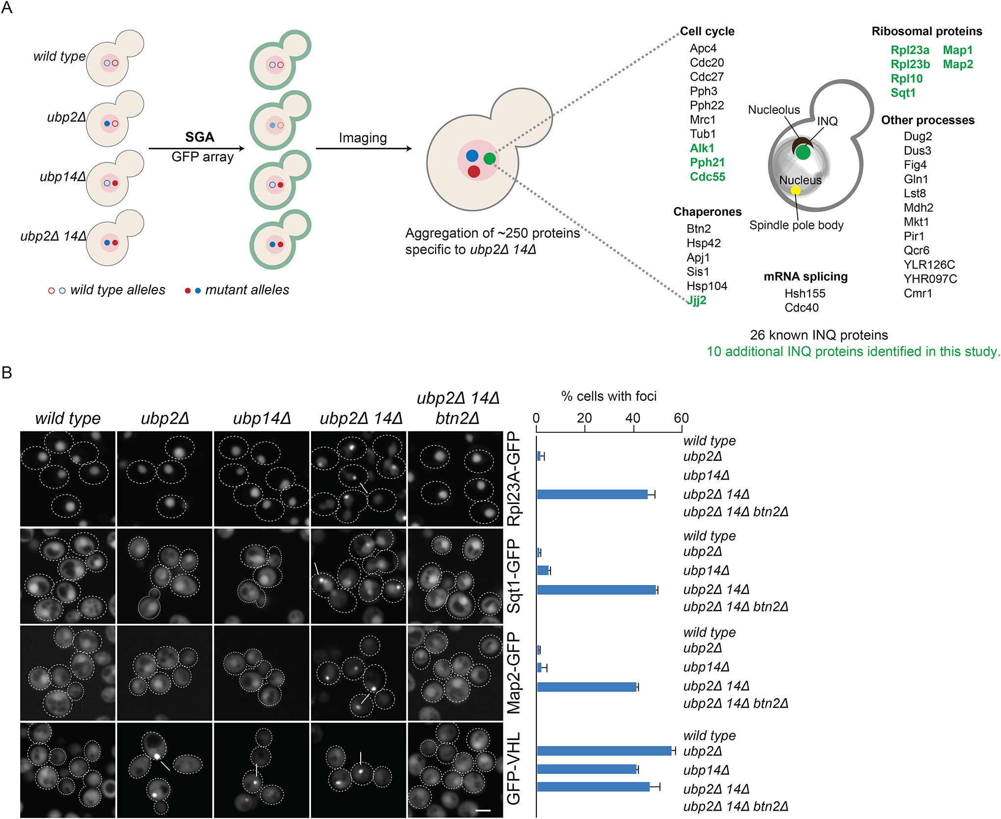
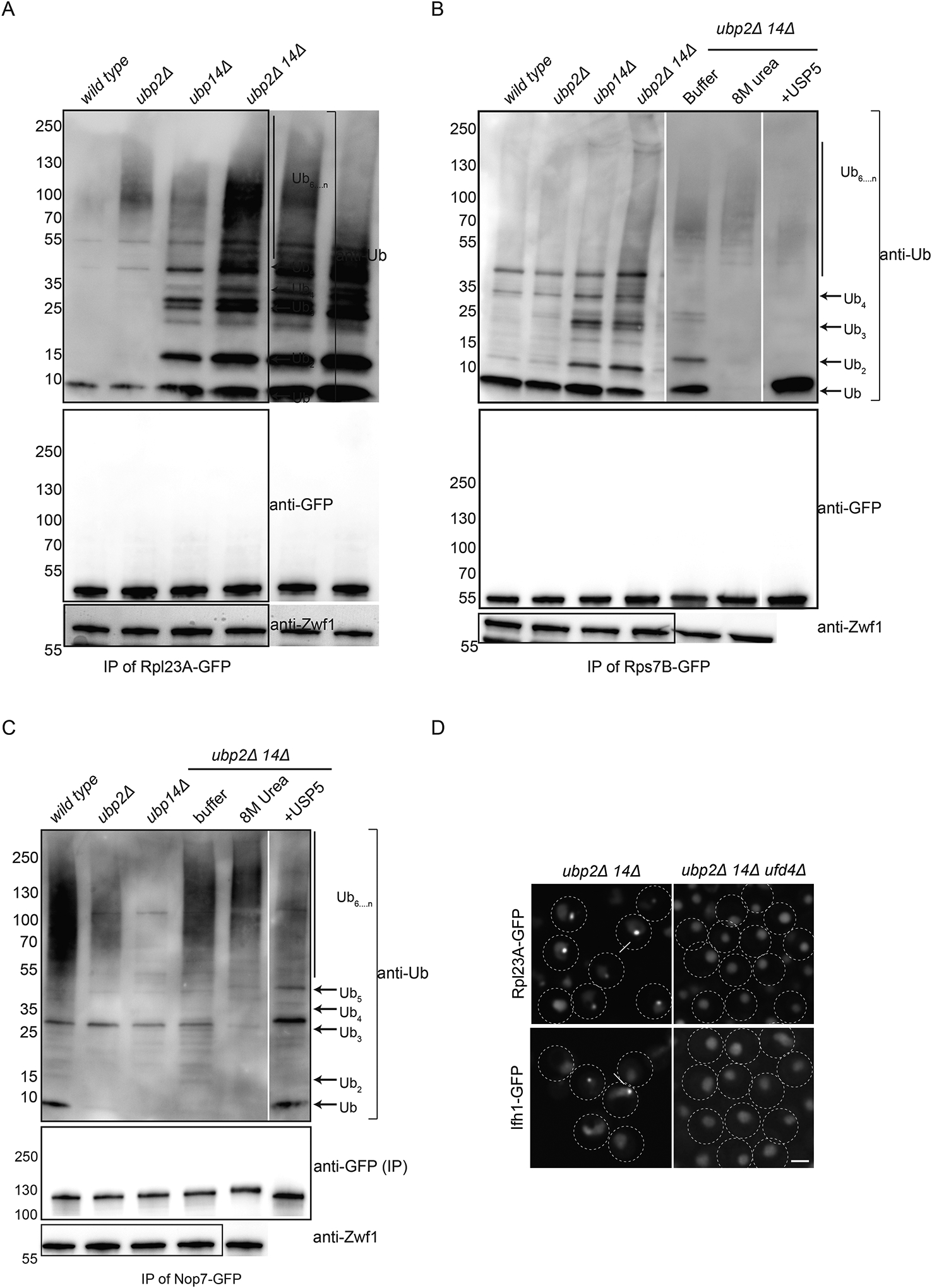
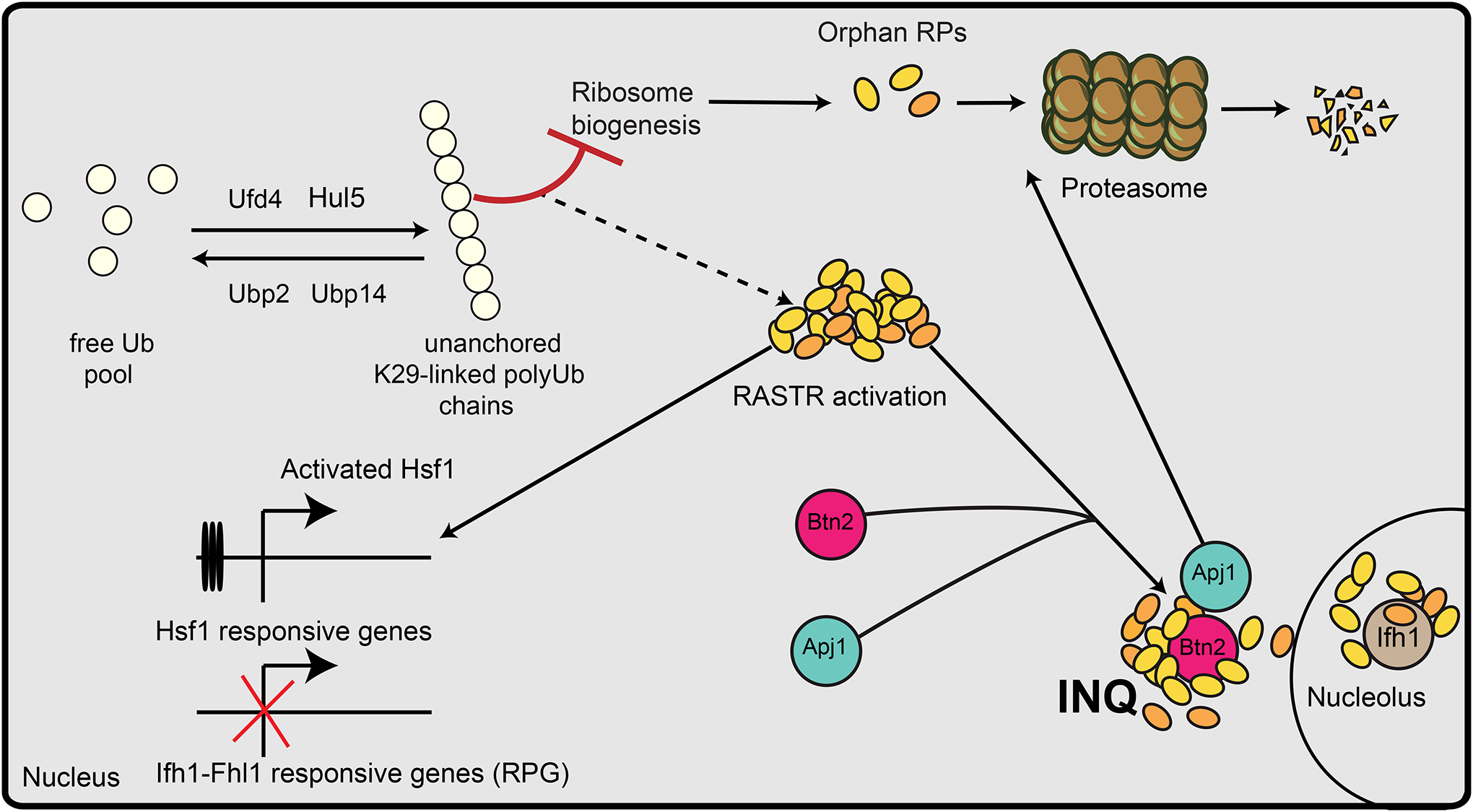
Ribosome assembly requires precise coordination between the production and assembly of ribosomal components. Mutations in ribosomal proteins that inhibit the assembly process or ribosome function are often associated with ribosomopathies, some of which are linked to defects in proteostasis. In this study, we examine the interplay between several yeast proteostasis enzymes, including deubiquitylases (DUBs) Ubp2 and Ubp14, and E3 ligases Ufd4 and Hul5, and we explore their roles in the regulation of the cellular levels of K29-linked unanchored polyubiquitin (polyUb) chains. Accumulating K29-linked unanchored polyUb chains associate with maturing ribosomes to disrupt their assembly, activate the ribosome assembly stress response (RASTR), and lead to the sequestration of ribosomal proteins at the intranuclear quality control compartment (INQ). These findings reveal the physiological relevance of INQ and provide insights into mechanisms of cellular toxicity associated with ribosomopathies.

Keywords: DUB-E3 ligase interplay; intranuclear quality control compartment; protein aggregation; ribosome assembly stress response; ribosomopathies; ubiquitin homeostasis; unconventional K29-linked unanchored polyubiquitin chains.


Conflict of interest statement

Declaration of interests The authors declare no competing interests.

Figures

Figure 1:
Figure 1:. Redundant DUBs Ubp2 and Ubp14 recycle K29-linked unanchored polyUb chains.
(A) Serial five-fold spot dilutions of DUB mutants. The indicated strains were plated onto minimal medium and incubated at 30°C or 37°C for 1 day (WT= wild type). (B) Immunoblot of the ubiquitinome of the indicated yeast strains enriched using Tandem Ubiquitin Binding Entity (TUBE) affinity matrix-based pull downs. Arrows indicate equidistant prominent bands of ubiquitin. Anti-Zwf1 serves as a loading control. (C) Label-free quantitative mass spectrometry on ubiquitylated peptides isolated using Lys-ℇ-Gly-Gly (diGly) antibodies (n=3). Fraction of proteins that are enriched (≥2-fold, P ≤ 0.05; red) or depleted (<2-fold, P ≤ 0.05; blue) for ubiquitin modification in ubp2Δ, ubp14Δ or ubp2Δ14Δ double mutant cells relative to wild type cells are shown. The fraction of proteins whose ubiquitylation status remained unchanged is shown in grey. (D) Graph summarizing the relative number of 7 different polyUb chains quantified in the indicated DUB mutants using label-free quantitative Lys-ℇ-Gly-Gly (diGly) proteomics mass spectrometry. Error bars indicate standard deviation (n=3) and p-values are derived from paired two-tail student’s t-test. (E) Graph showing the relative amount of free monomeric ubiquitin in the indicated mutants compared to a wild type strain. Error bars indicate standard deviation (n=3) and p-values are derived from unpaired two-tail student’s t-test. Representative immunoblots are shown in the upper panel. (F) Immunoblot with anti-Ub antibodies of unanchored polyUb chains purified from wild-type and the indicated yeast mutant extracts using Sepharose beads coupled to the ZnF-UBP domain of human Usp5. (G) Immunoblot of unanchored polyUb chains purified from the indicated yeast strain extracts using Sepharose-ZnF-UBP domain of human Usp5 and using sAB-K29 antibodies that specifically recognize K29-linked polyUb chains. (H) Co-immunoprecipitation of polyUb chains with the GFP-tagged NZF1 domain of TRABID using anti-GFP affinity matrices. Immunoprecipitates from the indicated yeast strains were probed with anti-Ub and anti-GFP antibodies. (I) Immunoblot with anti-Ub antibodies of the Ub variant UbG(75,76)V-HA purified using Sepharose-coupled anti-HA beads from lysates derived from the indicated strains. Arrows indicate equidistant prominent bands of ubiquitin.
Figure 2:
Figure 2:. Ufd4 and Hul5 E3 ubiquitin ligases are involved in the synthesis of K29-linked unanchored polyubiquitin chains.
(A) Serial five-fold spot dilutions of a wild-type strain (UBP2 UBP14) or a ubp2Δ 14Δ mutant expressing either wild type (Ub) or a UbK29R mutant as the sole source of ubiquitin. Plates were incubated at the indicated temperatures and imaged after 48 hours. An immunoblot monitoring free ubiquitin levels in ubp2Δ 14Δ mutants expressing either wild type Ub or the UbK29R mutant as the sole source of Ub is shown on the right (B) Five-fold serial spot dilutions of a wild type strain (WT) a ubp2Δ 14Δ double mutant (top two rows) and ubp2Δ 14Δ double mutants carrying deletions of the indicated genes. Plates were incubated at 30°C or 37°C as indicated. E3= E3 ligase; E4=E4 ligase; sHsp – small Heat Shock Protein. (C) Quantifications of immunoblots measuring the relative amount of free monomeric ubiquitin in the indicated mutants grown at 37°C (n=3). A representative anti-Ub immunoblot is shown above the graph. Quantifications are depicted with error bars indicating standard deviations and p-values derived from unpaired two-tail student’s t-test. The position of migration of relevant molecular markers is indicated on the left. (D) Relative abundance of 7 different polyUb chains quantified in the indicated yeast mutant strains using label-free quantitative diGlyproteomics mass spectrometry (n=3). Error bars indicate standard deviations and p-values were derived from paired two-tail student’s t-test.
Figure 3:
Figure 3:. Proteome-wide imaging screens reveal ubp2Δ ubp14Δ-dependent defects in ribosome homeostasis.
(A) Schematic depicting high-throughput imaging of the yeast GFP library in wild type and DUB mutant yeast strains. Proteins that accumulate at the Intranuclear quality control site (INQ) are highlighted with previously known (black) and those identified in this study (green). The spindle pole body (yellow), nucleolus (brown), and INQ (green) are depicted in the cell cartoon. (B) Example micrographs of yeast cells expressing specific GFP-fusion proteins in the indicated mutant strains. Arrows indicate GFP-protein aggregates. The percentage of cells with the indicated GFP-fusion protein inclusions is shown (right panel). Error bars indicate standard deviation (n=2). Scale bar: 3 μm
Figure 4:
Figure 4:. Ribosome biogenesis is disrupted in a ubp2Δ ubp14Δ double mutant.
(A) Northern blot of 35S rRNA processing. Levels of 35S, 33S/32S, 27SA2, 24S (upper and lower panels), and 7S (lower panel) rRNAs in the indicated yeast strains were measured in three independent biological replicates (R1-R3). The relative levels of various processed forms of 35S rRNA to its unprocessed form were quantified on the right. P-values are derived from unpaired two-tailed student’s t-test. ns=not significant (B) Polysome profiles (OD260) of wild type and the indicated DUB mutant strains are shown. Gradient fractions corresponding to 40S, 60S, 80S, and polysomes are indicated. The dotted box on each profile encompass the 40S and 60S subunits of the ribosome and those regions are enlarged to the right of each profile with arrows indicating the distance between the peaks of 40S and 60S subunits. Relative levels of 60S/40S subunits were quantified from 3 biological replicates of the polysome profiles and the results are depicted on the right. Error bars indicate standard deviations (n=3) and p-values are derived from unpaired two-tailed student’s t-test. ns=not significant; au=arbitrary units (C) Example micrographs of wild-type and DUB mutant strains expressing the indicated GFP-fusion proteins. Protein inclusions (red arrows) and perinucleolar protein localization (white arrows) are indicated. Scale bar: 3 μm (D) (upper panel) The ribosome assembly stress response (RASTR) induced by auxin-dependent depletion of Utp13, which is required for rRNA processing. Box plots show RNAPII binding (as measured by Rpb1 ChIP-seq) to promoters of ribosomal protein-encoding genes (RPG; n=139), HSF1 target genes (HSF1; n=19) or other genes (n = 4884). (lower panel) Box plots comparing RNAPII binding (as measured by Rpb1 ChIP-seq) in DUB mutant cells relative to wild type cells in the same gene categories described above. p-values are derived from the Wilcoxon test.
Figure 5:
Figure 5:. Unanchored polyubiquitin chains physically associate with ribosomes.
(A-C) Co-immunoprecipitation of polyUb with either Rpl23A-GFP (A), Rps7B-GFP (B) or Nop7-GFP (C). Anti-GFP immunoprecipitates of lysates derived from the indicated yeast strains were probed with anti-Ub and anti-GFP antibodies. Immunoprecipitates derived from ubp2Δ14Δ double mutant lysates were also treated with either purified Usp5, 8M urea, or buffer alone, as indicated above the relevant lanes. Ubiquitin species are indicated on the right and molecular weight markers are shown on the left. (D) Example micrographs of the indicated yeast mutant strains expressing Rpl23A-GFP or Ifh1-GFP. Arrows indicate GFP protein fusion aggregates. Scale bar: 3 μm
Figure 6:
Figure 6:. A model for ribosome biogenesis defects mediated by K29-linked polyubiquitin chains.
Opposing the activity of Ubiquitin E3 ligases (Ufd4 and Hul5), DUBs Ubp2 and Ubp14 recycle the K29-linked unanchored polyUb chains and maintain the free Ub pool. In the absence of Ubp2 and Ubp14, K29-linked unanchored polyUb chains accumulate to form inclusions and disrupt ribosome biogenesis, which triggers the Ribosome assembly stress response (RASTR) that is characterized by activation of Hsf1 target genes (such as BNT2 and APJ1) and accumulation of numerous proteins, including ribosomal proteins, their assembly and associated factors, at the Intranuclear Quality control compartment (INQ). INQ accumulation occurs in a Btn2-dependent manner along with sequestration of the transcription factor Ifh1 at the nucleolus.

Update of

Similar articles

Cited by

References

    1. Rape M (2018). Ubiquitylation at the crossroads of development and disease. Nat Rev Mol Cell Biol 19, 59–70. 10.1038/nrm.2017.83. - DOI - PubMed
    1. Komander D, and Rape M (2012). The ubiquitin code. Annu Rev Biochem 81, 203–229. 10.1146/annurev-biochem-060310-170328. - DOI - PubMed
    1. Clague MJ, Urbe S, and Komander D (2019). Breaking the chains: deubiquitylating enzyme specificity begets function. Nat Rev Mol Cell Biol 20, 338–352. 10.1038/s41580-019-0099-1. - DOI - PubMed
    1. Hanna J, Leggett DS, and Finley D (2003). Ubiquitin depletion as a key mediator of toxicity by translational inhibitors. Mol Cell Biol 23, 9251–9261. 10.1128/MCB.23.24.9251-9261.2003. - DOI - PMC - PubMed
    1. Stavreva DA, Kawasaki M, Dundr M, Koberna K, Muller WG, Tsujimura-Takahashi T, Komatsu W, Hayano T, Isobe T, Raska I, et al. (2006). Potential roles for ubiquitin and the proteasome during ribosome biogenesis. Mol Cell Biol 26, 5131–5145. 10.1128/MCB.02227-05. - DOI - PMC - PubMed

MeSH terms

LinkOut - more resources