. 2024 Jul 25;187(15):3888-3903.e18.
doi: 10.1016/j.cell.2024.05.026. Epub 2024 Jun 12.

Correction of age-associated defects in dendritic cells enables CD4+ T cells to eradicate tumors

Affiliations

Correction of age-associated defects in dendritic cells enables CD4+ T cells to eradicate tumors

Dania Zhivaki et al. Cell. .

Abstract

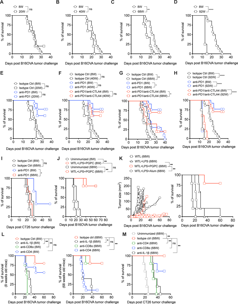
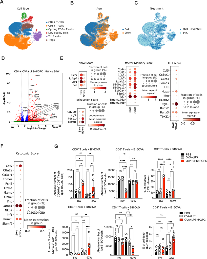
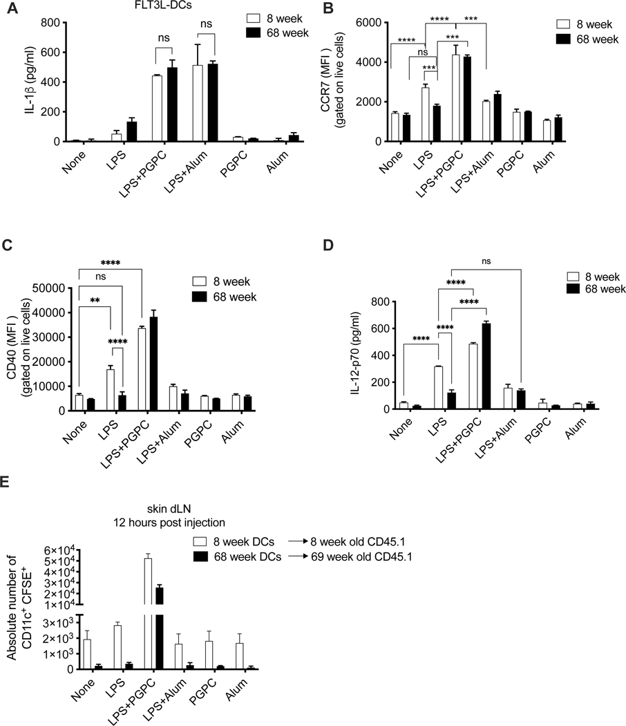
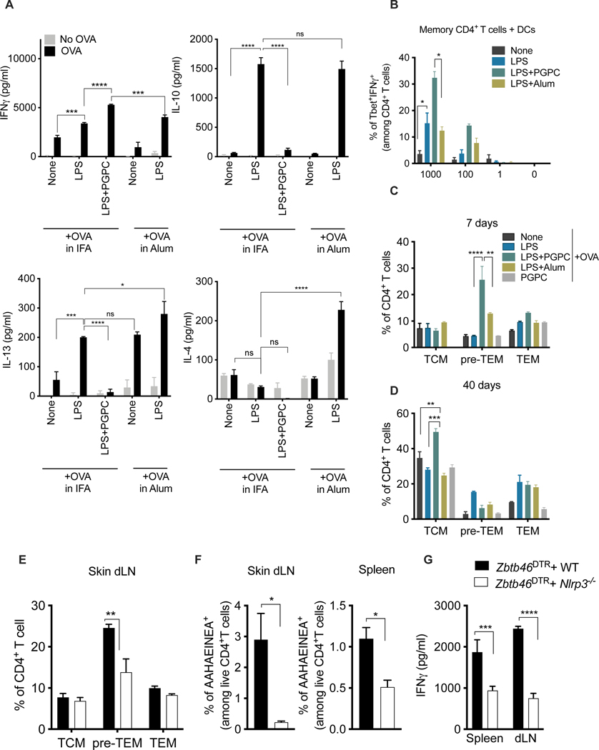
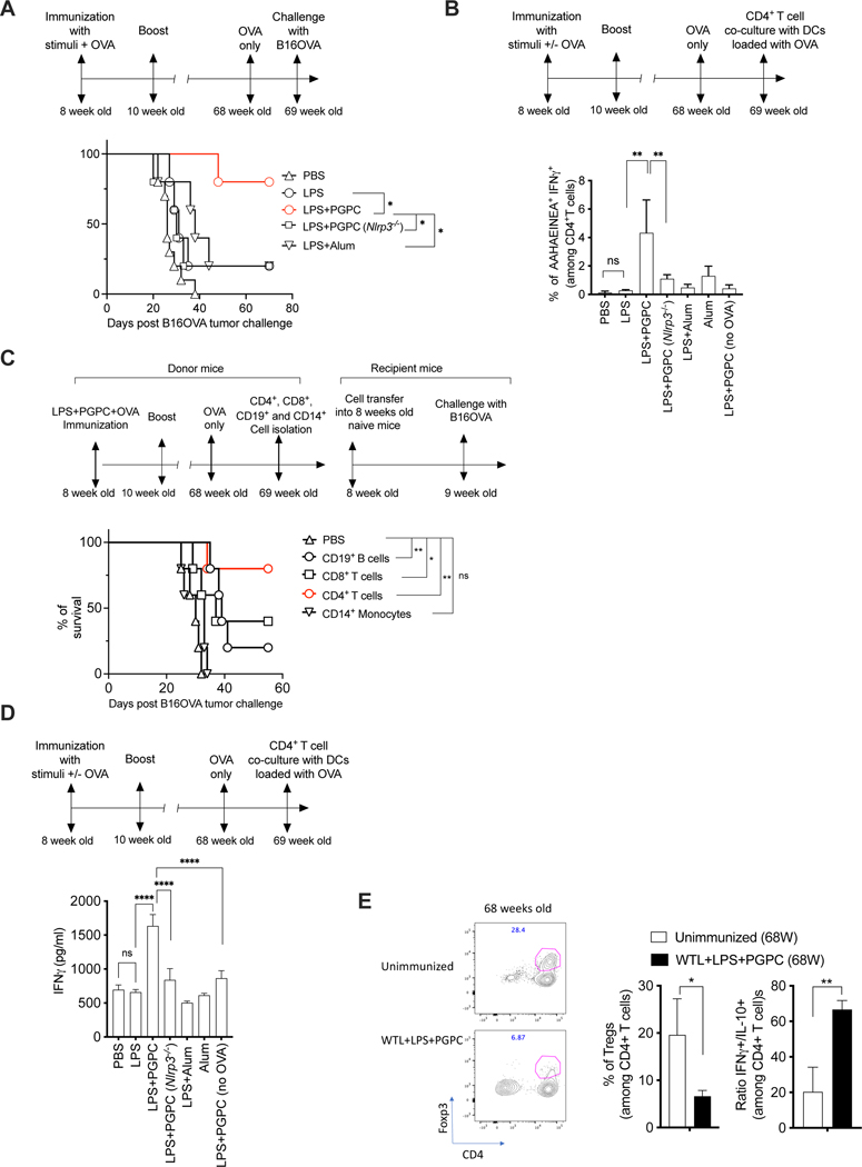
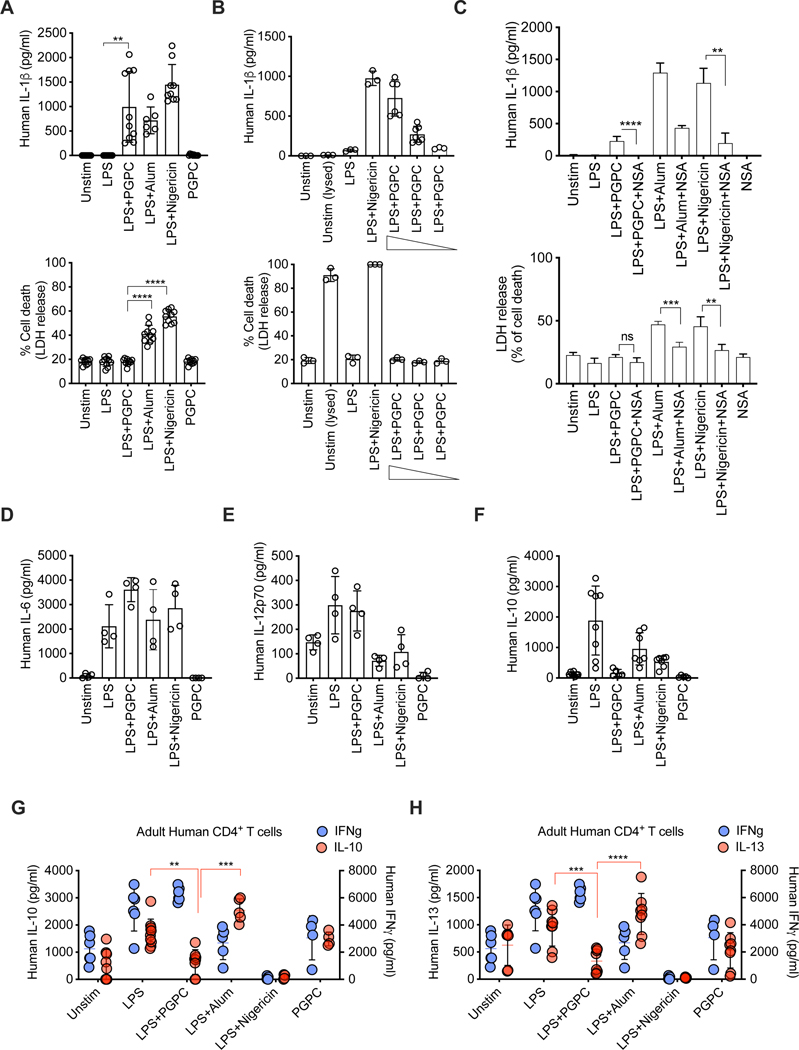
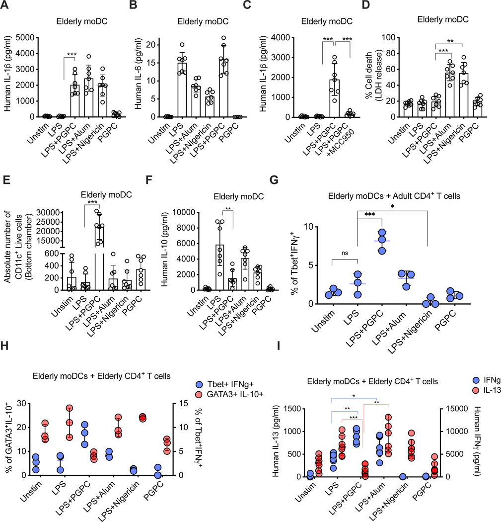
Defective host defenses later in life are associated with changes in immune cell activities, suggesting that age-specific considerations are needed in immunotherapy approaches. In this study, we found that PD-1 and CTLA4-based cancer immunotherapies are unable to eradicate tumors in elderly mice. This defect in anti-tumor activity correlated with two known age-associated immune defects: diminished abundance of systemic naive CD8+ T cells and weak migratory activities of dendritic cells (DCs). We identified a vaccine adjuvant, referred to as a DC hyperactivator, which corrects DC migratory defects in the elderly. Vaccines containing tumor antigens and DC hyperactivators induced T helper type 1 (TH1) CD4+ T cells with cytolytic activity that drive anti-tumor immunity in elderly mice. When administered early in life, DC hyperactivators were the only adjuvant identified that elicited anti-tumor CD4+ T cells that persisted into old age. These results raise the possibility of correcting age-associated immune defects through DC manipulation.

Keywords: PGPC; T cells; cancer; checkpoint inhibitor therapy; dendritic cells; elderly; hyperactivation; immunotherapy; innate immunity; tumor immunity; vaccine.


Conflict of interest statement

Declaration of interests J.C.K. consults and holds equity in Corner Therapeutics, Larkspur Biosciences, MindImmune Therapeutics, and Neumora Therapeutics. R.J.X. is a co-founder of Celsius and Jnana Therapeutics. None of these relationships impacted this study.

Figures

Figure 1.
Figure 1.. CD4+ T cells mediate anti-tumor immunity in old mice
(A-H) Mice were injected s.c. on the right flank with B16OVA cells. (A-D) Survival was monitored every 2 days and compared to 8 week old mice (n=5–14 mice per group). (E-H) When tumors reached 2–3 mm of size, mice were treated with the antibodies indicated. Survival was monitored as in (A) (n=5–14 mice per group). (I) Mice were injected s.c. on the right flank with CT26 cells. When tumors reached 2–3 mm of size, mice were either treated with the antibodies indicated. Survival was monitored as in (A). (J-K) Mice of the ages indicated were injected s.c. on the right flank with B16OVA cells. (J) When tumors reached 2 mm, B16OVA tumor-bearing mice were left unimmunized or immunized with B16OVA WTL+LPS+PGPC emulsified in IFA. Survival was monitored every 2 days. (K) 68 week old mice were immunized on the left flank WTL and the adjuvants indicated. Tumor size in mm2 was measured every two-three days (left panel). Survival was monitored every 2 days (right panel) (n=5 mice per group). Data are representitave of two independent experiments. (L) Mice of the ages indicated were injected s.c. on the right flank with B16OVA cells. When tumors reached 3mm in size, mice were immunized with WTL+LPS+PGPC and the antibodies indicated. Survival was monitored every 2 days (n=5 mice per group). (M) 68 week old Balbc mice were injected s.c. on the right flank with CT26 cells. When tumors reached 3mm in size, mice were either left unimmunized or were immunized with WTL+LPS+PGPC and the antibodies indicated. Survival was monitored every 2 days (n=5 mice per group).
Figure 2.
Figure 2.. CD4+ T cells acquire TH1 and cytotoxic anti-tumor activity in elderly mice following immunization with hyperactivators.
(A-G) Mice of the ages indicated were injected with PBS or immunized with OVA+LPS or OVA+LPS+ PGPC. Immunized mice received two boost injections. (A-H) 7 days after the last boost, skin dLNs were collected and CD3e+ T cells were enriched and sequenced (n=5 mice per group). (A) Uniform manifold approximation and projection (UMAP) visualization of T cell subsets in the dLN post leiden clustering. (B) UMAP visualization of T cells, color indicates the age of the mouse. (C) UMAP visualization of T cells, color indicates the immunization that the mouse received. (D) Volcano plot showing the differentially expressed genes comparing the CD4+ T cells of 90 week old mice immunized with OVA+LPS+PGPC to 8 week old mice. DESeq2 was used for differential gene expression analysis on the pseudobulked samples. (E-F) Dotplot visualization of gene expression for T cell state marker genes. Color indicates the mean expression of the log-normalized counts of each gene within the group. The size of the dot indicates the percentage of cells in each group that expressed the gene. (G) Splenic CD8+ T cells and CD4+ T cells from mice of the ages indicated were co-cultured with B16OVA cells (target cells) for 5 hours. T cell degranulation was assessed by monitoring the number of T cells expressing CD107a+. B16OVA cell viability was assessed by flow cytometry and LDH release (n=5 mice per group). Data are representitave of two independent experiments.
Figure 3.
Figure 3.. Hyperactivating stimuli correct migration defects in aging DCs.
(A-B) BMDCs from mice of the ages indicated were left untreated (“None”) or treated with LPS, Alum, or PGPC for 24h, or BMDCs were primed for 3h with LPS, then treated with indicated stimuli for 21h. (A) IL-1b release was monitored by ELISA. (B-C) The mean fluorescence intensity (MFI) of surface markers indicated (among CD11c+live cells) was measured by flow cytometry. (D) BMDCs were treated as described in A and cultured for 24 hours onto an anti-CD40 coated plate. IL-12p70 release was monitored by ELISA. Means and SDs from three replicates are shown. Data are representative of at least three independent experiments. (E) DCs were treated as in A on a rotator overnight. Cells were washed then labeled with CFSE and injected s.c. on the right flank in CD45.1 elderly or young mice as indicated. 12 hours post injection, the number of CD45.2+CFSE+ among CD11c+ live cells in the skin dLN was calculated by flow cytometry. Means and SDs from five mice are shown;data are representative of at least three independent experiments.
Figure 4.
Figure 4.. DC hyperactivators enhance CD4+ reactivation of TH1 cells and accelerate memory T generation.
(A) 8 week old mice were injected s.c. on the right flank with OVA either alone or with LPS, LPS+PGPC emulsified in either IFA or in Alum as indicated. At the days indicated post-immunization, skin dLN CD4+ T cells were co-cultured with BMDC loaded (or not) with OVA. Cytokine secretion was measured by ELISA. Means and SDs of 4 mice are shown and are representative of 3 independent experiments. (B) 8 week old mice received three injections s.c. with OVA emulsified in IFA every 7 days. 7 days after the last boost, skin dLN CD4+ T cells were co-cultured with BMDCs that were pretreated with indicated stimuli and loaded (or not) with OVA, as indicated. Five days post co-culture, the percentage of Tbet+IFNg+T cells was measured by flow cytometry. Means and SDs of five mice are shown and are representative of 2 independent experiments. (C-D) 8 week old mice were injected s.c. on the right flank with OVA and the adjuvants indicated. On the days indicated post immunization, the percentage of skin dLN cells were examined. Pre-TEM were identified as CD44loCD62Llo, T effector memory cells (TEM) as CD44hiCD62L, and T central memory cells (TCM) as CD44hi CD62Lhi are represented by gating on CD3+CD4+ live cells. (E-G) 8 week old CD45.1 mice were irradiated then reconstituted with mixed bone marrow (BM) of the genotypes indicated. Six weeks post-reconstitution, chimera mice were injected with DTx 3 times a week for a total of 9 DTx injections. Chimeric mice were then immunized s.c. on the right flank with OVA+LPS+PGPC. (E) Seven days post-immunization, the percentage of pre-TEM, TEM, TCM, and T naive cells in the skin dLN was measured by flow cytometry. (F) The percentage of AAHAEINEA+ among CD4+ live T cells from the spleen and dLN was measured using OVA peptide tetramer staining. Means and SDs of five mice are shown. (G) Skin dLN CD4+ T cells were co-cultured with BMDCs loaded with OVA at a ratio of 1:10 (DC:T cell) for 4 days. IFN-γ secretion was measured by ELISA. Means and SDs of five mice are shown.
Figure 5.
Figure 5.. Hyperactivating stimuli induce long-lived antigen specific CD4+ T cells that eradicate tumors.
(A-C) 8 week old WT or Nlrp3−/−mice mice were immunized s.c. on the right flank with OVA and the adjuvants, as indicated. Two weeks later mice then received a boost with OVA and the same adjuvant. (A) At 69 weeks, mice were injected with B16OVA cells on the left flank. Survival was monitored every 2 days (n=5 mice per group). (B) At 69 weeks, CD4+ T cells were co-cultured with BMDCs loaded with OVA for 4 days. The percentage of AAHAEINEA+ IFNγ+ among CD4+ live T cells was measured using OVA peptide tetramer staining followed by intracellular IFNγ staining. Means and SDs of five mice are shown. (C) 8 week old mice were immunized s.c. on the right flank with OVA+LPS+PGPC emulsified in IFA, as indicated. Two weeks later mice then received a boost with OVA and the same adjuvant. At 69 weeks, the cell types indicated were isolated and transferred intravenously into 8 week old naïve mice. Prior to cell transfer, host mice were injected i.p. with neutralizing antibodies for 5 consecutive injections to deplete host CD8+ or CD4+ or CD8+ plus CD4+ T cells. One week post cell transfer, mice were challenged with B16OVA cells on the left flank. Survival was monitored every 2 days (n=5 mice per group). (D) Mice were treated as in B. IFNγ release in the supernatant was measured by ELISA. Means and SDs of five mice are shown. (E) 68 week old mice were injected s.c. on the right flank with B16OVA cells. When tumors reached 2–3mm in size, mice were either left unimmunized or were immunized with LPS+PGPC. Fifteen days post immunization, intratumoral percentage of Tregs (CD4+Foxp3+ T cells) was measured by flow cytometry (left panel). CD45 TILs were activated for 24 hours with anti-CD3/CD28, then treated with PMA and ionomycin for 5 hours. The ratio of IFNγ over IL-10 producing CD4+ TILs was measured by intracellular staining.
Figure 6.
Figure 6.. Human hyperactive moDCs drive TH1-skewed immune responses.
(A-F) Human moDCs generated from healthy adultsweretreated as indicated.. PGPC concentrations in B were being 100μg/ml, 50 μg/ml, 25 μg/ml. Cytokines and LDH release were assessed, as indicated. Means and SDs of at least 3 donors is represented. (G-H) moDC were treated as above, then cultured with allogeneic T cells for 5 days. Cytokine secretion was measured by ELISA. Means and SDs of 4–8 donors is represented.
Figure 7.
Figure 7.. moDCs become hyperactive during aging and drive TH1-skewed immune responses.
(A-F) Human moDC generated from elderly donors were treated as indicated. (A-D, F). Cytokine or LDH release were measured from 7 donors (E) moDCs were cultured in the upper chamber of a 5μm transwell plate in the presence of CCL19. After 24h, the number of cells that migrated to the bottom chamber was determined by flow cytometry. (G-I) moDC from elderly donors were treated as above, then cultured for 5 days with allogeneic CD4+T cells from adult donors. (G) The percentage of IFNγ+Tbet+ or (H) GATA3+IL-10+ and IFNγ+Tbet+ were measured by flow cytometry. (I) Human IFNγ and IL-13 secretion was measured by ELISA. Means and SDs of 7 donors is represented.

Similar articles

Cited by

  • Elixir for DCs.
    Bird L. Bird L. Nat Rev Immunol. 2024 Aug;24(8):540. doi: 10.1038/s41577-024-01065-x. Nat Rev Immunol. 2024. PMID: 38997521 No abstract available.
  • Hematopoietic aging promotes cancer by fueling IL-1⍺-driven emergency myelopoiesis.
    Park MD, Le Berichel J, Hamon P, Wilk CM, Belabed M, Yatim N, Saffon A, Boumelha J, Falcomatà C, Tepper A, Hegde S, Mattiuz R, Soong BY, LaMarche NM, Rentzeperis F, Troncoso L, Halasz L, Hennequin C, Chin T, Chen EP, Reid AM, Su M, Cahn AR, Koekkoek LL, Venturini N, Wood-Isenberg S, D'souza D, Chen R, Dawson T, Nie K, Chen Z, Kim-Schulze S, Casanova-Acebes M, Swirski FK, Downward J, Vabret N, Brown BD, Marron TU, Merad M. Park MD, et al. Science. 2024 Oct 25;386(6720):eadn0327. doi: 10.1126/science.adn0327. Epub 2024 Oct 25. Science. 2024. PMID: 39236155 Free PMC article.
  • Restoring resident tissue macrophages to combat aging and cancer.
    Park MD, Yatim N, Zhang J, Cho BA, Yoo SK, Schaefer MM, Chowell D, Puleston DJ, Merad M. Park MD, et al. Nat Aging. 2025 Aug;5(8):1383-1392. doi: 10.1038/s43587-025-00898-y. Epub 2025 Aug 14. Nat Aging. 2025. PMID: 40813807 Review.
  • Development of therapeutic cancer vaccines based on cancer immunity cycle.
    Zhang J, Zheng Y, Xu L, Gao J, Ou Z, Zhu M, Wang W. Zhang J, et al. Front Med. 2025 Aug;19(4):553-599. doi: 10.1007/s11684-025-1134-6. Epub 2025 Jul 14. Front Med. 2025. PMID: 40653561 Review.
  • Genomic and the tumor microenvironment heterogeneity in multifocal hepatocellular carcinoma.
    Yang Y, Ni Q, Li H, Sun J, Zhou X, Qu L, Wang L, Zhao C, Zhang X. Yang Y, et al. Hepatology. 2025 Sep 1;82(3):582-598. doi: 10.1097/HEP.0000000000001191. Epub 2024 Dec 12. Hepatology. 2025. PMID: 40833995 Free PMC article.

References

    1. Akbar AN, and Gilroy DW (2020). Aging immunity may exacerbate COVID-19. Science (1979) 369, 256–257. - PubMed
    1. Goronzy JJ, and Weyand CM (2017). Successful and Maladaptive T Cell Aging. Immunity 46, 364. - PMC - PubMed
    1. Banchereau J, and Steinman RM (1998). Dendritic cells and the control of immunity. Nature 1998 392:6673 392, 245–252. - PubMed
    1. Bailey KL, Smith LM, Heires AJ, Katafiasz DM, Romberger DJ, and LeVan TD (2019). Aging leads to dysfunctional innate immune responses to TLR2 and TLR4 agonists. Aging Clin Exp Res 31, 1185–1193. - PMC - PubMed
    1. Montgomery RR, and Shaw AC (2015). Paradoxical changes in innate immunity in aging: recent progress and new directions. J Leukoc Biol 98, 937–943. - PMC - PubMed

LinkOut - more resources