. 2024 Jul 18;11(7):ENEURO.0117-24.2024.
doi: 10.1523/ENEURO.0117-24.2024. Print 2024 Jul.

Cortical Face-Selective Responses Emerge Early in Human Infancy

Affiliations

Cortical Face-Selective Responses Emerge Early in Human Infancy

Heather L Kosakowski et al. eNeuro. .

Abstract

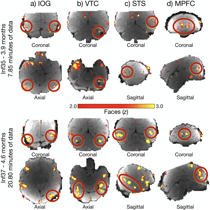
In human adults, multiple cortical regions respond robustly to faces, including the occipital face area (OFA) and fusiform face area (FFA), implicated in face perception, and the superior temporal sulcus (STS) and medial prefrontal cortex (MPFC), implicated in higher-level social functions. When in development, does face selectivity arise in each of these regions? Here, we combined two awake infant functional magnetic resonance imaging (fMRI) datasets to create a sample size twice the size of previous reports (n = 65 infants; 2.6-9.6 months). Infants watched movies of faces, bodies, objects, and scenes, while fMRI data were collected. Despite variable amounts of data from each infant, individual subject whole-brain activation maps revealed responses to faces compared to nonface visual categories in the approximate location of OFA, FFA, STS, and MPFC. To determine the strength and nature of face selectivity in these regions, we used cross-validated functional region of interest analyses. Across this larger sample size, face responses in OFA, FFA, STS, and MPFC were significantly greater than responses to bodies, objects, and scenes. Even the youngest infants (2-5 months) showed significantly face-selective responses in FFA, STS, and MPFC, but not OFA. These results demonstrate that face selectivity is present in multiple cortical regions within months of birth, providing powerful constraints on theories of cortical development.

Keywords: FFA; MPFC; OFA; STS; cerebral cortex; fMRI; faces; infant brain.


Conflict of interest statement

The authors declare no competing financial interests.

Figures

Figure 1.
Figure 1.
Awake infant fMRI scanning and stimuli. a, For each MRI visit, we swaddled the infant, applied hearing protection, and placed the infant in a custom 32-channel infant head coil. Movies were projected in a mirror over infants’ eyes. Photo credit for images in the middle and right to Caitlin Cunningham Photography. Photo credit for the image on the left to Kris Brewer. b, Example frames from dynamic face stimuli. c, Example frames from dynamic body, object, and scene stimuli. d, Infants that had enough usable data to compute a whole-brain contrast image are plotted on the x-axis, ordered by age in months. Data collected using Coil 2011 are indicated in brown; data collected using Coil 2021 are indicated in blue. The darker color indicates inclusion in fROI analyses (see Materials and Methods for inclusion criteria).
Figure 2.
Figure 2.
Data quality metrics for awake infant fMRI. a, Variable amounts of data were acquired from each 30 day participant session (dark purple) and included in subruns (light purple). Bars indicate mean minutes of data per participant session; error bars are standard deviation. b, The amount of data included in subruns for a participant session was correlated with age. c, For participants that had enough data to compute whole-brain contrast maps, the proportion of motion (number of volumes with greater than 0.5 mm or radians of frame-to-frame displacement) was positively correlated with age. d, For participants with enough data to be included in fROI analyses, motion was not correlated with age. Statistics supporting additional analyses of tSNR in Extended Data Figure 2-1.
Figure 3.
Figure 3.
Cortical responses to faces in individual infants. Similarly aged individual infants with variable amounts of data (top panel, Inf35, 3.9 months, 7.85 min of data; bottom panel, Inf37, 4.6 months, 20.80 min of data) have face activations (faces > nonfaces) in IOG (a), the approximate location of OFA in adults; VTC (b), the approximate location of FFA in adults; STS (c); and MPFC (d). Relevant activations for each region are circled in red. Whole-brain contrast maps (faces > nonfaces) are displayed on a template BOLD image, truncated, and thresholded at z = 2.0–3.0 to enable visualization of face activations and allow for a direct comparison of activations between the two infants. Slice view is indicated below each image; the left hemisphere is on the left. Activations for every infant are available on OSF (https://osf.io/h7rbv/). Information aligning infants to Kosakowski et al. (2022a) is available on OSF (https://osf.io/h7rbv/).
Figure 4.
Figure 4.
Group face responses in the infant cerebral cortex. Whole-brain group random effect analysis of Coil 2021 (n = 23) at a lenient threshold (p < 0.05) revealed face activations (faces > nonfaces) in (a) IOG, the approximate location of OFA in adults; (b) VTC, the approximate location of FFA in adults; (c) STS; and (d) MPFC. Hot colors indicate face activations; cool colors indicate average response to nonfaces. Activation clusters did not survive correction for multiple comparisons. Activations for each region are shown on infant template BOLD image in coronal (top row), axial (middle row), and sagittal (bottom row) views and highlighted with a red circle. Results for Coil 2011 data are visualized in Extended Data Figure 4-1.
Figure 5.
Figure 5.
Face-selective responses in the infant cortex. In analyses collapsed across Coil 2011 and Coil 2021 datasets, we identified an fROI in each participant as the top 5% of voxels that responded more to faces than nonfaces within a large anatomical search space (first column, blue, projected onto an infant anatomical image) of (a) IOG, the approximate location of OFA; (b) VTC, the approximate location of FFA; (c) STS; (d) MPFC; and (e) EVC. For each region, cross-validated fROI analyses were conducted in all infants (n = 37) and separately in younger (n = 19) and older (n = 18) infants. Bar charts show the mean response across participants in each fROI to faces (purple), bodies (pink), objects (yellow), and scenes (green). Error bars indicate within-subject standard error (Cousineau, 2005). Symbols indicate one-tailed statistics from linear mixed effect models: p < 0.1; *p < 0.05; **p < 0.01; ***p < 0.001. Additional statistics reported in Tables 1 and 2; post hoc analyses of the response to each condition as a function age are reported in Extended Data Figure 5-1 and Table 3.
Figure 6.
Figure 6.
Face selectivity is bilateral. In analyses collapsed across Coil 2011 and Coil 2021 datasets, we identified an fROI for each participant in each hemisphere in (a) IOG, the approximate location of OFA; (b) VTC, the approximate location of FFA; and (c) STS. For each region, cross-validated fROI analyses were conducted in all infants (n = 37). Bar charts show the mean response across participants in each fROI to faces (purple), bodies (pink), objects (yellow), and scenes (green). Error bars indicate within-subject standard error (Cousineau, 2005). Additional visualization reported in Extended Data Figure 6-1 and statistics reported in Table 4, and Extended Data Tables 4-1–4-5.

References

    1. Altvater-Mackensen N, Grossmann T (2018) Modality-independent recruitment of inferior frontal cortex during speech processing in human infants. Dev Cogn Neurosci 34:130–138. 10.1016/j.dcn.2018.10.002 - DOI - PMC - PubMed
    1. Arcaro MJ, Schade PF, Vincent JL, Ponce CR, Livingstone MS (2017) Seeing faces is necessary for face-domain formation. Nat Neurosci 20:1404–1412. 10.1038/nn.4635 - DOI - PMC - PubMed
    1. Arichi T, et al. (2012) Development of BOLD signal hemodynamic responses in the human brain. Neuroimage 63:663–673. 10.1016/j.neuroimage.2012.06.054 - DOI - PMC - PubMed
    1. Axelrod V, Rozier C, Malkinson TS, Lehongre K, Adam C, Lambrecq V, Navarro V, Naccache L (2019) Face-selective neurons in the vicinity of the human fusiform face area. Neurology 92:197–198. 10.1212/WNL.0000000000006806 - DOI - PubMed
    1. Aylward EH, Park JE, Field KM, Parsons AC, Richards TL, Cramer SC, Meltzoff AN (2005) Brain activation during face perception: evidence of a developmental change. J Cogn Neurosci 17:308–319. 10.1162/0898929053124884 - DOI - PubMed

LinkOut - more resources