. 2024 Jun 13;15(1):5070.
doi: 10.1038/s41467-024-49480-x.

MRI-based microthrombi detection in stroke with polydopamine iron oxide

Affiliations

MRI-based microthrombi detection in stroke with polydopamine iron oxide

Charlène Jacqmarcq et al. Nat Commun. .

Abstract

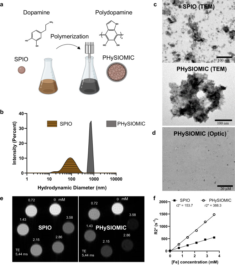
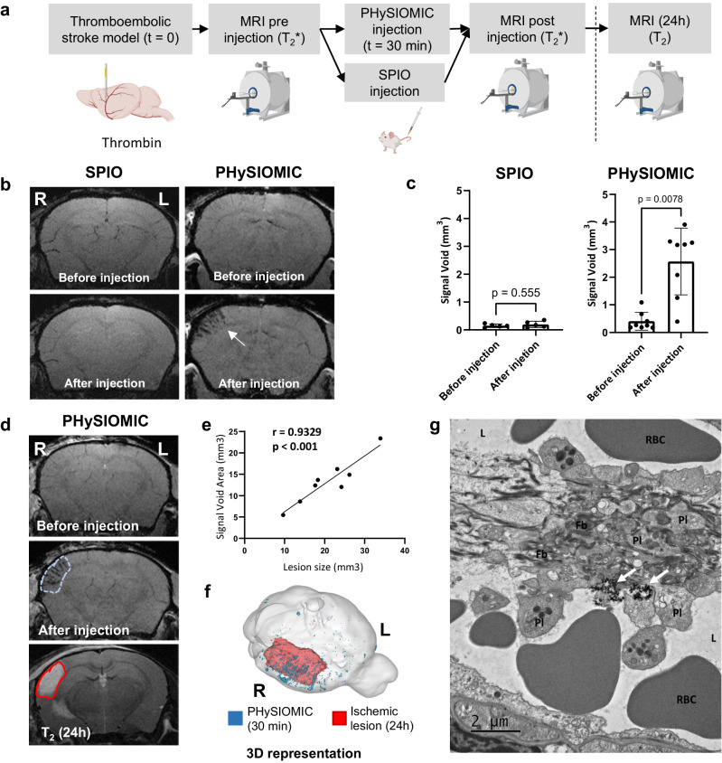
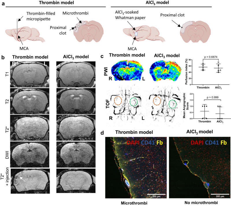
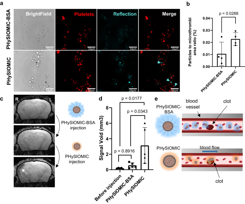
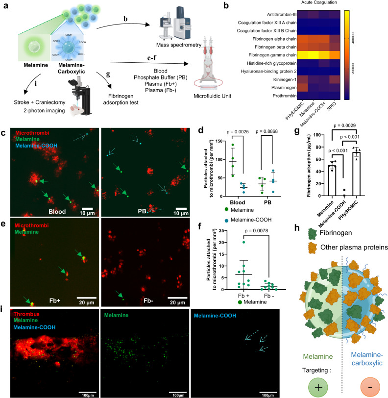
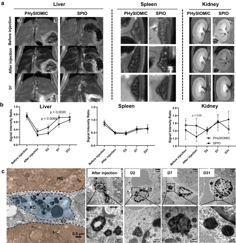
In acute ischemic stroke, even when successful recanalization is obtained, downstream microcirculation may still be obstructed by microvascular thrombosis, which is associated with compromised brain reperfusion and cognitive decline. Identifying these microthrombi through non-invasive methods remains challenging. We developed the PHySIOMIC (Polydopamine Hybridized Self-assembled Iron Oxide Mussel Inspired Clusters), a MRI-based contrast agent that unmasks these microthrombi. In a mouse model of thromboembolic ischemic stroke, our findings demonstrate that the PHySIOMIC generate a distinct hypointense signal on T2*-weighted MRI in the presence of microthrombi, that correlates with the lesion areas observed 24 hours post-stroke. Our microfluidic studies reveal the role of fibrinogen in the protein corona for the thrombosis targeting properties. Finally, we observe the biodegradation and biocompatibility of these particles. This work demonstrates that the PHySIOMIC particles offer an innovative and valuable tool for non-invasive in vivo diagnosis and monitoring of microthrombi, using MRI during ischemic stroke.


Conflict of interest statement

The authors declare the following competing interests: S.M.d.L., C.J., D.V., T.B., and M.G. have filed a patent application (WO2023007002A1) for the use of PHySIOMIC as a contrast agent to reveal microthrombi in ischemic stroke. The other authors declare that they have no competing interests.

Figures

Fig. 1
Fig. 1. PHySIOMIC synthesis and physico-chemical characteristics.
a Schematic diagram illustrating the PHySIOMIC synthesis process, which involves a simple polymerization step of dopamine to facilitate the clusterization of SPIO into PHySIOMIC. Created with BioRender.com. b DLS analysis of PHySIOMIC and SPIO, providing the mean hydrodynamic diameter (n = 3 particles preparations). c, d Morphological and core size observations conducted using transmission electron microscopy (TEM) showcasing the clusterization of SPIO into PHySIOMIC, and optical microscopy demonstrates the proper dispersion PHySIOMIC in a mannitol solution (n = 3 particles preparations). e, f MRI scans and relaxivity measurements obtained from a T2*-weighted sequence of PHySIOMIC and SPIO. Supplementary values of the DLS analysis are given in Table 1, and Supplementary Fig. 1. offers supplementary information on the r1 and r2 relaxivities (n = 1 particle preparation). Source data are provided as a Source Data file.
Fig. 2
Fig. 2. PHySIOMIC reveals microthrombosis during the acute phase of stroke in a thromboembolic stroke model, evidenced by a hyposignal that correlates with lesion size at 24 h.
a Protocol illustration of intravenous injection of PHySIOMIC or SPIO in a thrombin-induced stroke model, followed by acquisition of T2*-weighted images before, 30 minutes, and 24 hours after PHySIOMIC injection. Created with BioRender.com. b T2*-weighted MRI scans demonstrating the hypointense signal (arrow) induced by PHySIOMIC particles in the brain cortex, while no hypointense signal is observed with SPIO. c Quantification of signal void in mm3 before and after PHySIOMIC (n = 8 animals, paired t test, two-sided) or SPIO injection (n = 5 animals, paired t test, two-sided). Data are presented as mean ± SD. d The signal void area observed 30 min after the occlusion in a T2*-weighted sequence (blue dotted line) is predictive of the lesion size observed at 24 h after occlusion in a T2-weighted sequence (red continuous line). e Signal void area measures show a significant correlation with lesion size at 24 h (n = 8 animals, Pearson correlation coefficient, two-sided). Source data are provided as a Source Data file. f 3D representation of PHySIOMIC-induced hypointense signal and lesion hypersignal in MRI. R and L indicate right and left side of the presented brain. g TEM was performed on brain tissue collected after PHySIOMIC injection to visualize the presence of PHySIOMIC in the brain (observations in one animal). The images show PHySIOMIC particles (arrows) positioned around the clot composed of platelets (Pl) and fibrin (Fb) in the vessel lumen (L). RBC: Red Blood Cell. More images are shown in Supplementary Fig. 7.
Fig. 3
Fig. 3. Evaluation of MRI methods to detect differences between two MCAO models: thrombin model (with microthrombi) and AlCl3 model (without microthrombi).
a Description of the ‘thrombin model’ with thrombin injection in the MCA and ‘AlCl3 model’ with AlCl3 application on the MCA, and schematic coronal section of the brain in each model illustrating the presence or absence of microthrombi. Created with BioRender.com. b MRI images obtained using T1, T2, T2*-weighted, and diffusion-weighted imaging (DWI) after the induction of the MCAO models. Only the injection of PHySIOMIC in T2*-weighted imaging allows for the visualization of the microthrombi present in the thrombin model (indicated by the arrow). R and L indicate right and left side of the presented brain. c Study of cerebral perfusion using perfusion-weighted imaging (PWI, unpaired t test, two-sided) or angiography (TOF, Mann–Whitney, two-sided) in the models (n = 5 animals per group). Results are presented as mean ± SD. Source data are provided as a Source Data file. d Immunohistological images of the cerebral cortex were harvested 1 hour after the induction of each model. Microthrombi are identified through CD41 (platelet) and fibrin (yellow) staining, and nuclei are stained with DAPI (observations in 5 animals).
Fig. 4
Fig. 4. PHySIOMIC reveals a significant reduction in the number of microthrombi after thrombolysis.
a Protocol illustration of PHySIOMIC injection 10 min after occlusion, followed by T2*-weighted sequence acquisition before and after treatment with either rtPA or saline. Created with BioRender.com. b, c Representative T2*-weighted MRI scans and 3D reconstructed brain demonstrate microthrombosis in the ischemic cortex (arrows) and a significant decrease of the microthrombosis in the rtPA-treated group (asterisk). d Quantification of induced hyposignal reveals a diminution (p < 0.0001) in the treated group and no changes (p = 0.9732) for the saline group (n = 7 animals, two-way ANOVA with Sidak’s multiple comparisons test). e, f Magnetic resonance angiography displays no restoration of the blood flow in the MCA and downstream vasculature pre-treatment (red dotted lines), a partial restoration (yellow dashed line) for saline-treated mice, and a full restoration (green continuous line) for rtPA-treated mice (n = 7 animals, 2-way ANOVA). Data are presented as mean ± SD. R and L indicate right and left side of the presented brain. Source data are provided as a Source Data file.
Fig. 5
Fig. 5. PHySIOMIC failed to reveal microthrombi when saturated with albumin.
a BSA-coated PHySIOMIC were designed to prevent protein corona formation in blood. Both PHySIOMIC and PHySIOMIC-BSA were injected in vitro, into a microfluidic chamber along with blood. Images were acquired through a combination of bright field and fluorescence microscopy, enhanced by a reflection acquisition technique to observe PHySIOMIC interacting with platelets-composed microthrombi. PHySIOMIC-BSA shows fewer occurrences in microthrombi compared to PHySIOMIC. Scale bar 10 µm. b Quantitative results presented in mean ± SD (n = 4 microfluidic chambers, paired t test, two-sided). c, d MRI scans of mice injected first with PHySIOMIC-BSA, then PHySIOMIC and subsequent quantification showing no decrease in signal with PHYSIOMIC-BSA, but a clear hyposignal after PHySIOMIC injection marking microthrombosis (n = 5 animals, One-way ANOVA with Tukey’s multiple comparisons test). Results presented as mean ± SD. e Illustration of the proposed mechanism (Created with BioRender.com). PHySIOMIC-BSA forms a protein corona predominantly with BSA (in blue) and cannot target microthrombi, whereas PHySIOMIC develops a protein corona in vivo (in orange), enabling targeting to the clot. R and L indicate right and left side of the presented brain. Source data are provided as a Source Data file.
Fig. 6
Fig. 6. Surface functionalization effects on protein corona composition and impact of fibrinogen on targeting of microthrombi.
a Protocol illustration for mass spectrometry, microfluidic experiments, and two-photon microscopy with commercial fluorescent melamine microparticles with no functionalization (Melamine, equivalent to PHySIOMIC) or with carboxylic termination (Melamine-COOH). Created with BioRender.com. b Proteomic analysis performed with PHySIOMIC, melamine and melamine-COOH. Proteins from the ‘Acute coagulation’ subgroup are presented (n = 3 pooled plasma samples). c Melamine (green arrows) and melamine-COOH particles (blue dashed arrows) were injected either with Phosphate Buffer (PB) (no formation of protein corona) or whole blood (protein corona formation) in a microfluidic system where microthrombi were previously formed. d Quantification of the number of particles targeting microthrombi (expressed in the number per microthrombi surface). A statistical difference is found between melamine and melamine-COOH particles only when injected with blood. Results are expressed as mean ± SD (n = 4 microfluidic chambers, Student’s t test, two-sided). e Melamine particles were injected into the microfluidic system with either normal plasma (Fb+) or fibrinogen-depleted plasma (Fb−). f Quantification of the targeting of particles to microthrombi is higher in normal plasma than in fibrinogen-depleted plasma. Results are expressed as mean ± SD (n = 9 microfluidic chambers, Mann–Whitney test, two-sided). g Fibrinogen adsorption on the surface on melamine, melamine-COOH and PHySIOMIC particles presented as mean ± SD (n = 5 replicated sample measurements, One-way ANOVA with Tukey’s multiple comparisons), after a 30 minutes exposition to a fibrinogen solution at 37 °C. h Diagram illustrating the potential impact of fibrinogen on protein corona composition in relation to microthrombi passive targeting. Created with BioRender.com. i Two-photon observations of melamine and melamine-COOH particles injected intravenously into a mouse after a thrombin-induced stroke, showing the specific melamine attachment around thrombi in vivo (n = 3 animals). Source data are provided as a Source Data file.
Fig. 7
Fig. 7. Similar biodistribution and biodegradation of PHySIOMIC and SPIO in liver and spleen, evidenced by TEM and MRI.
a Longitudinal T2-weighted images acquired before and after injection of PHySIOMIC and SPIO (4 mg kg−1), and at 2 and 7 days post injection. b Quantification of T2 intensity values in the liver, spleen, and kidneys. Faster biodegradation is observed in the Liver with PHySIOMIC, and a part of injected SPIO is eliminated through the kidney. Results are expressed as mean ± SD (n = 5 animals, Two-way ANOVA with Sidak’s multiple comparisons test). c TEM images of the degradation of PHySIOMIC in the Kupffer cells (KC, blue) located inside the Space of Diss (SD, gray) between the hepatocytes (HC, brown) of the liver. PHySIOMIC (arrows) are observed in lysosomes of the Kupffer cells where the structure of clusters observed within lysosomes undergoes significant alteration after one month (n = 2 animals). Source data are provided as a Source Data file.

References

    1. Ma H, et al. Thrombolysis guided by perfusion imaging up to 9 hours after onset of stroke. N. Engl. J. Med. 2019;380:1795–1803. doi: 10.1056/NEJMoa1813046. - DOI - PubMed
    1. de los Ríos la Rosa F, et al. Eligibility for intravenous recombinant tissue-type plasminogen activator within a population. Stroke. 2012;43:1591–1595. doi: 10.1161/STROKEAHA.111.645986. - DOI - PMC - PubMed
    1. Berkhemer OA, et al. A randomized trial of intraarterial treatment for acute ischemic stroke. N. Engl. J. Med. 2015;372:11–20. doi: 10.1056/NEJMoa1411587. - DOI - PubMed
    1. Goyal M, et al. Randomized assessment of rapid endovascular treatment of ischemic stroke. N. Engl. J. Med. 2015;372:1019–1030. doi: 10.1056/NEJMoa1414905. - DOI - PubMed
    1. Molina CA, et al. REVASCAT: a randomized trial of revascularization with SOLITAIRE FR device vs. best medical therapy in the treatment of acute stroke due to anterior circulation large vessel occlusion presenting within eight-hours of symptom onset. Int. J. Stroke. 2015;10:619–626. doi: 10.1111/ijs.12157. - DOI - PubMed

MeSH terms