. 2024 Jun 13;11(1):624.
doi: 10.1038/s41597-024-03479-7.

Neurophysiological recordings from parietal areas of macaque brain during an instructed-delay reaching task

Affiliations

Neurophysiological recordings from parietal areas of macaque brain during an instructed-delay reaching task

S Diomedi et al. Sci Data. .

Abstract

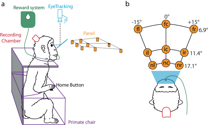
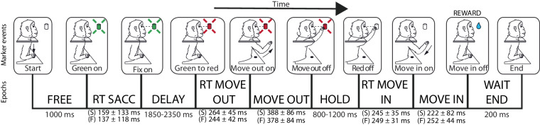
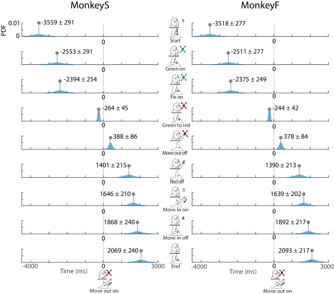
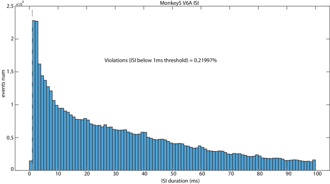
Facilitating data sharing in scientific research, especially in the domain of animal studies, holds immense value, particularly in mitigating distress and enhancing the efficiency of data collection. This study unveils a meticulously curated collection of neural activity data extracted from six electrophysiological datasets recorded from three parietal areas (V6A, PEc, PE) of two Macaca fascicularis during an instructed-delay foveated reaching task. This valuable resource is now accessible to the public, featuring spike timestamps, behavioural event timings and supplementary metadata, all presented alongside a comprehensive description of the encompassing structure. To enhance accessibility, data are stored as HDF5 files, a convenient format due to its flexible structure and the capability to attach diverse information to each hierarchical sub-level. To guarantee ready-to-use datasets, we also provide some MATLAB and Python code examples, enabling users to quickly familiarize themselves with the data structure.


Conflict of interest statement

The authors declare no competing interests.

Figures

Fig. 1
Fig. 1
Anatomical locations of areas V6A, PEc, PE, and position of the recording chambers. (a) Posterolateral view of a right hemisphere and posteromesial view of a left hemisphere of M. fascicularis brain showing location and extent of areas V6A, PEc and PE of SPL (magenta, green and orange dashed lines, respectively). The right hemisphere is partially dissected to show the areas hidden in the parieto-occipital sulcus. Colored areas represent the reconstructions of the recording regions within these areas as a mean of four hemispheres from two animals (modified from). (b,c) Mesial views of the three-dimensional reconstruction of the monkey brains, displaying the diameter and inclination of the recording chamber in each of the 2 monkeys. (d,e) Dorsal views of three-dimensional reconstructions of the monkey brains, displaying the location of the recording chamber in each of the 2 monkeys. The reconstructions were made using Caret software (http://brainvis.wustl.edu/wiki/index.php/Caret:About), starting from the histological sections,. The black circles provide an estimate of the chamber position and dimension. Abbreviations: as, arcuate sulcus; cal, calcarine sulcus; cs, central sulcus; ios, inferior occipital sulcus; ips, intraparietal sulcus; lf, lateral fissure; ls, lunate sulcus; ots, occipito-temporal sulcus; pos, parieto-occipital sulcus; ps, principal sulcus; sts, superior temporal sulcus.
Fig. 2
Fig. 2
Experimental apparatus. (a) Lateral view of the experimental apparatus. Eye tracking system with cold mirror (Cyan), Reward system (Green), Recording chamber (Red), Primate chair (Purple), the Panel with the 9 targets (Orange). The Home Button embedded in the primate chair was used as the initial position for the reaching movements. (b) Top view of experimental apparatus. Spatial coordinates of targets are indicated as vergence (depth) and version (direction) angles in relation to the eyes. Abbreviations: fl, far left; fc, far central; fr, far right; il, intermediate left; ic, intermediate central; ir, intermediate right; nl, near left; nc, near central; nr, near right.
Fig. 3
Fig. 3
Task sequence. Top: task events from left to right: trial start (Start), target appearance (Green on), fixation onset (Fix on), go signal for the outward movement (Green to red), start of outward movement (Move out on), touch and beginning of the target holding (Move out off), go signal for the inward movement (Red off), release of the target and start of the inward movement (Move in on), end of the inward movement (Move in off), end of the trial (End). Bottom: placed between two adjacent event markers, there are the names of the task epochs and their duration. (S): MonkeyS, and (F): MonkeyF.
Fig. 4
Fig. 4
Schematic view of the HDF5 file organization. In each file, each group contains other groups up to the last hierarchical level in which datasets contain the actual data (similarly to the system of folders, subfolders, and files on a computer). Each group / dataset can be independently labelled with metadata (attribute).
Fig. 5
Fig. 5
Event markers’ timing. In each row, the blue profiles represent the Gaussian probability distribution for each event timing relative to the alignment (Move out on, gray vertical line). Left column for MonkeyS, right column for MonkeyF.
Fig. 6
Fig. 6
Raster plot example MonkeyS V6A unit_11. Example of raster plot of one neuron obtained with the script H5_raster. X-axis represents time and y-axis depicts the number of trials recorded. Each black line represents an action potential; each colored circle represents event markers, as in the legend.
Fig. 7
Fig. 7
ISI plot example of MonkeyS V6A whole population using H5_ISI.m script. X-axis represents the distance between two consecutive spikes and y-axis shows the count of ISIs.

References

    1. Vines TH, et al. Mandated data archiving greatly improves access to research data. FASEB J. 2013;27:1304–1308. doi: 10.1096/fj.12-218164. - DOI - PubMed
    1. Russel, W. M. S. & Burch, R. L. The principles of humane experimental technique. Methuen (1959).
    1. eBioMedicine The 3Rs of Animal Research. eBioMedicine. 2022;76:103900. doi: 10.1016/j.ebiom.2022.103900. - DOI - PMC - PubMed
    1. Federer LM, Lu Y-L, Joubert DJ, Welsh J, Brandys B. Biomedical Data Sharing and Reuse: Attitudes and Practices of Clinical and Scientific Research Staff. PLOS ONE. 2015;10:e0129506. doi: 10.1371/journal.pone.0129506. - DOI - PMC - PubMed
    1. Krawczyk M, Reuben E. (Un)Available upon Request: Field Experiment on Researchers’ Willingness to Share Supplementary Materials. Account. Res. 2012;19:175–186. doi: 10.1080/08989621.2012.678688. - DOI - PubMed