

Subretinal fluid in macular edema secondary to branch retinal vein occlusion
- PMID: 38871805
- PMCID: PMC11176314
- DOI: 10.1038/s41598-024-64047-y
Subretinal fluid in macular edema secondary to branch retinal vein occlusion
Abstract
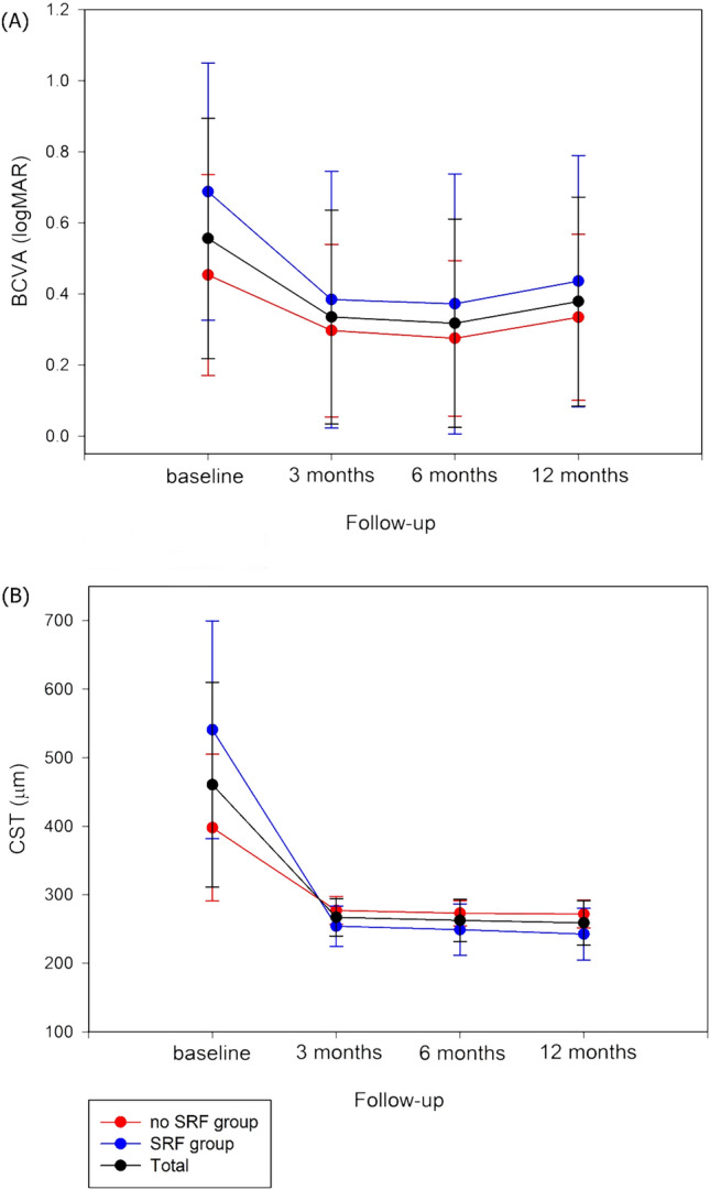
We identified characteristics of patients with subretinal fluid (SRF) in macular edema (ME) secondary to branch retinal vein occlusion (BRVO) and determined their clinical outcomes after anti-vascular endothelial growth factor (VEGF) treatment. Fifty-seven eyes of BRVO patients with ME were divided into two groups according to the presence or absence of SRF at diagnosis. We compared the aqueous profiles, ocular and systemic characteristics at baseline, and the clinical outcomes. The SRF group had significantly greater central subfield thickness (CST) values and poorer best-corrected visual acuity (BCVA) at baseline compared to the non-SRF group. The former group had significantly higher aqueous levels of interleukin-8, VEGF, and placental growth factor. CST reduction and BCVA improvement during treatment were significantly greater in the SRF group than in the non-SRF group. Consequently, CST values were significantly lower in the SRF group than in the non-SRF group at 12 months, when BCVA did not differ significantly between the two groups. The SRF group required more frequent anti-VEGF treatment over 12 months and exhibited a higher rate of macular atrophy. Based on the aqueous profiles and the number of treatments required, the presence of SRF in BRVO patients appears to be associated with higher disease activity.
© 2024. The Author(s).
Conflict of interest statement
The authors declare no competing interests.
Figures
Similar articles
-
Characteristics of major and macular branch retinal vein occlusion.Sci Rep. 2022 Aug 18;12(1):14103. doi: 10.1038/s41598-022-18414-2. Sci Rep. 2022. PMID: 35982117 Free PMC article.
-
Predictive factors for functional improvement after intravitreal bevacizumab therapy for macular edema due to branch retinal vein occlusion.Graefes Arch Clin Exp Ophthalmol. 2011 Feb;249(2):183-92. doi: 10.1007/s00417-010-1470-2. Epub 2010 Aug 18. Graefes Arch Clin Exp Ophthalmol. 2011. PMID: 21337042 Free PMC article.
-
Prospective study of intravitreal triamcinolone acetonide versus bevacizumab for macular edema secondary to central retinal vein occlusion.Retina. 2011 May;31(5):838-45. doi: 10.1097/IAE.0b013e3181f4420d. Retina. 2011. PMID: 21293319 Clinical Trial.
-
Anti-vascular endothelial growth factor for macular oedema secondary to branch retinal vein occlusion.Cochrane Database Syst Rev. 2020 Jul 7;7(7):CD009510. doi: 10.1002/14651858.CD009510.pub3. Cochrane Database Syst Rev. 2020. PMID: 32633861 Free PMC article.
-
Comparison between Ozurdex and intravitreal anti-vascular endothelial growth factor treatment for retinal vein occlusion-related macular edema: A systematic review and meta-analysis of randomized controlled trials.Indian J Ophthalmol. 2019 Nov;67(11):1800-1809. doi: 10.4103/ijo.IJO_382_19. Indian J Ophthalmol. 2019. PMID: 31638037 Free PMC article.
Cited by
-
Short and long-term impact of intravitreal anti-VEGF therapy interruption in retinal vein occlusion during the COVID-19 pandemic: functional outcomes and AI-based fluid analysis of macular edema.Int J Retina Vitreous. 2025 Aug 5;11(1):92. doi: 10.1186/s40942-025-00717-x. Int J Retina Vitreous. 2025. PMID: 40765020 Free PMC article.
References
MeSH terms
Substances
Grants and funding
LinkOut - more resources
Full Text Sources
Miscellaneous
