Review
. 2024 Jul;24(7):448-460.
doi: 10.1038/s41568-024-00700-y. Epub 2024 Jun 13.

The path to leptomeningeal metastasis

Affiliations
Review

The path to leptomeningeal metastasis

Jan Remsik et al. Nat Rev Cancer. 2024 Jul.

Abstract

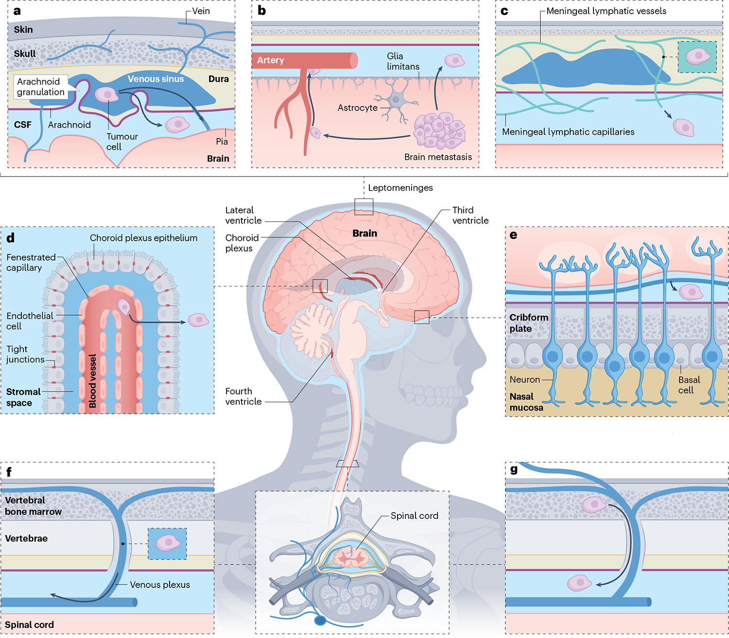
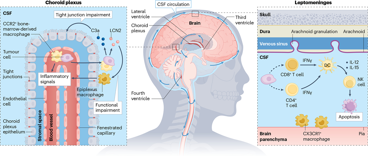
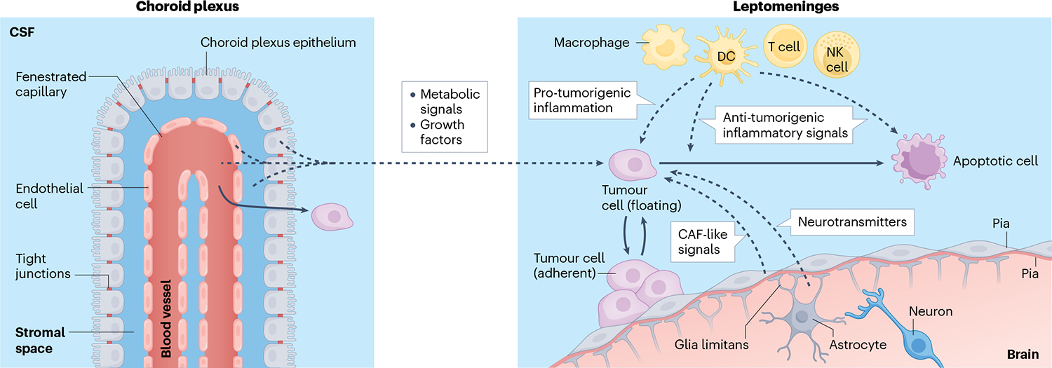
The leptomeninges, the cerebrospinal-fluid-filled tissues surrounding the central nervous system, play host to various pathologies including infection, neuroinflammation and malignancy. Spread of systemic cancer into this space, termed leptomeningeal metastasis, occurs in 5-10% of patients with solid tumours and portends a bleak clinical prognosis. Previous, predominantly descriptive, clinical studies have provided few insights. Recent development of preclinical leptomeningeal metastasis models, alongside genomic, transcriptomic and proteomic sequencing efforts, has provided groundwork for mechanistic understanding and identification of long-needed therapeutic targets. Although previously understood as an anatomically isolated compartment, the leptomeninges are increasingly appreciated as a major conduit of communication between the systemic circulation and the central nervous system. Despite the unique nature of the leptomeningeal microenvironment, the general principles of metastasis hold true: cells metastasizing to the leptomeninges must gain access to the new environment, survive within the space and evade the immune system. The study of leptomeningeal metastasis has the potential to uncover novel site-specific metastatic principles and illuminate the physiology of the leptomeningeal space. In this Review, we provide a biology-focused overview of how metastatic cells reach the leptomeninges, thrive in this nutritionally sparse environment and evade the detection of the omnipresent immune system.


Conflict of interest statement

Competing interests

J.R. and A.B. are inventors on the provisional US patent applications 63/449,817 and 63/449,823 and international patent application PCT/US24/18343. A.B. holds an unpaid position on the scientific advisory board for Evren Scientific and is an inventor on the US patents 62/258,044, 10/413,522 and 63/052,139.

Figures

Fig. 1 |
Fig. 1 |. Anatomical routes of cancer cell entry into the leptomeninges.
The leptomeninges consist of the pia and arachnoid membranes, contain the cerebrospinal fluid (CSF) and encase the entire central nervous system. Cancer cells outside the central nervous system may access the leptomeningeal compartment through various anatomical ports of entry. a, Cancer cells within the venous sinuses may breach the arachnoid granulations. b, Tumours or single cells within the brain parenchyma may invade and breach the glia limitans to reach the CSF-filled perivascular (Virchow-Robin) spaces. c, Cancer cells within the dura may invade the meningeal lymphatic vessels to access the CSF. d, Cancer cells within the arterial circulation slip through fenestrated vessels to arrive in the well-perfused choroid plexi. Crossing the choroid plexus epithelial cell tight junctions provides access to the CSF-filled ventricles. e, Cancer cells may migrate along cranial nerves, making use of pre-existing tissue planes. f, Cancer cells within the low-pressure, valveless, Batson venous plexus of the spinal cord are well positioned to breach the spinal arachnoid to gain entry into the CSF. g, Communicating bridging veins provide leptomeningeal access for cancer cells within the bone marrow.
Fig. 2 |
Fig. 2 |. Leptomeningeal immune response to cancer.
In the absence of malignancy, the cerebrospinal fluid (CSF) is nearly acellular with few CD4+ T cells. Invasive cancer cells provoke a wide variety of inflammatory responses. Cancer cells generate the acute-phase reactants complement component 3 (C3) and lipocalin 2 (LCN2). C3 signalling reduces choroid plexus epithelial cell tight junction integrity, whereas LCN2 enables cancer cells to outcompete CSF macrophages for iron. CCR2+ bone-marrow-derived macrophages migrate into the leptomeninges where together with CX3CR1+-resident macrophages, they mediate anticancer activity. Leptomeningeal cancer cells recruit CD8+ T cells and CD4+ T cells; however, mechanisms for this remain unknown (as indicated by the dashed lines). CD8+ T and CD4+ T cells generate interferon-γ (IFNγ) in response to cancer cells, activating leptomeningeal dendritic cells. Dendritic cells generate IL-12 and IL-15 to prompt natural killer (NK) cells to initiate cancer cell apoptosis.
Fig. 3 |
Fig. 3 |. Integration of microenvironmental signals by leptomeningeal immune cells.
Extensive profiling efforts of leptomeningeal metastasis suggest that cancer cells in this compartment receive a wide spectrum of extracellular signals. Although comprehensive work in this area has not yet been completed, several hypotheses can be envisioned such that cancer cell–microenvironmental interactions improve cancer cell growth within the nutrient-poor and inflamed leptomeninges. Signals supported through experimental evidence are indicated with solid arrows, and those hypothesized are indicated with dashed arrows. Cancer cells within the leptomeningeal space exist in an equilibrium between adherent and floating states receiving various local signals. In response to local inflammation, choroid plexus epithelial cells lose barrier function, enabling transport of nutrients and growth factors from the blood, stroma, endothelium and epithelium into the cerebrospinal fluid (CSF), supporting cancer growth. Infiltrating leukocytes also promote tumour growth through induction of signal transducer and activator of transcription (STAT) proteins and nuclear factor-κB (NF-κB) pathways in the cancer cell. By contrast, these inflammatory signals can also induce cancer cell death. Pial and arachnoid fibroblasts may alter their secretome in response to cancer within the space, potentially taking on a cancer-associated fibroblast (CAF)-like role. Finally, the homeostasis of the brain parenchyma is likely perturbed by leptomeningeal metastasis promoting the accumulation of neurotransmitters and their metabolites within the local microenvironment. DC, dendritic cell; NK cell, natural killer cell.

References

    1. Vanderah TW & Gould DJ Nolte’s the Human Brain : An Introduction to its Functional Anatomy 8th edn (Elsevier, 2021).
    1. Remsik J et al. Leptomeningeal metastatic cells adopt two phenotypic states. Cancer Rep. 5, e1236 (2022). - PMC - PubMed
    1. Subira D et al. Diagnostic and prognostic significance of flow cytometry immunophenotyping in patients with leptomeningeal carcinomatosis. Clin. Exp. Metastasis 32, 383–391 (2015). - PubMed
    1. Scott BJ & Kesari S Leptomeningeal metastases in breast cancer. Am. J. Cancer Res 3, 117–126 (2013). - PMC - PubMed
    1. Magbanua MJ et al. Molecular profiling of tumor cells in cerebrospinal fluid and matched primary tumors from metastatic breast cancer patients with leptomeningeal carcinomatosis. Cancer Res. 73, 7134–7143 (2013). - PubMed

LinkOut - more resources