. 2024 Jun 13;21(1):155.
doi: 10.1186/s12974-024-03149-w.

Delayed plasma kallikrein inhibition fosters post-stroke recovery by reducing thrombo-inflammation

Affiliations

Delayed plasma kallikrein inhibition fosters post-stroke recovery by reducing thrombo-inflammation

Steffen Haupeltshofer et al. J Neuroinflammation. .

Abstract

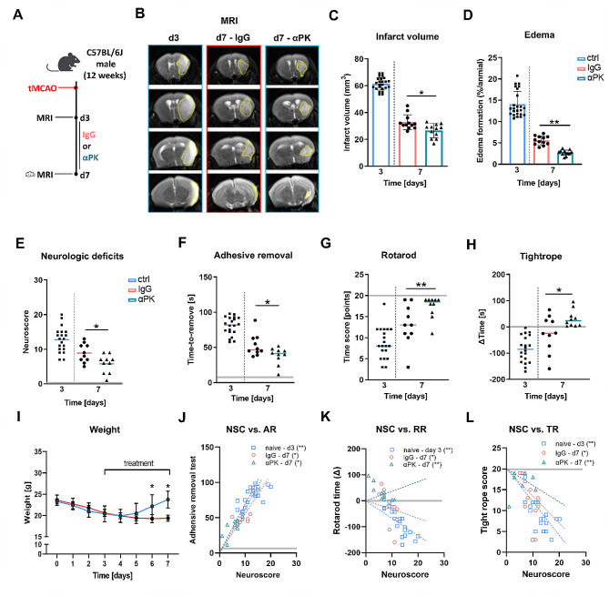
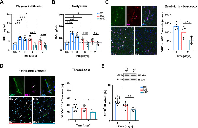
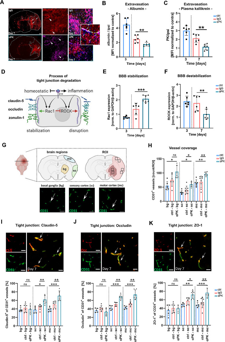
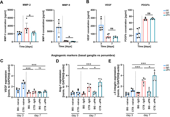
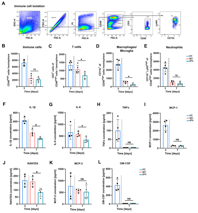
Activation of the kallikrein-kinin system promotes vascular leakage, inflammation, and neurodegeneration in ischemic stroke. Inhibition of plasma kallikrein (PK) - a key component of the KKS - in the acute phase of ischemic stroke has been reported to reduce thrombosis, inflammation, and damage to the blood-brain barrier. However, the role of PK during the recovery phase after cerebral ischemia is unknown. To this end, we evaluated the effect of subacute PK inhibition starting from day 3 on the recovery process after transient middle artery occlusion (tMCAO). Our study demonstrated a protective effect of PK inhibition by reducing infarct volume and improving functional outcome at day 7 after tMCAO. In addition, we observed reduced thrombus formation in cerebral microvessels, fewer infiltrated immune cells, and an improvement in blood-brain barrier integrity. This protective effect was facilitated by promoting tight junction reintegration, reducing detrimental matrix metalloproteinases, and upregulating regenerative angiogenic markers. Our findings suggest that PK inhibition in the subacute phase might be a promising approach to accelerate the post-stroke recovery process.

Keywords: Blood-brain barrier; Extravasation; Inflammation; Ischemic stroke; Kallikrein-kinin system; Plasma kallikrein; Recovery; Subacute; Thrombo-inflammation; Thrombosis.


Conflict of interest statement

The authors declare no competing interests.

Figures

Fig. 1
Fig. 1
Delayed anti-plasma kallikrein (αPK) treatment transiently improved focal deficits and reduced edema formation. (A) Mice were treated with isotype immunoglobulin G (IgG) or αPK at days 3 and 4 (400 µg/kg) and days 5 to 7 (200 µg/kg) after 60-min transient middle artery occlusion (tMCAO). Cerebral damage was visualized using magnetic resonance imaging (MRI) at days 3 and 7. (B) Representative images of T2 MRI scans 3 and 7 days after tMCAO. The yellow dotted lines depict infarct areas. Quantification of (C) brain infarct volume and (D) brain edema (n = 24/12/12). (E) Assessment of general and focal deficits at 3 and 7 days of tMCAO mice treated with IgG or αPK. Evaluation of sensorimotor function of tMCAO with (F) adhesive removal test (AR), (G) rotarod test (RR), and (H) tightrope test (TR) (n = 20/10/10). (I) Quantification of body weight at days 0 to 7 after tMCAO (TR) (n = 10/10). (JL) Spearman correlation of sensorimotor deficits with neurological score evaluation. Two-way ANOVA and post hoc Sídák test. *P < 0.05, **P < 0.01, ***P < 0.001 for αPK vs. IgG; grey lines represent the baseline values measured in healthy mice
Fig. 2
Fig. 2
Subacute inhibition of plasma kallikrein (PK) protected against persistent thrombosis in microvasculature transient middle artery occlusion (tMCAO). (A) Quantification of activated PK and (B) its respective downstream target bradykinin in the blood at day − 1 (baseline) and days 1, 3, 5, and 7 after tMCAO of mice treated with immunoglobulin G (IgG) or anti-PK (αPK) (n = 8). (C) Histological staining of CD31+ blood vessels and the bradykinin-1 receptor (B1R+) at days 3 and 7 (white arrows) (n = 5). (D) Histological staining of CD31+ blood vessels and GPIX+ platelet adhesion molecules showing thrombi at days 3 and 7 after tMCAO (white arrows) (n = 9/5/5). (E) Protein density analysis of platelet adhesion molecule GP1b at days 3 and 7 after tMCAO (n = 10/8/8). One-way ANOVA and post hoc Dunn’s test. *P < 0.05, **P < 0.01, ***P < 0.001 for αPK vs. IgG
Fig. 3
Fig. 3
Blockade of plasma kallikrein (PK) was essential for blood-brain barrier (BBB) stabilization and attenuation of extravasation after transient middle artery occlusion (tMCAO). (A) Exemplary staining of albumin and PK (white arrows) in the ischemic lesion. Extravasation of (B) albumin and (C) blood-resident PK into the brain parenchyma of immunoglobulin G (IgG)- and anti-PK (αPK)-treated mice (n = 6). (D) Schematic pathway of tight junction (TJ) protein degradation in endothelial cells bradykinin-1 receptor (B1R) activation under pathophysiological conditions. (E) Ras-related C3 botulinum toxin substrate 1 (Rac1)-axis activation led to stabilization of the BBB (n = 8). (F) Intracellular Rho-associated kinases (ROCK)-axis activation mediated degradation of the BBB after ischemic stroke (n = 8). (G) Representative image of anti-CD31 histological staining and determination of region of interest (ROI) in the basal ganglia and sensory and motor cortex. (H) Quantitative analysis of histological CD31+ vessel density from respective brain regions at days 3 and 7 (IgG vs. αPK) (n = 6–8). (IK) Representative images of TJ protein staining and analysis of histological staining for CD31 vessel density and co-expression of claudin-5, occludin, and ZO-1 at days 3 and 7 after tMCAO in the respective IgG and αPK groups (n = 5–6). One-way ANOVA and post hoc Dunn’s test. *P < 0.05, **P < 0.01, ***P < 0.001 for αPK vs. IgG
Fig. 4
Fig. 4
Delayed plasma kallikrein (PK) inhibition blocked destabilization of the blood-brain barrier (BBB), leading to enhanced angiogenic marker expression after transient middle artery occlusion (tMCAO). Secretion patterns on the ipsilateral side of ischemic brains at day 3 and day 7. Amount of disruptive (A) matrix metalloproteinases (MMP)-2 and − 9, (B) vascular endothelial growth factor (VEGF), and platelet-derived growth factor subunit B (PDGFb) angiogenic markers (n = 6). Expression analysis of angiogenic-supporting factors (C) VEGF, (D) angiopoietin-1 (Ang-1), and (E) integrin-5 (α5) subunit from the basal ganglia (BG; infarct core) and cortex (CTX) from ischemic mouse brains (n = 5). One-way ANOVA and post hoc Dunn’s test. *P < 0.05, **P < 0.01, ***P < 0.001 for αPK vs. IgG
Fig. 5
Fig. 5
Plasma kallikrein (PK) inhibition induced alteration of immune cell influx and ameliorated inflammatory cytokines after transient middle artery occlusion (tMCAO). (A + B) Representative dot plot of brain-isolated immune cells and quantitative analysis of infiltrated immune cells into the ischemic brain at days 3 and 7 after immunoglobulin G (IgG) and anti-PK (αPK) in tMCAO mice (n = 5). Analysis of (C) CD3+ lymphocytes (adaptive immune system), (D) macrophages/microglia, and (E) neutrophil granulocytes (innate immune system) from IgG- and αPK-treated tMCAO mice (n = 5). Inflammatory cytokines (F) interleukin (IL)-1β, (G) IL-6, (H) tumor necrosis factor alpha (TNFα), (I) monocyte chemoattractant protein (MCP)-1, (J) regulated and normal T cell expressed and secreted (RANTES), (K) MCP-2, and (L) granulocyte-macrophage colony-stimulating factor (GM-CSF) in the brain parenchyma (n = 4). One-way ANOVA and post hoc Dunn’s test. *P < 0.05 for αPK vs. IgG

References

    1. GBD 2019 Stroke Collaborators Global, regional, and national burden of stroke and its risk factors, 1990–2019: a systematic analysis for the global burden of Disease Study 2019. Lancet Neurol. 2021;20:795–820. doi: 10.1016/S1474-4422(21)00252-0. - DOI - PMC - PubMed
    1. Kim J-T, Fonarow GC, Smith EE, Reeves MJ, Navalkele DD, Grotta JC, et al. Treatment with tissue plasminogen activator in the Golden Hour and the shape of the 4.5-Hour time-benefit curve in the National United States get with the guidelines-Stroke Population. Circulation. 2017;135:128–39. doi: 10.1161/CIRCULATIONAHA.116.023336. - DOI - PubMed
    1. Jadhav AP, Desai SM, Jovin TG. Indications for mechanical thrombectomy for Acute ischemic stroke: current guidelines and Beyond. Neurology. 2021;97:S126–36. doi: 10.1212/WNL.0000000000012801. - DOI - PubMed
    1. De Meyer SF, Langhauser F, Haupeltshofer S, Kleinschnitz C, Casas AI. Thromboinflammation in Brain Ischemia: recent updates and future perspectives. Stroke. 2022;53:1487–99. doi: 10.1161/STROKEAHA.122.038733. - DOI - PubMed
    1. Albert-Weißenberger C, Sirén A-L, Kleinschnitz C. Ischemic stroke and traumatic brain injury: the role of the kallikrein-kinin system. Prog Neurobiol. 2013;101–102:65–82. doi: 10.1016/j.pneurobio.2012.11.004. - DOI - PubMed

Substances

LinkOut - more resources