

Imaging at the nexus: how state of the art imaging techniques can enhance our understanding of cancer and fibrosis
- PMID: 38872212
- PMCID: PMC11177383
- DOI: 10.1186/s12967-024-05379-1
Imaging at the nexus: how state of the art imaging techniques can enhance our understanding of cancer and fibrosis
Abstract
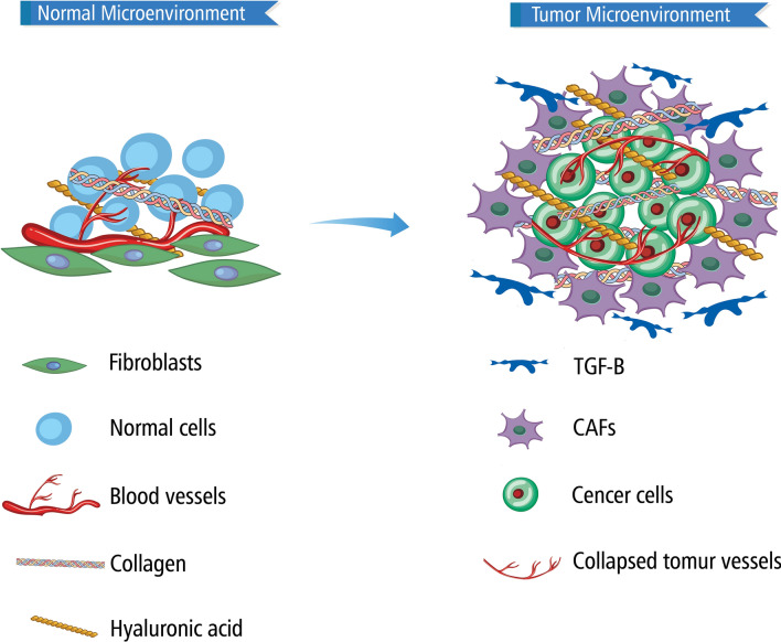
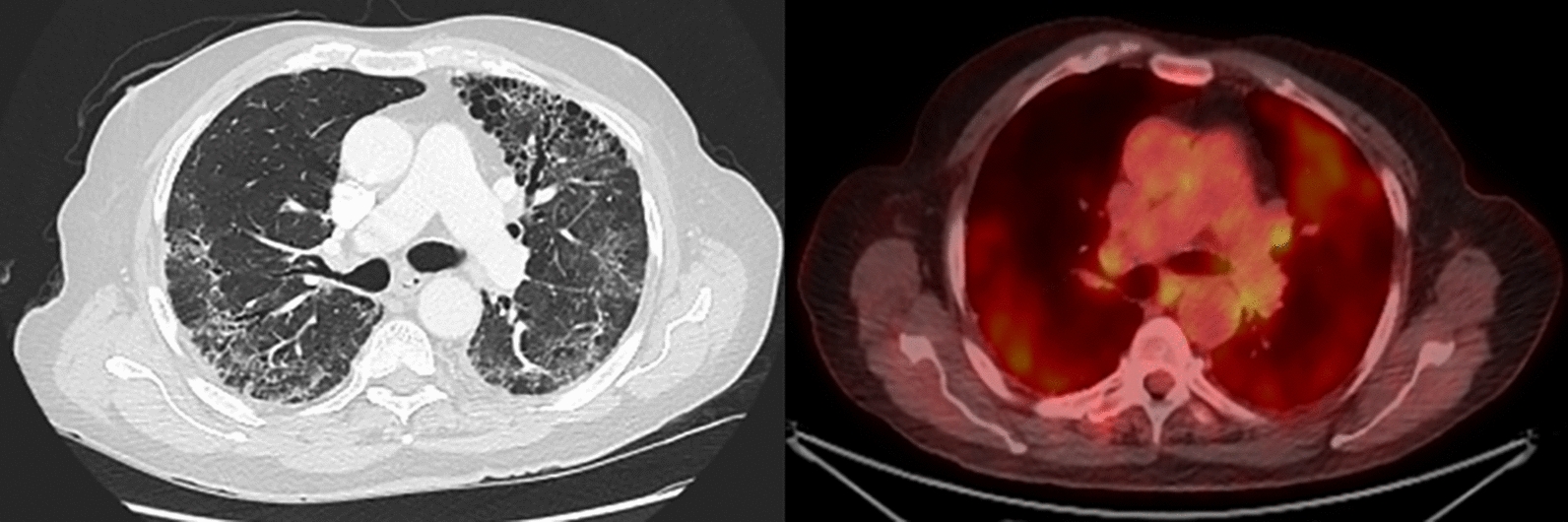
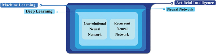
Both cancer and fibrosis are diseases involving dysregulation of cell signaling pathways resulting in an altered cellular microenvironment which ultimately leads to progression of the condition. The two disease entities share common molecular pathophysiology and recent research has illuminated the how each promotes the other. Multiple imaging techniques have been developed to aid in the early and accurate diagnosis of each disease, and given the commonalities between the pathophysiology of the conditions, advances in imaging one disease have opened new avenues to study the other. Here, we detail the most up-to-date advances in imaging techniques for each disease and how they have crossed over to improve detection and monitoring of the other. We explore techniques in positron emission tomography (PET), magnetic resonance imaging (MRI), second generation harmonic Imaging (SGHI), ultrasound (US), radiomics, and artificial intelligence (AI). A new diagnostic imaging tool in PET/computed tomography (CT) is the use of radiolabeled fibroblast activation protein inhibitor (FAPI). SGHI uses high-frequency sound waves to penetrate deeper into the tissue, providing a more detailed view of the tumor microenvironment. Artificial intelligence with the aid of advanced deep learning (DL) algorithms has been highly effective in training computer systems to diagnose and classify neoplastic lesions in multiple organs. Ultimately, advancing imaging techniques in cancer and fibrosis can lead to significantly more timely and accurate diagnoses of both diseases resulting in better patient outcomes.
Keywords: Cancer; Diagnosis; Fibrosis; Imaging techniques; Tumor microenvironment.
© 2024. The Author(s).
Conflict of interest statement
Dr. Capaccione has served as an advisor for Cardinal Health. The remaining authors have nothing to disclose.
Figures

Similar articles
-
Imaging Evaluation of Peritoneal Metastasis: Current and Promising Techniques.Korean J Radiol. 2024 Jan;25(1):86-102. doi: 10.3348/kjr.2023.0840. Korean J Radiol. 2024. PMID: 38184772 Free PMC article. Review.
-
Deep learning techniques in PET/CT imaging: A comprehensive review from sinogram to image space.Comput Methods Programs Biomed. 2024 Jan;243:107880. doi: 10.1016/j.cmpb.2023.107880. Epub 2023 Oct 21. Comput Methods Programs Biomed. 2024. PMID: 37924769 Review.
-
Application of Artificial Intelligence in Oncologic Molecular PET-Imaging: A Narrative Review on Beyond [18F]F-FDG Tracers - Part I. PSMA, Choline, and DOTA Radiotracers.Semin Nucl Med. 2024 Jan;54(1):171-180. doi: 10.1053/j.semnuclmed.2023.08.004. Epub 2023 Sep 24. Semin Nucl Med. 2024. PMID: 37752032 Review.
-
Comparative analysis of [18F]F-FAPI PET/CT, [18F]F-FDG PET/CT and magnetization transfer MR imaging to detect intestinal fibrosis in Crohn's disease: A prospective animal model and human cohort study.Eur J Nucl Med Mol Imaging. 2024 Jun;51(7):1856-1868. doi: 10.1007/s00259-024-06644-7. Epub 2024 Feb 15. Eur J Nucl Med Mol Imaging. 2024. PMID: 38355741
-
More advantages in detecting bone and soft tissue metastases from prostate cancer using 18F-PSMA PET/CT.Hell J Nucl Med. 2019 Jan-Apr;22(1):6-9. doi: 10.1967/s002449910952. Epub 2019 Mar 7. Hell J Nucl Med. 2019. PMID: 30843003
Cited by
-
Myofibroblasts persist through immune privilege mechanisms to mediate oral submucous fibrosis: Uncovering the pathogenesis.J Oral Biol Craniofac Res. 2024 Nov-Dec;14(6):773-781. doi: 10.1016/j.jobcr.2024.10.008. Epub 2024 Oct 20. J Oral Biol Craniofac Res. 2024. PMID: 39502133 Free PMC article. Review.
-
The Development of Al18F-NOTA-FAP-2286 as an FAP-Targeted PET Tracer and the Translational Application in the Diagnosis of Acquired Drug Resistance in Progressive Prostate Cancer.Pharmaceutics. 2025 Apr 23;17(5):552. doi: 10.3390/pharmaceutics17050552. Pharmaceutics. 2025. PMID: 40430845 Free PMC article.
References
Publication types
MeSH terms
Grants and funding
LinkOut - more resources
Full Text Sources
Medical
