

The neural substrates of social cognition deficits in newly diagnosed multiple sclerosis patients
- PMID: 38872257
- PMCID: PMC11251485
- DOI: 10.1002/acn3.52085
The neural substrates of social cognition deficits in newly diagnosed multiple sclerosis patients
Abstract
Objective: Cognitive and affective symptoms in multiple sclerosis (MS) can be independently impaired and have different pathways of progression. Cognitive alterations have been described since the earliest MS stages; by contrast, the social cognition (SC) domain has never been investigated in the first year from MS diagnosis. We aimed to evaluate SC and unravel its neural bases in newly diagnosed MS patients.
Methods: Seventy MS patients underwent at diagnosis a 3 T-MRI and a neuropsychological/SC assessment (median time between diagnosis and MRI/cognitive evaluation = 0 months). We tested two matched reference samples: 31 relapsing-remitting MS patients with longer course (mean ± SD disease duration = 7.0 ± 4.5 years) and 38 healthy controls (HCs). Cortical thicknesses (CTh) and volumes of brain regions were calculated.
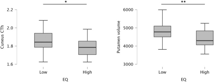
Results: Newly diagnosed MS patients performed significantly lower than HCs in facial emotion recognition (global: p < 0.001; happiness: p = 0.041, anger: p = 0.007; fear: p < 0.001; disgust: p = 0.004) and theory of mind (p = 0.005), while no difference was found between newly diagnosed and longer MS patients. Compared to lower performers, higher performers in facial emotion recognition showed greater volume of amygdala (p = 0.032) and caudate (p = 0.036); higher performers in theory of mind showed greater CTh in lingual gyrus (p = 0.006), cuneus (p = 0.024), isthmus cingulate (p = 0.038), greater volumes of putamen (p = 0.016), pallidum (p = 0.029), and amygdala (p = 0.032); patients with higher empathy showed lower cuneus CTh (p = 0.042) and putamen volume (p = 0.007).
Interpretations: SC deficits are present in MS patients since the time of diagnosis and remain persistent along the disease course. Specific basal, limbic, and occipital areas play a significant role in the pathogenesis of these alterations.
© 2024 The Authors. Annals of Clinical and Translational Neurology published by Wiley Periodicals LLC on behalf of American Neurological Association.
Conflict of interest statement
The authors have no conflict of interest to declare for the present manuscript.
Figures


Similar articles
-
Social cognition deficits and the role of amygdala in relapsing remitting multiple sclerosis patients without cognitive impairment.Mult Scler Relat Disord. 2019 Apr;29:118-123. doi: 10.1016/j.msard.2019.01.030. Epub 2019 Jan 23. Mult Scler Relat Disord. 2019. PMID: 30710839
-
Social Cognition Abilities in Patients With Different Multiple Sclerosis Subtypes.J Int Neuropsychol Soc. 2017 Sep;23(8):653-664. doi: 10.1017/S1355617717000510. Epub 2017 Jun 28. J Int Neuropsychol Soc. 2017. PMID: 28656885
-
Is impairment of facial emotion recognition independent of cognitive dysfunction in multiple sclerosis?Neurol Sci. 2024 Jun;45(6):2791-2800. doi: 10.1007/s10072-024-07314-0. Epub 2024 Jan 22. Neurol Sci. 2024. PMID: 38246940 Free PMC article.
-
Pathologic and imaging correlates of cognitive deficits in multiple sclerosis: changing the paradigm of diagnosis and prognosis.Cogn Behav Neurol. 2014 Mar;27(1):1-7. doi: 10.1097/WNN.0000000000000023. Cogn Behav Neurol. 2014. PMID: 24674960 Review.
-
Social Cognition in Multiple Sclerosis: a Meta-Analysis.Neuropsychol Rev. 2016 Jun;26(2):160-72. doi: 10.1007/s11065-016-9320-6. Epub 2016 Jun 20. Neuropsychol Rev. 2016. PMID: 27324894 Review.
References
-
- Calabrese M, Magliozzi R, Ciccarelli O, Geurts JJG, Reynolds R, Martin R. Exploring the origins of grey matter damage in multiple sclerosis. Nat Rev Neurosci. 2015;16:147‐158. - PubMed
-
- Portaccio E, Amato MP. Cognitive impairment in multiple sclerosis: an update on assessment and management. NeuroSci. 2022;3:667‐676.
MeSH terms
LinkOut - more resources
Full Text Sources
Medical