. 2024 Sep 4;32(9):3025-3041.
doi: 10.1016/j.ymthe.2024.06.005. Epub 2024 Jun 12.

The TRIM28/miR133a/CD47 axis acts as a potential therapeutic target in pancreatic necrosis by impairing efferocytosis

Affiliations

The TRIM28/miR133a/CD47 axis acts as a potential therapeutic target in pancreatic necrosis by impairing efferocytosis

Qingtian Zhu et al. Mol Ther. .

Abstract

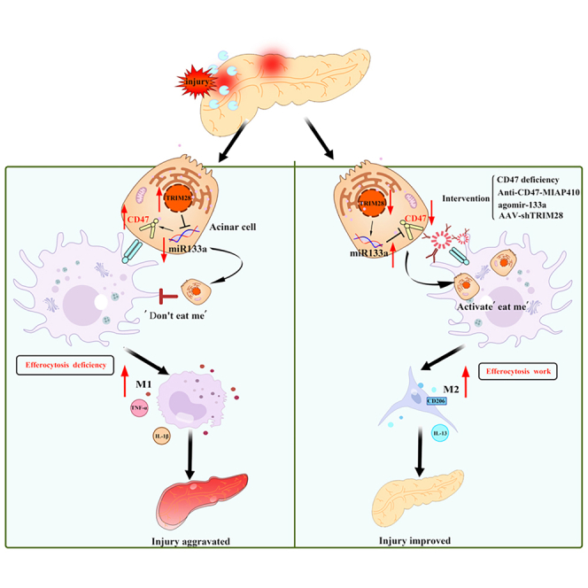
Efferocytosis, the clearance of apoptotic cells by macrophages, plays a crucial role in inflammatory responses and effectively prevents secondary necrosis. However, the mechanisms underlying efferocytosis in acute pancreatitis (AP) remain unclear. In this study, we demonstrated the presence of efferocytosis in injured human and mouse pancreatic tissues. We also observed significant upregulation of CD47, an efferocytosis-related the "do not eat me" molecule in injured acinar cells. Subsequently, we used CRISPR-Cas9 gene editing, anti-adeno-associated virus (AAV) gene modification, and anti-CD47 antibody to investigate the potential therapeutic role of AP. CD47 expression was negatively regulated by upstream miR133a, which is controlled by the transcription factor TRIM28. To further investigate the regulation of efferocytosis and reduction of pancreatic necrosis in AP, we used miR-133a-agomir and pancreas-specific AAV-shTRIM28 to modulate CD47 expression. Our findings confirmed that CD47-mediated efferocytosis is critical for preventing pancreatic necrosis and suggest that targeting the TRIM28-miR133a-CD47 axis is clinically relevant for the treatment of AP.

Keywords: acute pancreatitis; apoptotic cell clearance; clinical translation; immune microenvironment.


Conflict of interest statement

Declaration of interests The authors declare no competing interests.

Figures

None
Graphical abstract
Figure 1
Figure 1
The do not eat me ligand CD47 is progressively upregulated in injured PACs (A and B) Gene set enrichment analysis demonstrates differential pathways associated with cell death pathways, among them, the phagocytosis pathways is significant changed (NES = 2.0922, p = 0.0012). (C) RNA-seq analysis reveals the expression levels of representative genes, including CD47, CD24, CD31, and MHC I, in the phagocytosis-related pathways. (D) The mRNA expression level of CD24, CD31, MHC I, and CD47 of pancreas on time course AP mice by qPCR detection. (E) Expression and relative quantification of CD47 in pancreatic tissue homogenates obtained from time course AP mice by WB (n = 3). (F) Immunofluorescent staining images (scale bar, 25 μm) of amylase and CD47 on the pancreas of AP mice and normal control mice. ns, not significant, ∗p<0.05, ∗∗p<0.01, ∗∗∗p<0.001.
Figure 2
Figure 2
Efferocytosis is present in injured pancreatic tissue of mice (A and B) Hematoxylin and eosin staining images (scar bar, 25 μM), TEM (scar bar, 25 μM) and high-resolution immunofluorescent imaging (scar bar, 25 μM; taken with 40× and 100× objectives) of mice on time course AP (n = 5) confirms that efferocytosis is present on the injury acinar cells. (C) Infiltrating macrophages and efferocytosis quantified. (D) Detailed description of in vitro co-culture trial of BMDM and 266-6 cells or PACs. (E) FACS for the detection of efferocytosis in vitro that stimulated by CCK in time gradients (0 h, 1 h, and 2 h; n = 4), which the right upper square frame (red) includes efferocytosis cells. (F and G) Quantification of macrophage rate and LDH release level of co-culture trial (n = 4). ∗p<0.05, ∗∗p<0.01, ∗∗∗p<0.001.
Figure 3
Figure 3
Conventional CD47 KO improves efferocytosis and protects AP in vivo (A and B) Hematoxylin and eosin-stained images and quantification showed that CD47 gene deficiency prevents the severity of AP mice (scar bar, 25 μm, n = 6). (C and D) Serum enzymology and inflammatory factors of AP mice. (E) Additionally, we quantify efferocytosis index with 'uncleared apoptotic cells' (yellow stars) and efferocytosis (yellow arrows) on high-resolution immunofluorescent (scar bar, 25 μm; taken with 40× and 100× objectives). Representative TEM images suggest phagocytic rate by macrophages on CD47 KO mice and WT mice after Cae-induced (scar bar, 25 μm; n = 4). (F) High-resolution fluorescence image quantification (macrophage count and efferocytosis rate) of pancreatic tissues on AP mice (n = 4). (G and H) Representative flowchart images and quantification of CD206+ macrophages (M2) and TNF-α+ macrophages (M1) in pancreatic tissues of AP mice (n = 4). ∗p<0.05, ∗∗p<0.01, ∗∗∗p<0.001.
Figure 4
Figure 4
Pancreas-specific CD47 overexpression aggravates pancreatic necrosis AAV-CD47 or AAV-Con wrapped with AAV is injected intrapancreatically into C57BL/6, and then establish AP mice model. (A and B) Expression and relative quantification of CD47 in pancreatic tissue homogenates obtained from AAV-Con and AAV-CD47 overexpression mice by WB. (C) The mRNA expression level of CD47 of pancreas on AAV-Con and AAV-CD47 overexpression mice by qPCR. (D and E) Hematoxylin and eosin staining images and quantification show that the overexpression of CD47 aggravates the severity of Cae-AP mice (scar bar, 25 μm; n = 5). (F and G) Overexpression of CD47 elevates the levels of serum enzymes and the serum inflammatory factors. ∗p<0.05, ∗∗p<0.01, ∗∗∗p<0.001.
Figure 5
Figure 5
Anti-CD47 Ab improves efferocytosis to protect against pancreatic necrosis in AP (A and B) Hematoxylin and eosin-stained images (scale bar, 25 μM; taken with 40× and 100× objectives) and histological scores (edema, inflammation, and necrosis) of pancreatic tissues. (C and D) Serum enzymology and inflammatory factors of IgG and anti-CD47 mice induced by Cae (n = 6). (E) Representative TEM images and high-resolution immunofluorescent images of IgG and anti-CD47 AP mice. (F) High-resolution fluorescence image quantification (macrophage count and efferocytosis rate) of pancreatic tissues on IgG and anti-CD47 AP mice (n = 4). (G and H) Representative flowchart images and quantification of CD206+ macrophages (M2) and TNF-α+ macrophages (M1) in pancreatic tissues of AP mice (n = 4). ∗p<0.05, ∗∗p<0.01, ∗∗∗p<0.001.
Figure 6
Figure 6
MiR133a promotes efferocytosis efficiency by negatively regulating CD47 (A) qPCR detected the miR133a mRNA levels of pancreatic tissue of time gradient Cae-induced AP mice (n = 4). (B) Immunofluorescent images (scar bar, 25 μM; taken with 40× and 100× objectives) and miR133a quantification of pancreatic tissue on Cae-induced AP mice and normal control mice. (C) Hematoxylin and eosin (H&E)-stained images and histological scores (edema, inflammation, and necrosis) of pancreatic tissues. (D) Serum enzymology of AP mice (n = 6). Similar to anti-CD47 Ab, agomir-miR133a improves the severity of pancreatitis, decreases the levels of serum enzymes and decreases the serum inflammatory factors. (E) H&E-stained images (scar bar, 25 μM), TEM (scar bar, 25 μM) and high-resolution immunofluorescent imaging (scar bar, 25 μM; taken with 40× and 100× objectives) of mice. Correspondingly, agomir-miR133a has a lower number of apoptotic cells (TUNEL in green), together with fewer free apoptotic bodies (yellow stars). Apoptotic cells are commonly close to macrophages (Mac-3 in red) and are considered as efferocytosis if they are co-localized (yellow arrows), and representative images of TEM suggests phagocytic content by macrophages in pancreatic tissues after agomir-miR133a treatment; however, the above phenomena are counteracted in CD47 KO mice. (F and G) High-resolution fluorescence image quantification (macrophage count and efferocytosis rate) of pancreatic tissues on AP mice (n = 4). ∗p<0.05, ∗∗p<0.01, ∗∗∗p<0.001.
Figure 7
Figure 7
TRIM28 down-regulates miR133a to repress efferocytosis (A) The mRNA expression level of TRIM28 of pancreas on time course AP mice. (B–D) Expression and relative quantification of CD47 in pancreatic tissue homogenates and the mRNA expression level of CD47 by WB or qPCR obtained from AAV-shCon and AAV-shTRIM28 mice. (E) IHC staining images of TRIM28/CD47 and IF staining images (scar bar, 25 μM) of miR133a on AAV-shCon and AAV-shTRIM28 mice. (F) Quantification of TRIM28, miR133a and CD47. (G and H) Hematoxylin and eosin-stained images and quantification show that AAV-shTRIM28 prevents the severity of AP mice (scar bar, 25 μm, n = 6). (I and J) Serum enzymology and inflammatory factors of AP mice (n = 6). (K) Representative TEM images and high-resolution immunofluorescent images of AAV-shCon and AAV-shTRIM28 AP mice. (L) High-resolution fluorescence image quantification (macrophage count and efferocytosis rate) of pancreatic tissues on AAV-shCon and AAV-shTRIM28 AP mice (n = 4). ∗p<0.05, ∗∗p<0.01, ∗∗∗p<0.001.
Figure 8
Figure 8
The regulatory role of TRIM28-miR133a-CD47 axis and efferocytosis in human tissue specimens (A) Representative hematoxylin and eosin-stained images, immunofluorescent images of miR133a and IHC staining images of TRIM28/CD47 on normal pancreatic tissues (n = 6) and pancreatitis patients tissue array (n = 17). (B) Histological scores (edema, inflammation, and necrosis) and quantification of TRIM28, miR133a, and CD47 on pancreatic tissues. (C) Correlation analysis confirms that the expression of the above three are positively correlated with necrotic acinar cells in human clinical AP samples; TRIM28 and CD47 expression are positively correlated. (D) High-resolution immunofluorescent imaging (scar bar, 25 μM; taken with 40× and 100× objectives) of human pancreatic tissues. (E) High-resolution fluorescence image quantification (macrophage count and efferocytosis rate) of pancreatic tissues on normal control and AP pancreas (n = 4). (F) The graphical abstract of our research. ∗p<0.05, ∗∗p<0.01, ∗∗∗p<0.001.

References

    1. Mederos M.A., Reber H.A., Girgis M.D. Acute Pancreatitis: A Review. Jama. 2021;325:382–390. - PubMed
    1. Sah R.P., Garg P., Saluja A.K. Pathogenic mechanisms of acute pancreatitis. Curr. Opin. Gastroenterol. 2012;28:507–515. - PMC - PubMed
    1. Talukdar R., Sareen A., Zhu H., Yuan Z., Dixit A., Cheema H., George J., Barlass U., Sah R., Garg S.K., et al. Release of Cathepsin B in Cytosol Causes Cell Death in Acute Pancreatitis. Gastroenterology. 2016;151:747–758.e5. - PMC - PubMed
    1. Sendler M., Dummer A., Weiss F.U., Krüger B., Wartmann T., Scharffetter-Kochanek K., van Rooijen N., Malla S.R., Aghdassi A., Halangk W., et al. Tumour necrosis factor α secretion induces protease activation and acinar cell necrosis in acute experimental pancreatitis in mice. Gut. 2013;62:430–439. - PubMed
    1. Sendler M., Weiss F.U., Golchert J., Homuth G., van den Brandt C., Mahajan U.M., Partecke L.I., Döring P., Gukovsky I., Gukovskaya A.S., et al. Cathepsin B-Mediated Activation of Trypsinogen in Endocytosing Macrophages Increases Severity of Pancreatitis in Mice. Gastroenterology. 2018;154:704–718.e10. - PMC - PubMed

MeSH terms

LinkOut - more resources