Case Reports
. 2024 May 14;16(5):e60273.
doi: 10.7759/cureus.60273. eCollection 2024 May.

Port-Site Metastasis After Laparoscopic Gastrectomy Extending to the Thigh: A Case Report

Affiliations
Case Reports

Port-Site Metastasis After Laparoscopic Gastrectomy Extending to the Thigh: A Case Report

Kentaro Goto et al. Cureus. .

Abstract

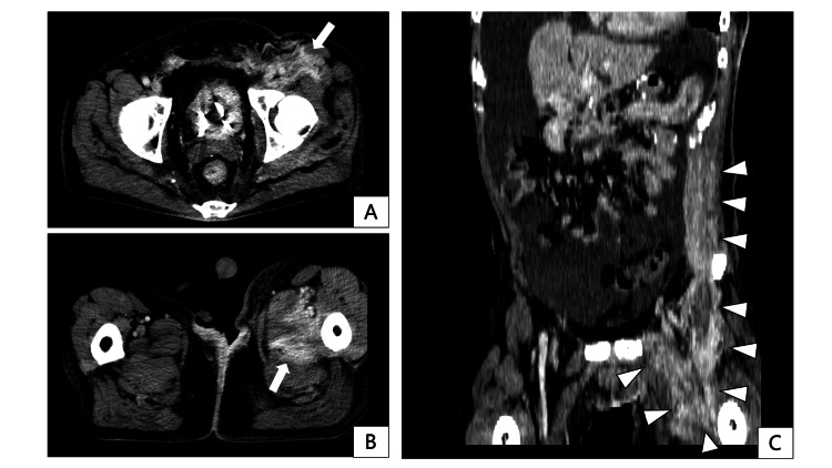
Port-site metastasis (PSM) is rare following laparoscopic gastrectomy for gastric cancer. Previous reports focused on localized lesions treated with excision; contrastingly, case reports describing extensive invasion into the lower extremity skeletal muscles causing deterioration in activities of daily living are nonexistent. A 55-year-old male underwent a laparoscopic distal gastrectomy for gastric cancer. The pathological findings revealed a stage IIIA tumor. Two years later, skin hardening was observed on the left upper abdominal wall. Computed tomography displayed a 13-cm-long, flat tumor along the skeletal muscle around the left upper 12 mm port site and right hydronephrosis. The patient was diagnosed with PSM and retroperitoneal recurrence. Despite chemotherapy, three years postoperatively, PSM widely spread from the left upper abdomen to the left thigh, eventually inducing opioid-resistant leg pain and subsequent walking difficulties. Palliative radiotherapy could not improve these symptoms. The patient died three years and five months postoperatively. Extensively invasive PSM can induce refractory cancer pain and physical disorders. Therefore, early detection and palliative resection of PSM may help maintain the quality of life of patients with gastric cancer.

Keywords: abdominal wall recurrence; gastric cancer; laparoscopic surgery; muscle metastasis; port-site recurrence.


Conflict of interest statement

The authors have declared that no competing interests exist.

Figures

Figure 1
Figure 1. Endoscopic findings of the primary tumor.
Figure 2
Figure 2. Histological findings from hematoxylin-eosin-stained specimen.
Figure 3
Figure 3. Preoperative contrast-enhanced computed tomography.
The tumor is shown as a contrast-enhanced lesion in the posterior gastric wall (white arrow).
Figure 4
Figure 4. Trocar placement at surgery.
The umbilical incision is extended for specimen retrieval. An Information drain is placed at the suprapancreatic region from the upper right 5 mm port. Image created by the authors.
Figure 5
Figure 5. Pathological findings of resected specimen.
A: macroscopic findings; B: microscopic findings
Figure 6
Figure 6. Contrast-enhanced computed tomography and positron emission tomography-computed tomography (PET-CT) at the time of port-site recurrence, taken two years postoperatively.
A, B: A flat, contrast-enhancing tumor measuring 13 cm in maximum diameter is identified in the subcutaneous tissue, external/internal oblique muscle, and transversus abdominis muscles around the left upper abdominal 12 mm port site (indicated by the white arrow). Concurrent thickening of the right renal pelvis wall (white arrowheads in A), as well as right hydronephrosis and thickening of the anterior renal fascia (white arrowheads in B), are also noted. C: PET-CT showing an accumulation with a max-standardized uptake value (max-SUV) of 3.68 in the abdominal wall lesion.
Figure 7
Figure 7. Contrast-enhanced computed tomography at the time of port-site metastasis extension to the left thigh, taken three years postoperatively.
A, B: Axial images. High-density areas with contrast enhancement are observed subcutaneously at the left inguinal region, extending to the left thigh quadriceps and adductor muscles (indicated by the white arrow). C: Coronal image. High-density areas in the left inguinal subcutaneous and thigh muscles appear continuous with the known left abdominal wall port-site metastasis (indicated by the white arrowheads).
Figure 8
Figure 8. Photograph of the skin infiltration from port-site metastasis (indicated by the black arrowheads).
The lesion had a hyperpigmented area of nodular aspect.

References

    1. Global Cancer Statistics 2020: GLOBOCAN estimates of incidence and mortality worldwide for 36 cancers in 185 countries. Sung H, Ferlay J, Siegel RL, Laversanne M, Soerjomataram I, Jemal A, Bray F. CA Cancer J Clin. 2021;71:209–249. - PubMed
    1. Five-year survival outcomes of laparoscopy-assisted vs open distal gastrectomy for advanced gastric cancer: the JLSSG0901 randomized clinical trial. Etoh T, Ohyama T, Sakuramoto S, et al. JAMA Surg. 2023;158:445–454. - PMC - PubMed
    1. Port-site recurrence after laparoscopy-assisted gastrectomy: report of the first case. Lee YJ, Ha WS, Park ST, Choi SK, Hong SC. J Laparoendosc Adv Surg Tech A. 2007;17:455–457. - PubMed
    1. Port site metastasis after laparoscopic-assisted distal gastrectomy (LADG) Sakurai K, Tanaka H, Lee T, et al. Int Surg. 2013;98:363–366. - PMC - PubMed
    1. Long-term survival following port-site metastasectomy in a patient with laparoscopic gastrectomy for gastric cancer: a case report. Kim SH, Kim DJ, Kim W. J Gastric Cancer. 2015;15:209–213. - PMC - PubMed

Publication types

LinkOut - more resources