

Atypical presentation of an epiphrenic esophageal diverticulum 20 years post fundoplication: a case report and review
- PMID: 38872729
- PMCID: PMC11138112
- DOI: 10.1093/jscr/rjae316
Atypical presentation of an epiphrenic esophageal diverticulum 20 years post fundoplication: a case report and review
Abstract
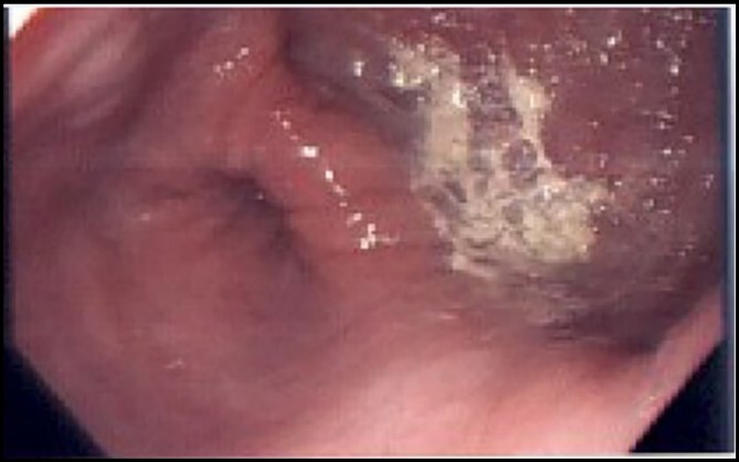
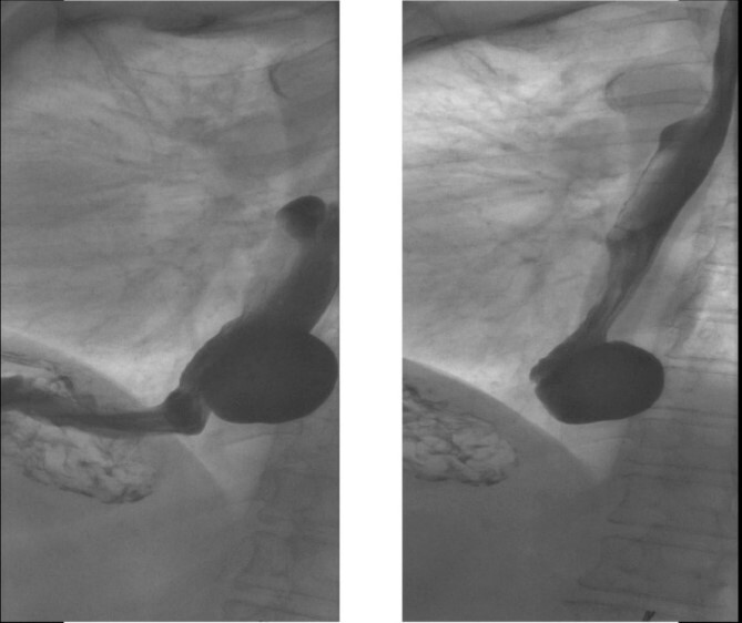
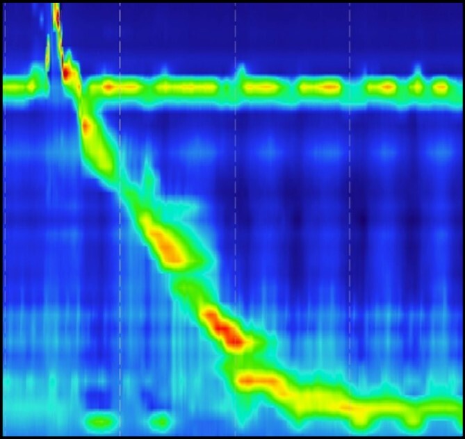
Esophageal diverticulum is a rare condition characterized by the herniation of the esophageal mucosa outside the esophageal wall. Here, we explore the prevalence of ED and its associated esophageal dysmotility. We also shed light on the potential impact of previous surgical interventions, such as Nissen's fundoplication, on the development of ED. This manuscript presents the case of a 72-year-old woman with a history of Nissen's fundoplication surgery who experienced worsening symptoms of dysphagia, heartburn and postprandial cough. Despite exhibiting a normal motility pattern, upper endoscopy revealed a large epiphrenic esophageal diverticulum. The patient underwent successful surgical resection with myotomy, resulting in the resolution of symptoms with no complications. This case highlights the rarity of symptomatic ED and the need to recognize it while choosing the optimal treatment modality.
Keywords: diverticulectomy; epiphrenic diverticulum; esophageal diverticulum; myotomy.
Published by Oxford University Press and JSCR Publishing Ltd. © The Author(s) 2024.
Conflict of interest statement
None declared.
Figures

Similar articles
-
Laparoscopic Approach to Epiphrenic Diverticula Under Endoscopic Assistance: A Technical Report.J Laparoendosc Adv Surg Tech A. 2024 Feb;34(2):162-166. doi: 10.1089/lap.2023.0469. Epub 2023 Dec 18. J Laparoendosc Adv Surg Tech A. 2024. PMID: 38109227 Review.
-
Myotomy-First Approach to Epiphrenic Esophageal Diverticula.J Laparoendosc Adv Surg Tech A. 2019 Jun;29(6):726-729. doi: 10.1089/lap.2019.0239. Epub 2019 Apr 29. J Laparoendosc Adv Surg Tech A. 2019. PMID: 31034339
-
Esophageal epiphrenic diverticulum treated with laparoscopic surgery in a patient with systemic sclerosis: A rare case report.Int J Surg Case Rep. 2023 May;106:108136. doi: 10.1016/j.ijscr.2023.108136. Epub 2023 Apr 7. Int J Surg Case Rep. 2023. PMID: 37080142 Free PMC article.
-
Combined thoracoscopic and laparoscopic surgery for epiphrenic diverticulum with associated gastroesophageal reflux disease: a case report.Surg Case Rep. 2024 Jan 15;10(1):17. doi: 10.1186/s40792-024-01813-0. Surg Case Rep. 2024. PMID: 38221572 Free PMC article.
-
Laparoscopic Treatment of a Huge Epiphrenic Esophageal Diverticulum: A Case Report and Literature Review.Ann Ital Chir. 2025;96(3):322-328. doi: 10.62713/aic.3802. Ann Ital Chir. 2025. PMID: 40090848 Review.
References
Publication types
Grants and funding
LinkOut - more resources
Full Text Sources
