

Enhancing volumetric muscle loss (VML) recovery in a rat model using super durable hydrogels derived from bacteria
- PMID: 38872731
- PMCID: PMC11170101
- DOI: 10.1016/j.bioactmat.2024.04.006
Enhancing volumetric muscle loss (VML) recovery in a rat model using super durable hydrogels derived from bacteria
Abstract
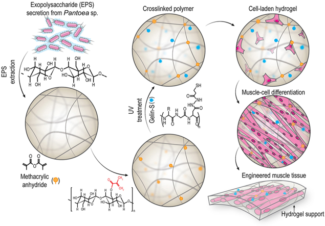
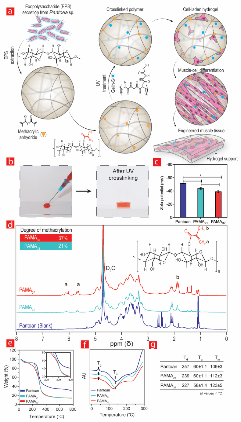
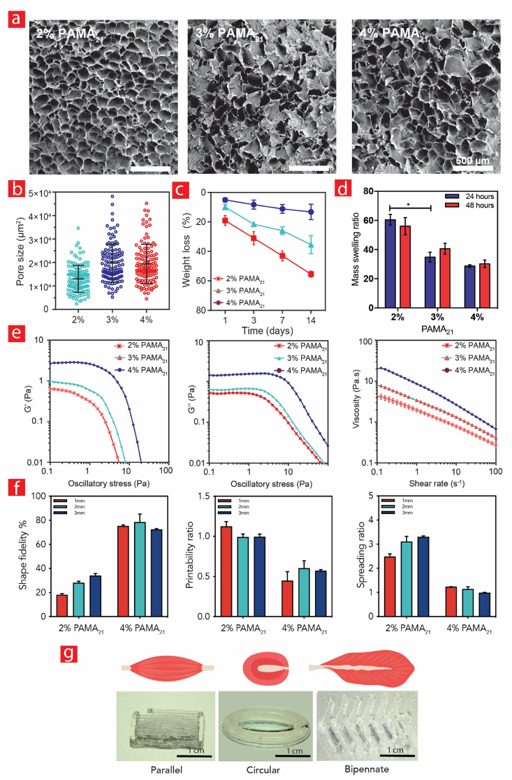
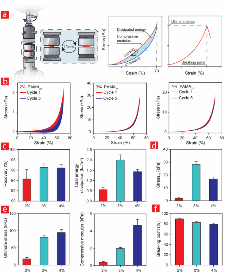
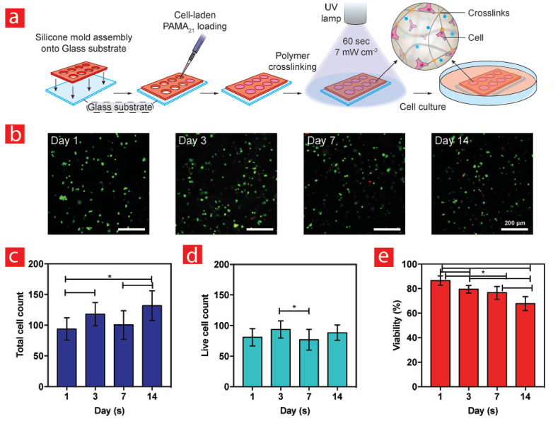
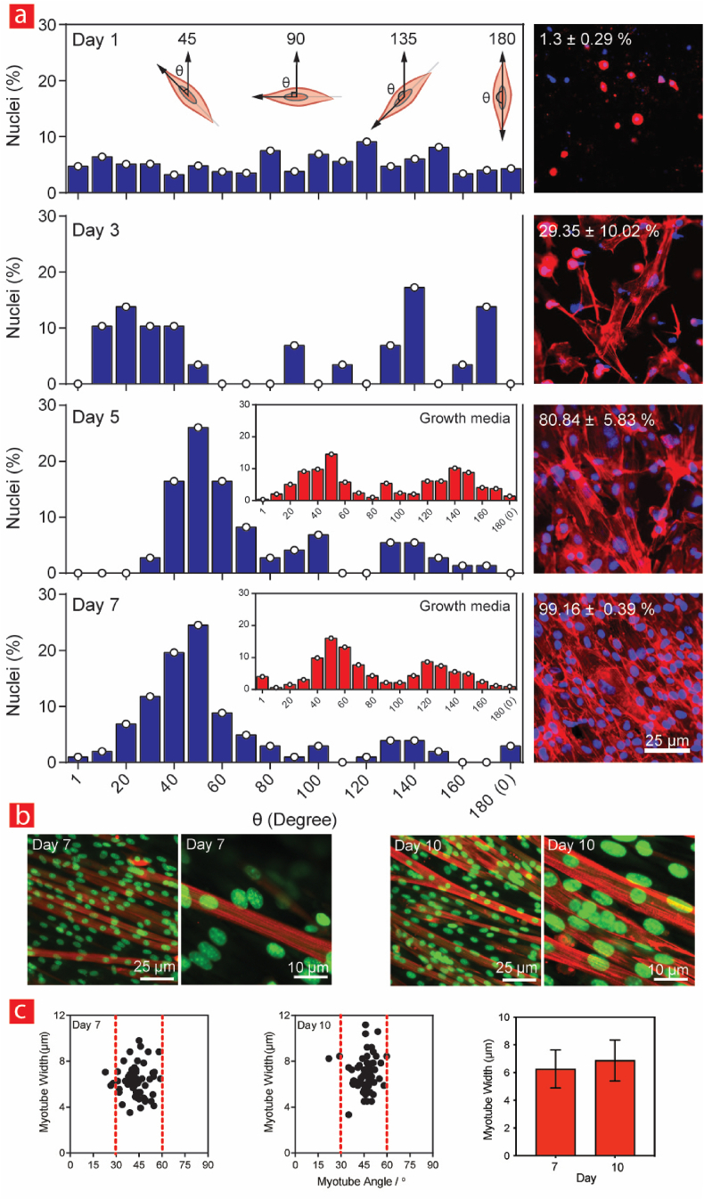
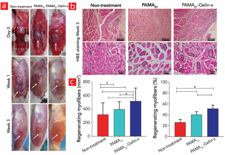
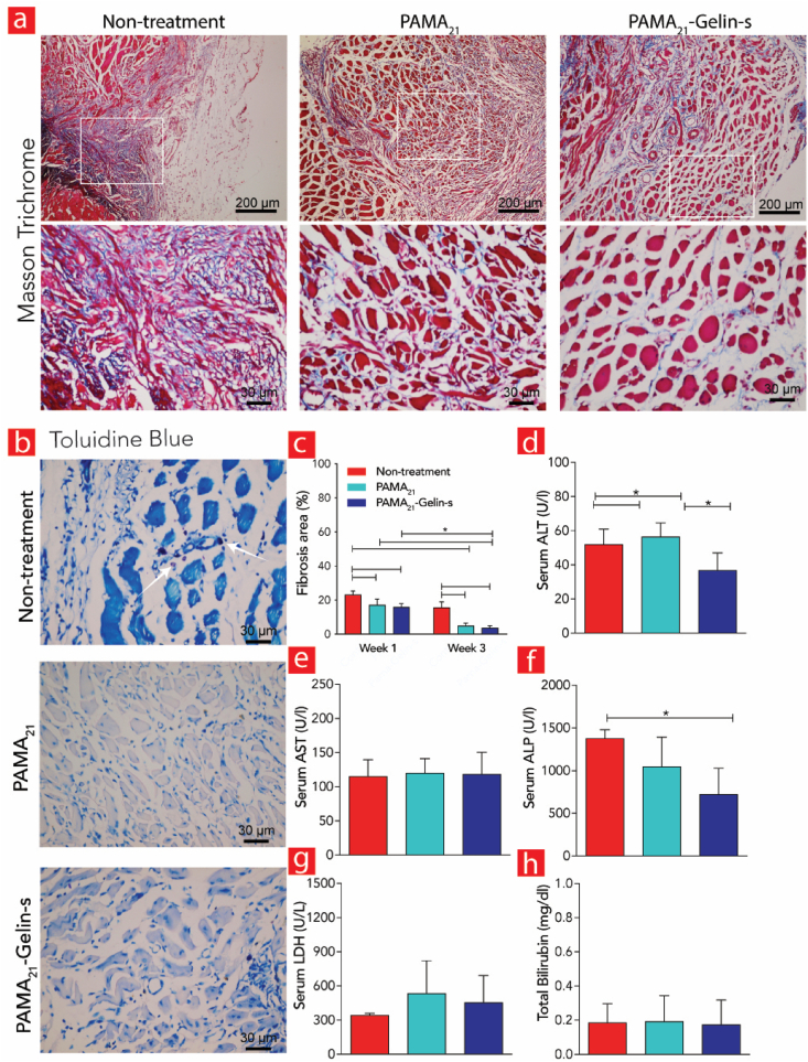
Bacteria can be programmed to deliver natural materials with defined biological and mechanical properties for controlling cell growth and differentiation. Here, we present an elastic, resilient and bioactive polysaccharide derived from the extracellular matrix of Pantoea sp. BCCS 001. Specifically, it was methacrylated to generate a new photo crosslinkable hydrogel that we coined Pantoan Methacrylate or put simply PAMA. We have used it for the first time as a tissue engineering hydrogel to treat VML injuries in rats. The crosslinked PAMA hydrogel was super elastic with a recovery nearing 100 %, while mimicking the mechanical stiffness of native muscle. After inclusion of thiolated gelatin via a Michaelis reaction with acrylate groups on PAMA we could also guide muscle progenitor cells into fused and aligned tubes - something reminiscent of mature muscle cells. These results were complemented by sarcomeric alpha-actinin immunostaining studies. Importantly, the implanted hydrogels exhibited almost 2-fold more muscle formation and 50 % less fibrous tissue formation compared to untreated rat groups. In vivo inflammation and toxicity assays likewise gave rise to positive results confirming the biocompatibility of this new biomaterial system. Overall, our results demonstrate that programmable polysaccharides derived from bacteria can be used to further advance the field of tissue engineering. In greater detail, they could in the foreseeable future be used in practical therapies against VML.
© 2024 The Authors.
Conflict of interest statement
The authors declare that they have no known competing financial interests or personal relationships that could have appeared to influence the work reported in this paper.
Figures
Similar articles
-
Photoreactive Hydrogel Stiffness Influences Volumetric Muscle Loss Repair.Tissue Eng Part A. 2022 Apr;28(7-8):312-329. doi: 10.1089/ten.TEA.2021.0137. Epub 2022 Jan 4. Tissue Eng Part A. 2022. PMID: 34409861 Free PMC article.
-
Keratin Hydrogel Enhances In Vivo Skeletal Muscle Function in a Rat Model of Volumetric Muscle Loss.Tissue Eng Part A. 2017 Jun;23(11-12):556-571. doi: 10.1089/ten.TEA.2016.0458. Epub 2017 Apr 14. Tissue Eng Part A. 2017. PMID: 28169594 Free PMC article.
-
Bioprinted anisotropic scaffolds with fast stress relaxation bioink for engineering 3D skeletal muscle and repairing volumetric muscle loss.Acta Biomater. 2023 Jan 15;156:21-36. doi: 10.1016/j.actbio.2022.08.037. Epub 2022 Aug 21. Acta Biomater. 2023. PMID: 36002128
-
Skeletal Muscle Tissue Engineering: Biomaterials-Based Strategies for the Treatment of Volumetric Muscle Loss.Bioengineering (Basel). 2020 Jul 31;7(3):85. doi: 10.3390/bioengineering7030085. Bioengineering (Basel). 2020. PMID: 32751847 Free PMC article. Review.
-
Extrusion 3D (Bio)Printing of Alginate-Gelatin-Based Composite Scaffolds for Skeletal Muscle Tissue Engineering.Materials (Basel). 2022 Nov 10;15(22):7945. doi: 10.3390/ma15227945. Materials (Basel). 2022. PMID: 36431432 Free PMC article. Review.
Cited by
-
Hyaluronic Acid-Coated Melt Electrowritten Scaffolds Promote Myoblast Attachment, Alignment, and Differentiation.bioRxiv [Preprint]. 2025 Mar 10:2025.03.06.641880. doi: 10.1101/2025.03.06.641880. bioRxiv. 2025. PMID: 40161586 Free PMC article. Preprint.
-
Biomedical engineering utilizing living photosynthetic cyanobacteria and microalgae: Current status and future prospects.Mater Today Bio. 2024 Jul 14;27:101154. doi: 10.1016/j.mtbio.2024.101154. eCollection 2024 Aug. Mater Today Bio. 2024. PMID: 39113912 Free PMC article. Review.
References
-
- Sorensen J.R., Mcfaline-Figueroa J., Call J.A. Pathophysiology of volumetric muscle loss and targets for regenerative rehabilitation. Regenerative Rehabilitation: From Basic Science to the Clinic. 2022:177–225. Springer.
-
- Carleton M.M., Sefton M.V. Injectable and degradable methacrylic acid hydrogel alters macrophage response in skeletal muscle. Biomaterials. 2019;223 - PubMed
LinkOut - more resources
Full Text Sources
