

Antenatal imaging diagnosis of thoraco-omphalopagus conjoined twins
- PMID: 38872747
- PMCID: PMC11169063
- DOI: 10.1016/j.radcr.2024.05.003
Antenatal imaging diagnosis of thoraco-omphalopagus conjoined twins
Abstract
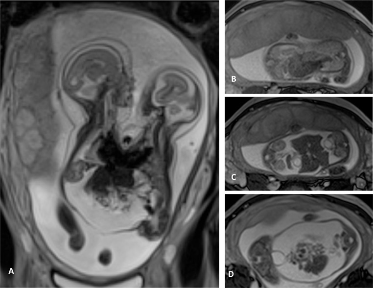
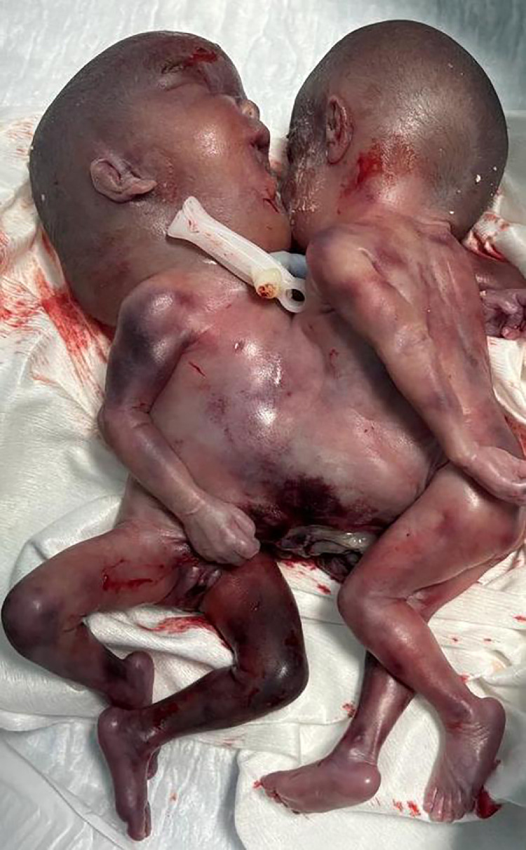
Conjoined twins occur in an estimated one in every 200,000 live births. The etiology remains largely speculative, with genetic and environmental factors being considered. The thoraco-omphalopagus type accounts for approximately 40% of cases, making it a focal point for clinical and radiological research. Radiological imaging plays a pivotal role in delineating anatomical details, offering insights into the feasibility of surgical interventions and informing parental counselling regarding prognosis and management options. We present a case of thoracoomphalogus conjoined twins diagnosed during the third trimester of pregnancy in a 19-year-old woman. The detailed radiological assessment using ultra-sound and MRI provided crucial information on organ sharing and vascular anatomy, which is critical for management strategies. This case underscores the critical role of prenatal imaging in detecting complex congenital anomalies, facilitating informed decision-making by healthcare providers and families.
Keywords: Antenatal diagnosis; Conjoined twins; MRI; Prognosis; Ultrasound.
© 2024 The Authors. Published by Elsevier Inc. on behalf of University of Washington.
Figures


Similar articles
-
Thoraco-omphalopagus conjoined twins: comprehensive evaluation with sonography and MRI in first trimester-a rare imaging diagnosis.BJR Case Rep. 2024 Nov 23;11(1):uaae045. doi: 10.1093/bjrcr/uaae045. eCollection 2025 Jan. BJR Case Rep. 2024. PMID: 39759171 Free PMC article.
-
A Case of Thoraco-Omphalopagus Conjoined Twins: Clinical Imaging and Anatomical Classification.Cureus. 2023 Dec 13;15(12):e50452. doi: 10.7759/cureus.50452. eCollection 2023 Dec. Cureus. 2023. PMID: 38222228 Free PMC article.
-
Thoraco-omphalopagus conjoined twin: A rare case report.Int J Surg Case Rep. 2022 Oct;99:107683. doi: 10.1016/j.ijscr.2022.107683. Epub 2022 Sep 19. Int J Surg Case Rep. 2022. PMID: 36137433 Free PMC article.
-
Successful dilation and evacuation for second trimester conjoined twin: a case report and review of the literature.J Med Case Rep. 2021 May 21;15(1):298. doi: 10.1186/s13256-021-02815-4. J Med Case Rep. 2021. PMID: 34020695 Free PMC article. Review.
-
Conjoined twins in a spontaneous monochorionic triplet pregnancy: A case report and literature review.Medicine (Baltimore). 2021 Jan 29;100(4):e24490. doi: 10.1097/MD.0000000000024490. Medicine (Baltimore). 2021. PMID: 33530268 Free PMC article. Review.
Cited by
-
Successful Termination of Conjoined Twins in the Second Trimester: A Case Report.Cureus. 2025 Jun 3;17(6):e85309. doi: 10.7759/cureus.85309. eCollection 2025 Jun. Cureus. 2025. PMID: 40621229 Free PMC article.
-
Thoraco-omphalopagus conjoined twins: comprehensive evaluation with sonography and MRI in first trimester-a rare imaging diagnosis.BJR Case Rep. 2024 Nov 23;11(1):uaae045. doi: 10.1093/bjrcr/uaae045. eCollection 2025 Jan. BJR Case Rep. 2024. PMID: 39759171 Free PMC article.
References
-
- Pajkrt E, Jauniaux E. First-trimester diagnosis of conjoined twins. Prenat Diagn. 2005;25:820–826. - PubMed
Publication types
LinkOut - more resources
Full Text Sources
