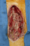
Deep Sternal Wound Infection Caused by Rhizopus Species After Coronary Artery Bypass Graft
- PMID: 38872847
- PMCID: PMC11170494
- DOI: 10.1093/ofid/ofae302
Deep Sternal Wound Infection Caused by Rhizopus Species After Coronary Artery Bypass Graft
Abstract
Deep sternal wound infection is a rare complication of cardiac surgery that is typically caused by skin resident flora, such as species of Staphylococcus and Streptococcus. Infections caused by fungi are less common and are generally caused by Candida species. Regardless of etiology, these infections are associated with significant morbidity and mortality. We present a case of postoperative mediastinitis that occurred following a 5-vessel coronary artery bypass graft and was caused by a filamentous fungus of the Rhizopus genus. The patient was treated with serial debridement, liposomal amphotericin B, and isavuconazonium and was discharged from the hospital in stable condition. Fungal mediastinitis is a rare entity, and clinicians must maintain a high level of suspicion to make the diagnosis. A fungal cause of postoperative mediastinitis should be considered in patients with negative bacterial cultures, uncontrolled diabetes, or current immunosuppression or those who present weeks after surgery with a subacute onset of symptoms.
Keywords: Mucorales; Rhizopus; cardiac surgery; deep sternal wound infection; mediastinitis.
© The Author(s) 2024. Published by Oxford University Press on behalf of Infectious Diseases Society of America.
Conflict of interest statement
Potential conflicts of interest. All authors: No reported conflicts.
Figures


Similar articles
-
Successful Treatment of Sternal Osteomyelitis and Mediastinitis Caused by Rhizopus Following Cardiac Surgery.Cureus. 2024 Jun 13;16(6):e62312. doi: 10.7759/cureus.62312. eCollection 2024 Jun. Cureus. 2024. PMID: 39006712 Free PMC article.
-
A Rare Case of Aspergillus Mediastinitis After Coronary Artery Bypass Surgery: A Case Report and Literature Review.Am J Case Rep. 2021 Dec 15;22:e933193. doi: 10.12659/AJCR.933193. Am J Case Rep. 2021. PMID: 34907149 Free PMC article. Review.
-
Major bleeding complicating deep sternal infection after cardiac surgery.J Thorac Cardiovasc Surg. 2003 Mar;125(3):554-8. doi: 10.1067/mtc.2003.31. J Thorac Cardiovasc Surg. 2003. PMID: 12658197
-
Postoperative mediastinitis in cardiac surgery - microbiology and pathogenesis.Eur J Cardiothorac Surg. 2002 May;21(5):825-30. doi: 10.1016/s1010-7940(02)00084-2. Eur J Cardiothorac Surg. 2002. PMID: 12062270
-
Candida mediastinitis after a cardiac operation.Ann Thorac Surg. 1990 Jan;49(1):157-63. doi: 10.1016/0003-4975(90)90382-g. Ann Thorac Surg. 1990. PMID: 2404472 Review.
References
-
- Meszaros K, Fuehrer U, Grogg S, et al. . Risk factors for sternal wound infection after open heart operations vary according to type of operation. Ann Thorac Surg 2016; 101:1418–25. - PubMed
-
- Lu JC, Grayson AD, Jha P, Srinivasan AK, Fabri BM. Risk factors for sternal wound infection and mid-term survival following coronary artery bypass surgery. Eur J Cardiothorac Surg 2003; 23:943–9. - PubMed
-
- Phoon PHY, Hwang NC. Deep sternal wound infection: diagnosis, treatment and prevention. J Cardiothorac Vasc Anesth 2020; 34:1602–13. - PubMed
-
- Kreter B, Woods M. Antibiotic prophylaxis for cardiothoracic operations: meta-analysis of thirty years of clinical trials. J Thorac Cardiovasc Surg 1992; 104:590–9. - PubMed
-
- Sommerstein R, Atkinson A, Kuster SP, et al. . Antimicrobial prophylaxis and the prevention of surgical site infection in cardiac surgery: an analysis of 21,007 patients in Switzerland. Eur J Cardiothorac Surg 2019; 56:800–6. - PubMed

