Review
. 2024 May 30:39:456-478.
doi: 10.1016/j.bioactmat.2024.04.026. eCollection 2024 Sep.

An overview of magnesium-based implants in orthopaedics and a prospect of its application in spine fusion

Affiliations
Review

An overview of magnesium-based implants in orthopaedics and a prospect of its application in spine fusion

Xuan He et al. Bioact Mater. .

Abstract

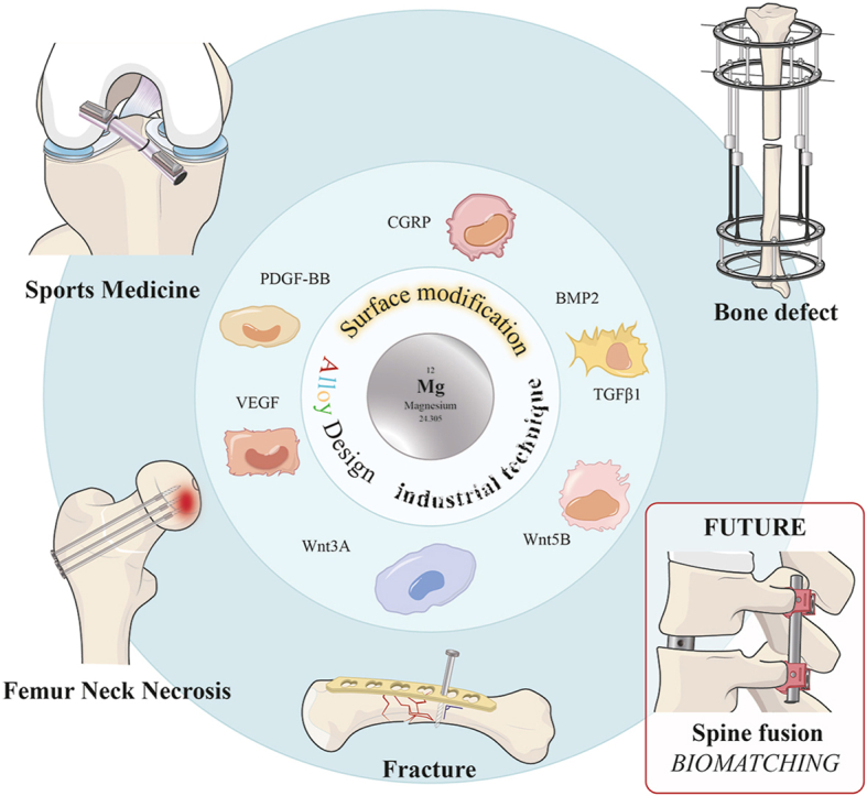
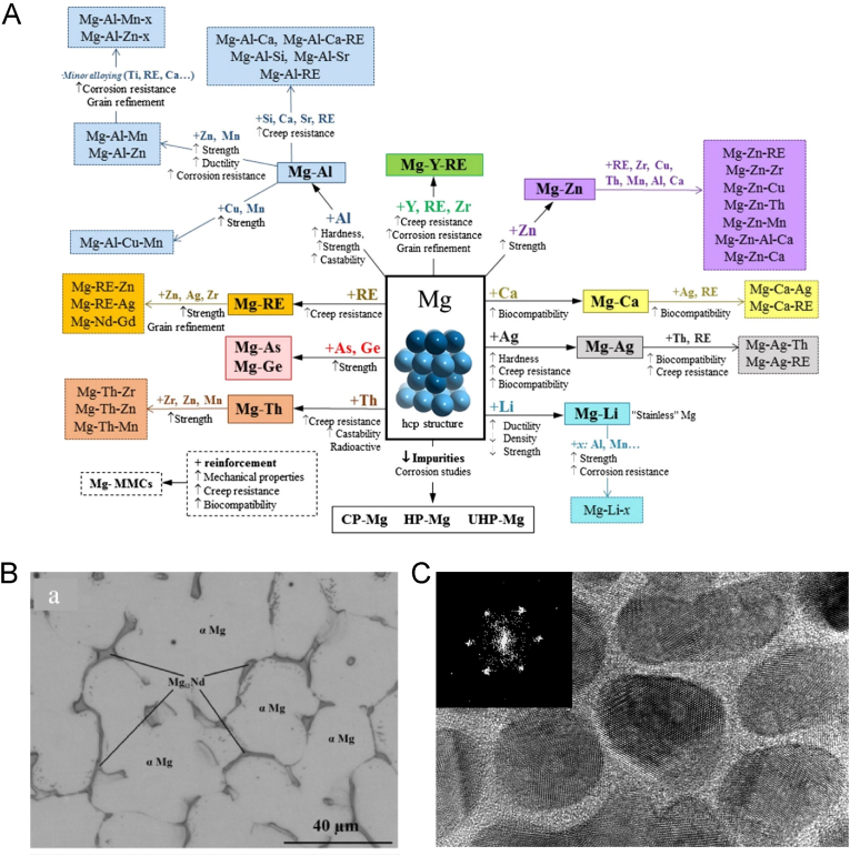
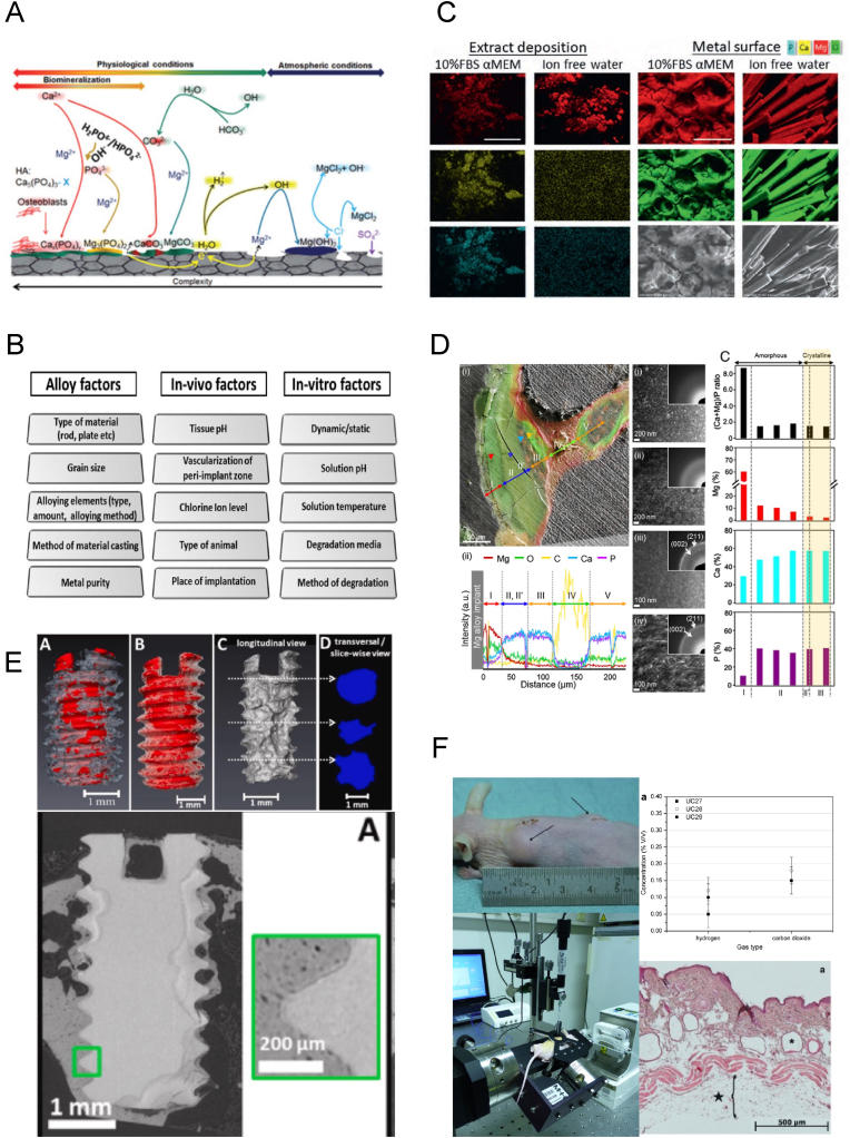
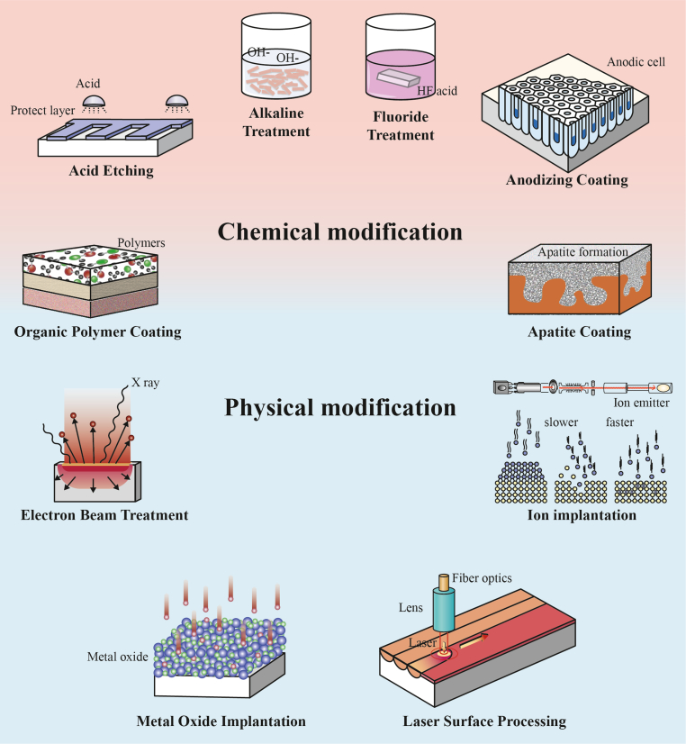
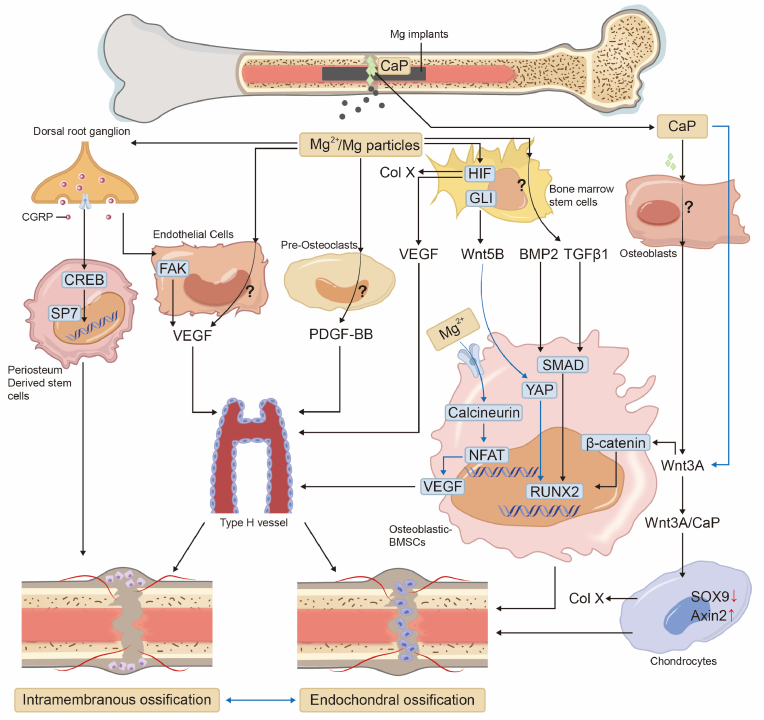
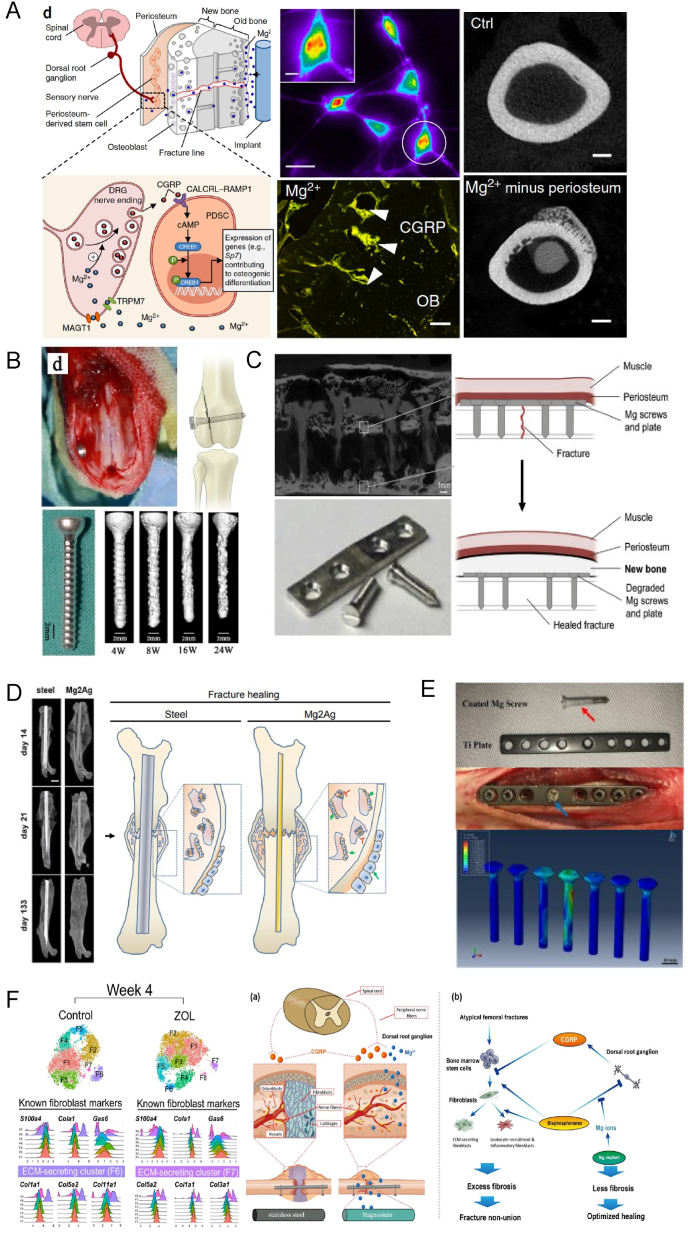
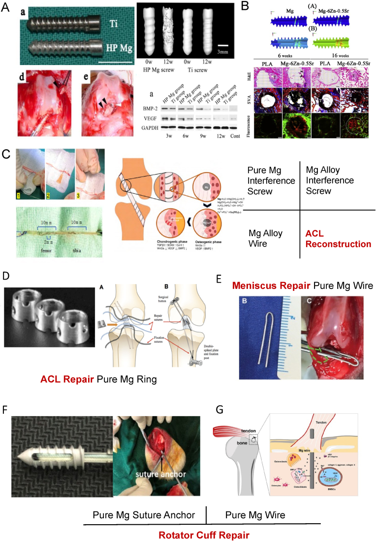
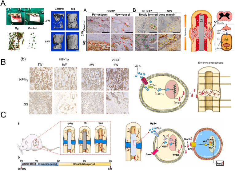
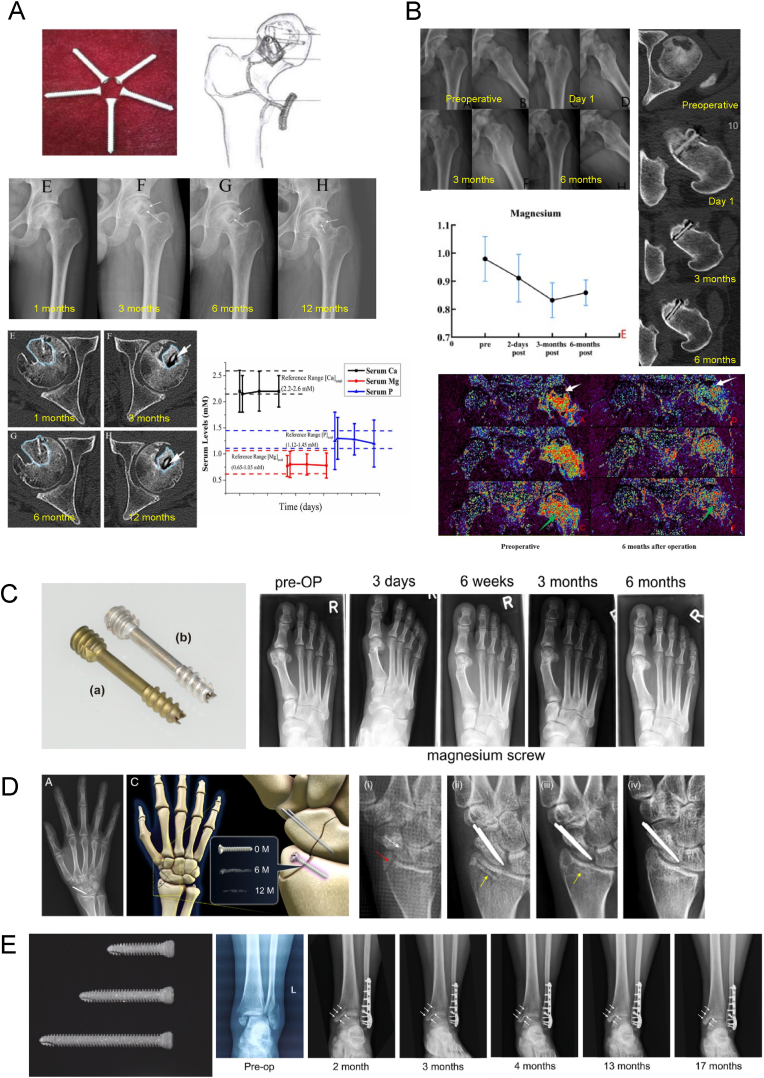
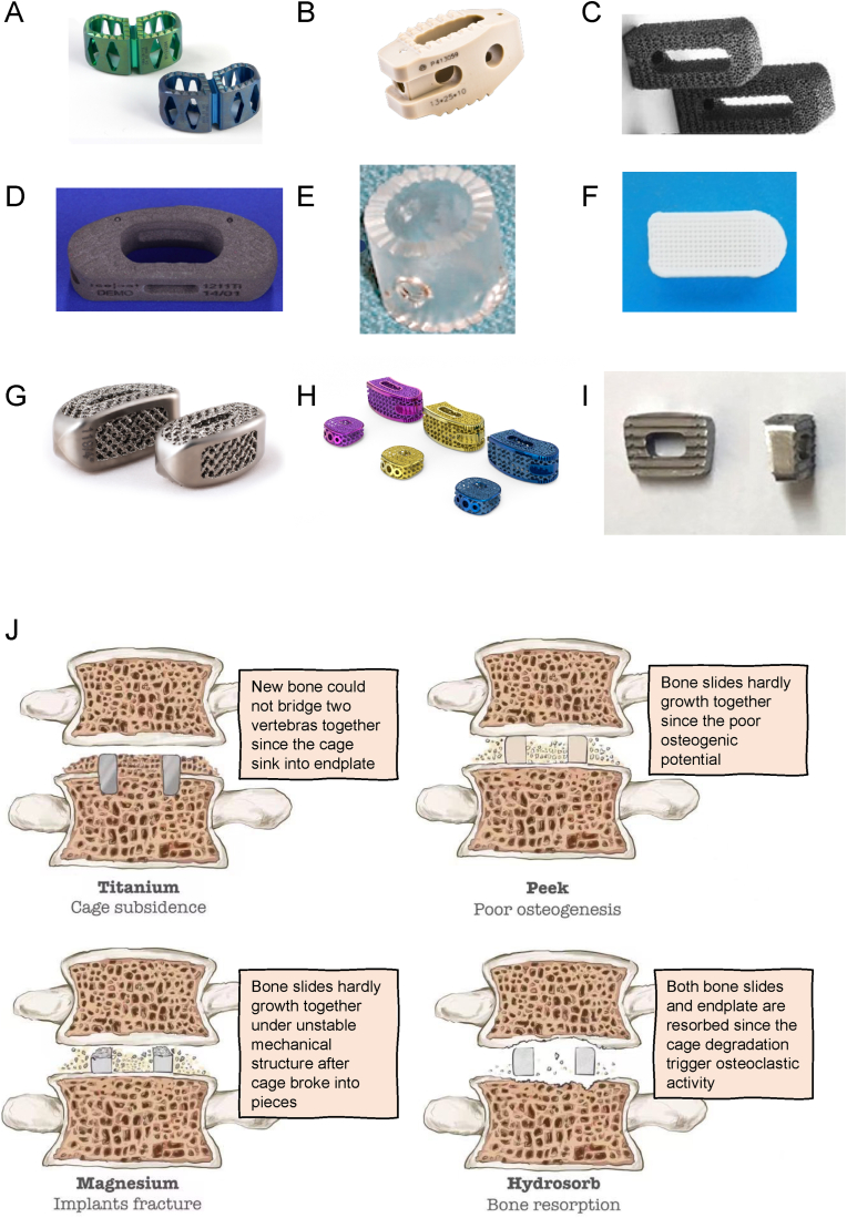
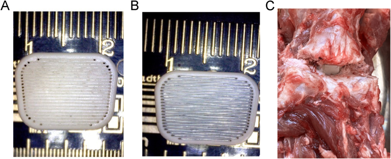
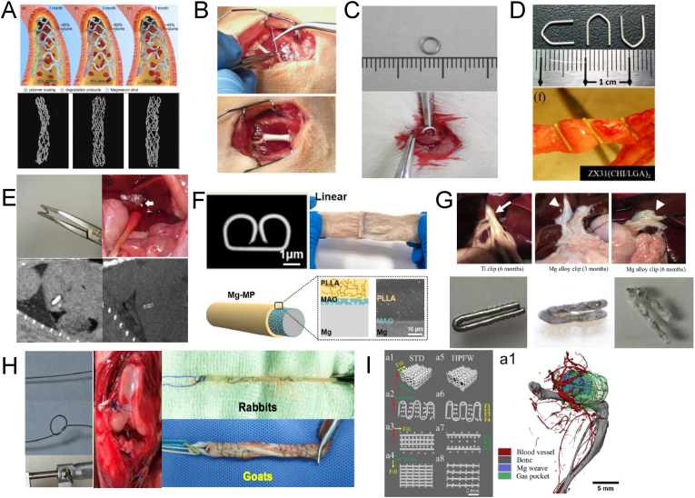
Due to matching biomechanical properties and significant biological activity, Mg-based implants present great potential in orthopedic applications. In recent years, the biocompatibility and therapeutic effect of magnesium-based implants have been widely investigated in trauma repair. In contrast, the R&D work of Mg-based implants in spinal fusion is still limited. This review firstly introduced the general background for Mg-based implants. Secondly, the mechanical properties and degradation behaviors of Mg and its traditional and novel alloys were reviewed. Then, different surface modification techniques of Mg-based implants were described. Thirdly, this review comprehensively summarized the biological pathways of Mg degradation to promote bone formation in neuro-musculoskeletal circuit, angiogenesis with H-type vessel formation, osteogenesis with osteoblasts activation and chondrocyte ossification as an integrated system. Fourthly, this review followed the translation process of Mg-based implants via updating the preclinical studies in fracture fixation, sports trauma repair and reconstruction, and bone distraction for large bone defect. Furthermore, the pilot clinical studies were involved to demonstrate the reliable clinical safety and satisfactory bioactive effects of Mg-based implants in bone formation. Finally, this review introduced the background of spine fusion surgeryand the challenges of biological matching cage development. At last, this review prospected the translation potential of a hybrid Mg-PEEK spine fusion cage design.

Keywords: Magnesium; Magnesium alloys; Magnesium implants; Magnesium surface modification; Spine fusion.


Conflict of interest statement

This study was supported by 10.13039/501100009592Beijing Municipal Science and Technology Project (Z201100005520073), Key Clinical projects of Peking University Third hospital (BYSY2022064), 10.13039/501100002858China Postdoctoral Science Foundation (M2023740146), and 10.13039/501100001809National Natural Science Foundation of China (82302731). Yufeng Zheng is editor-in-chief for Bioactive Materials and was not involved in the editorial review or the decision to publish this article. All authors declare that there are no competing interests.

Figures

Image 1
Graphical abstract
Fig. 1
Fig. 1
A. Alloying elements additions affect Mg mechanical properties and degradation behavior, Esmaily et al. [58], copyright 2017, ELSEVIER, Creative Commons Attribution License; B. Microstructure of JDBM alloy, Zhang et al. [59], copyright 2012, ELSEVIER; C. Microstructure of Mg-based SNDP-CG alloy, Wu et al. [17], copyright 2017, Springer Nature.
Fig. 2
Fig. 2
A. The degradation process of Mg, Gonzalez et al. [60], copyright 2018, Ke Ai, Creative Commons Attribution License; B. Factors affect Mg degradation rate, Pogorielov et al. [61], copyright 2017, AK journals, Creative Commons Attribution License; C. Mg in vitro degradation products analysis, He et al. [62], copyright 2024, Ke Ai, Creative Commons Attribution License; D. Mg in vivo degradation products analysis, Lee et al. [63], copyright 2016, PNAS, open access; E. Mg implants in vivo corrosion evaluation by using a synchrotron-radiation micro CT (SRμCT), Krüger et al. [64], copyright 2022, Ke Ai, Creative Commons Attribution License; F. Hydrogen concentration in tissue cavities after Mg implantation, top left: subcutaneous gas accumulation, top right: hydrogen concentration in cavities, bottom left: amperometric hydrogen sensor and mass spectrometric measurements, bottom right: subcutaneous gas cavities, H&E staining, Kuhlmann et al. [65], copyright 2013, ELSEVIER.
Fig. 3
Fig. 3
Surface modification techniques of Mg and its alloys.
Fig. 4
Fig. 4
The synergetic and cross talking net among various Mg degradation triggered osteogenic pathways, CaP: calcium phosphate crystal; CGRP: calcitonin gene-related peptide; CREB: cAMP-response element binding protein; Sp7: Osterix; FAK: focal adhesion kinase; VEGF: vascular endothelial growth factor; PDGF: platelet-derived growth factor; GLI: glioma-associated oncogene homolog; HIF: hypoxia inducible factor; WNT: wingless/integrated; BMP2: bone morphogenetic protein; TGF: transforming growth factor; SMAD: small mothers against decapentaplegic; YAP: hippo/yes-associated protein; NFAT: nuclear factor of activated T cells; SOX: sex determining region Y; RUNX: runt-related transcription factor; Axin: axis inhibitor; symbol “?”: the potential mechanisms require further investigation or confirmation.
Fig. 5
Fig. 5
Mg implants application in fracture fixation, A. Mg2+ from IMN triggered CGRP releasing to promote osteogenesis, Zhang et al. [208], copyright 2016, Springer Nature; B. Mg screw fixed distal femur fracture, Han et al. [225], copyright 2015, ELSVIER; C. Mg screw and plate fixed ulna fracture, Chaya et al. [226], copyright 2015, ELSVIER; D. Mg–Ag alloy IMN fixed femur shaft fracture, Jähn et al. [227], copyright 2016, ELSVISER; E. coated Mg screw fixed femur shaft fracture, Tian et al. [209], copyright 2018, ELSVISER; F. Mg2+ form IMN triggered CGRP releasing to suppress local fibrosis to promote atypical fracture healing, Zheng et al. [210], copyright 2022, ELSEVIER.
Fig. 6
Fig. 6
Mg implants application in sports medicine, A. Mg interference screw promoted bone tendon healing via upregulating BMP2 and VEGF expression, Cheng et al. [220], copyright 2016, ELSEVIER; B. Mg–Zn–Sr alloy interference screw promoted tunnel healing, Wang et al. [102], copyright 2018, ELSEVIER; C. Mg–Zn-Gd alloy wire suture tendon graft mediated fibrocartilages regeneration, He et al. [62], copyright 2024, Ke Ai, Creative Commons Attribution License; D. Mg ring repaired ACL rupture, Farraro et al. [230], copyright 2016, Wiley, free access; E. Mg wire enhanced meniscus regeneration, Zhang et al. [232], copyright 2019, SAGE; F. Mg suture anchor to enhanced bone tendon healing for reducing rotator cuff re-tear rate, Chen et al. [233], copyright 2022, ELSEVIER, Creative Commons Attribution License. G. Mg wire repaired teared rotator cuff, Zhang et al. [231], copyright 2022, ELSEVIER, Creative Commons Attribution License.
Fig. 7
Fig. 7
Mg implants application in bone distraction, A. Mg2+ from IMN enhanced H type vessel formation with detraction process via CGRP-FAK-VEGF axis, Li et al. [113], copyright 2021, ELSEVIER; B. Mg IMN degradation elevate HIF-1α and VEGF expression to facilitate bone formation via inhibiting VHL, Hamushan et al. [235], copyright 2020, SAGE; C. Mg IMN degradation upregulated Wnt5b expression to promote osteogenesis with detraction, Hamushan et al. [234], copyright 2021, KE AI, Creative Commons Attribution License.
Fig. 8
Fig. 8
Mg implants application in clinical scenario, A. 1-year-follow up study observed Mg screw fixed bone slides to treat femur head necrosis, Zhao et al. [236], copyright 2016, ELSEVIER; B. 6-month-follow up study observed Mg screw fixed bone slide to treat femur head necrosis, Sun et al. [237], copyright 2023, Wiley, open access; C. MAGNEZIX® screw applied for hallux valgus surgery, Windhagen et al. [238], copyright 2013, BMC, Creative Commons Attribution License; D. Mg screw fixed distal radius fracture, Lee et al. [63], copyright 2016, PNAS, open access; E. Mg JDBM alloy screw fixed medial ankle fracture, Xie et al. [239], copyright 2016, ELSVIER, Creative Commons Attribution License.
Fig. 9
Fig. 9
Development and limitation of lumbar interbody fusion cages, A. Ti cage; B. PEEK cage; C. Ta cage, Lebhar et al. [255], copyright 2020, ELSEVIER, Elsevier user license; D. Carbon-Fibere Reinforced Polyether Ether Ketone (CFRP) cage, Burkhardt et al. [256], copyright 2021, ELSEVIER; E. Hydrosorb cage, Laubach et al. [258], copyright 2022, ELSEVIER; F. Polycaprolactone/β-tricalcium phosphate cage, Liu et al. [259], copyright 2023, WILEY, Creative Commons Attribution License; G. first generation of biomechanical matching cage (Ti6Al4V); H. Osteo Match cage (Ti6Al4V); I. Mg cage, Guo et al. [271]. copyright 2020, ATM, Creative Commons Attribution License; J. the disadvantages of different cages potentially failing in spine fusion.
Fig. 10
Fig. 10
The first generation of Mg-PEEK spine fusion cage, A. PEEK-based cage with processed embedding canals; B. PEEK-based cage embedding with Mg wire; C. the goat cadaver test for Mg-PEEK cage.
Fig. 11
Fig. 11
A. Mg based coronary artery stent, Zong et al. [276], copyright 2022, ELSVIER, Creative Commons Attribution License; B. Mg wire induced nerve regeneration, Vennemeyer et al. [280], copyright 2015, SAGE; C. Mg wire ligated bladder, Okamura et al. [281], copyright 2021, Springer; D. Mg wire ligated urinary tract, Chang et al. [282], copyright 2020, MDPI, Creative Commons Attribution License; E. Mg wire used for hepatectomy, Urade et al. [279], copyright 2019, BMC, Creative Commons Attribution 4.0 International License; F. Mg wire anastomosed intestine, Zhang et al. [277], copyright 2023, Ke Ai, Creative Commons Attribution License; G. Mg wire used for cholecystectomy, Yoshida et al. [278], copyright, 2017, ELSEVIER; H. Mg wire weaving ACL tendon graft in reconstruction surgery, He et al. [62,91], copyright 2022, MDPI, Creative Commons Attribution License and 2024, Ke Ai, Creative Commons Attribution License; I. Mg based scaffold based on Mg wire to treat defect, Xue et al. [283], copyright 2022, IOP, Creative Commons Attribution License.

Similar articles

Cited by

References

    1. Manam N., Harun W., Shri D., Ghani S., Kurniawan T., Ismail M.H., Ibrahim M. Study of corrosion in biocompatible metals for implants: a review. J. Alloys Compd. 2017;701:698–715.
    1. Reith G., Schmitz-Greven V., Hensel K.O., Schneider M.M., Tinschmann T., Bouillon B., Probst C. Metal implant removal: benefits and drawbacks–a patient survey. BMC Surg. 2015;15(1):1–8. - PMC - PubMed
    1. Williams B.R., McCreary D.L., Chau M., Cunningham B.P., Pena F., Swiontkowski M.F. Functional outcomes of symptomatic implant removal following ankle fracture open reduction and internal fixation. Foot Ankle Int. 2018;39(6):674–680. - PubMed
    1. Lovald S., Mercer D., Hanson J., Cowgill I., Erdman M., Robinson P., Diamond B. Complications and hardware removal after open reduction and internal fixation of humeral fractures. J. Trauma. 2011;70(5):1273–1277. ; discussion 1277-8. - PubMed
    1. Ostergaard P.J., Hall M.J., Xiong G., Zhang D., Earp B.E. Risk factors for implant removal after surgical fixation of midshaft clavicle fractures. Orthopedics. 2022;45(4):e201–e206. - PubMed

LinkOut - more resources