

Melt electrowritten poly-lactic acid /nanodiamond scaffolds towards wound-healing patches
- PMID: 38873104
- PMCID: PMC11170272
- DOI: 10.1016/j.mtbio.2024.101112
Melt electrowritten poly-lactic acid /nanodiamond scaffolds towards wound-healing patches
Abstract
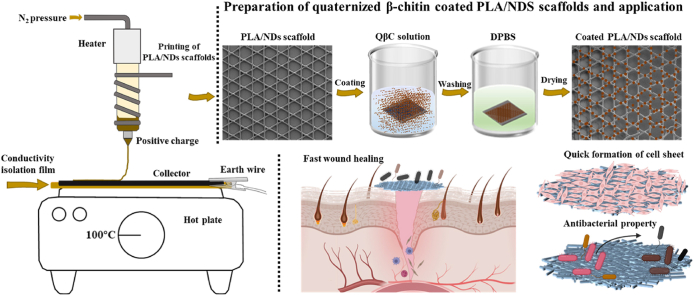
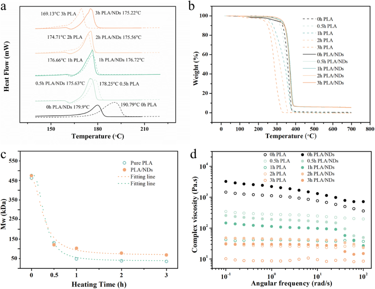
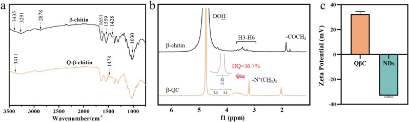
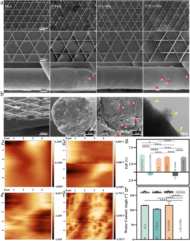
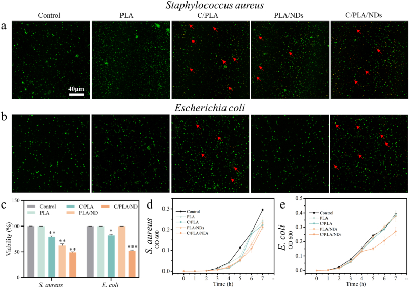
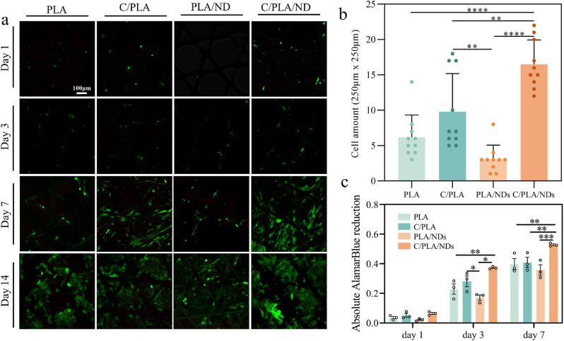
Multifunctional wound dressings, enriched with biologically active agents for preventing or treating infections and promoting wound healing, along with cell delivery capability, are highly needed. To address this issue, composite scaffolds with potential in wound dressing applications were fabricated in this study. The poly-lactic acid/nanodiamonds (PLA/ND) scaffolds were first printed using melt electrowriting (MEW) and then coated with quaternized β-chitin (QβC). The NDs were well-dispersed in the printed filaments and worked as fillers and bioactive additions to PLA material. Additionally, they improved coating effectiveness due to the interaction between their negative charges (from NDs) and positive charges (from QβC). NDs not only increased the thermal stability of PLA but also benefitted cellular behavior and inhibited the growth of bacteria. Scaffolds coated with QβC increased the effect of bacteria growth inhibition and facilitated the proliferation of human dermal fibroblasts. Additionally, we have observed rapid extracellular matrix (ECM) remodeling on QβC-coated PLA/NDs scaffolds. The scaffolds provided support for cell adhesion and could serve as a valuable tool for delivering cells to chronic wound sites. The proposed PLA/ND scaffold coated with QβC holds great potential for achieving fast healing in various types of wounds.
Keywords: Melt electrowriting; Nanodiamonds; Quaternized β-chitin; Wound healing.
© 2024 The Authors.
Conflict of interest statement
The authors declare that they have no known competing financial interests or personal relationships that could have appeared to influence the work reported in this paper.
Figures


Similar articles
-
Electrospun Nanodiamond-Silk Fibroin Membranes: A Multifunctional Platform for Biosensing and Wound-Healing Applications.ACS Appl Mater Interfaces. 2020 Oct 28;12(43):48408-48419. doi: 10.1021/acsami.0c15612. Epub 2020 Oct 13. ACS Appl Mater Interfaces. 2020. PMID: 33047948
-
Melt Electrowritten Sandwich Scaffold Technique Using Sulforhodamine B to Monitor Stem Cell Behavior.Tissue Eng Part C Methods. 2020 Oct;26(10):519-527. doi: 10.1089/ten.TEC.2020.0240. Tissue Eng Part C Methods. 2020. PMID: 32977739
-
Development of Super-Paramagnetic Iron Oxide Nanoparticle-Coated Melt Electrowritten Scaffolds for Biomedical Applications.Macromol Biosci. 2024 Mar;24(3):e2300397. doi: 10.1002/mabi.202300397. Epub 2023 Nov 13. Macromol Biosci. 2024. PMID: 37902248
-
Unveiling the potential of melt electrowriting in regenerative dental medicine.Acta Biomater. 2023 Jan 15;156:88-109. doi: 10.1016/j.actbio.2022.01.010. Epub 2022 Jan 10. Acta Biomater. 2023. PMID: 35026478 Free PMC article. Review.
-
Tissue scaffolds for skin wound healing and dermal reconstruction.Wiley Interdiscip Rev Nanomed Nanobiotechnol. 2010 Sep-Oct;2(5):510-25. doi: 10.1002/wnan.100. Wiley Interdiscip Rev Nanomed Nanobiotechnol. 2010. PMID: 20607703 Review.
Cited by
-
Integrating melt electrowriting (MEW) PCL scaffolds with fibroblast-laden hydrogel toward vascularized skin tissue engineering.Mater Today Bio. 2025 Feb 19;31:101593. doi: 10.1016/j.mtbio.2025.101593. eCollection 2025 Apr. Mater Today Bio. 2025. PMID: 40104645 Free PMC article.
-
Advanced biomaterial strategies for overcoming age-associated wound healing impairments.APL Bioeng. 2025 Jun 6;9(2):021501. doi: 10.1063/5.0251889. eCollection 2025 Jun. APL Bioeng. 2025. PMID: 40488106 Free PMC article. Review.
-
Smart Polymeric 3D Microscaffolds Hosting Spheroids for Neuronal Research via Quantum Metrology.Adv Healthc Mater. 2025 Mar;14(7):e2403875. doi: 10.1002/adhm.202403875. Epub 2025 Jan 15. Adv Healthc Mater. 2025. PMID: 39815162 Free PMC article.
-
Quantum Sensing Unravels Antioxidant Efficacy Within PCL/Matrigel Skin Equivalents.Small. 2024 Dec;20(49):e2403729. doi: 10.1002/smll.202403729. Epub 2024 Sep 9. Small. 2024. PMID: 39246220 Free PMC article.
-
Nanodiamond: a multifaceted exploration of electrospun nanofibers for antibacterial and wound healing applications.J Nanobiotechnology. 2025 Apr 9;23(1):285. doi: 10.1186/s12951-025-03351-9. J Nanobiotechnology. 2025. PMID: 40205555 Free PMC article. Review.
References
LinkOut - more resources
Full Text Sources
