Gastric epithelial neoplasm of fundic-gland mucosa lineage: representative of the low atypia differentiated gastric tumor and Ki67 may help in their identification
- PMID: 38873175
- PMCID: PMC11169639
- DOI: 10.3389/pore.2024.1611734
Gastric epithelial neoplasm of fundic-gland mucosa lineage: representative of the low atypia differentiated gastric tumor and Ki67 may help in their identification
Abstract
Background: Gastric epithelial neoplasm of the fundic-gland mucosa lineages (GEN-FGMLs) are rare forms of gastric tumors that encompass oxyntic gland adenoma (OGA), gastric adenocarcinoma of the fundic-gland type (GA-FG), and gastric adenocarcinoma of the fundic-gland mucosa type (GA-FGM). There is no consensus on the cause, classification, and clinicopathological features of GEN-FGMLs, and misdiagnosis is common because of similarities in symptoms.
Methods: 37 cases diagnosed with GEN-FGMLs were included in this study. H&E-stained slides were reviewed and clinicopathological parameters were recorded. Immunohistochemical staining was conducted for MUC2, MUC5AC, MUC6, CD10, CD56, synaptophysin, chromograninA, p53, Ki67, pepsinogen-I, H+/K+-ATPase and Desmin.
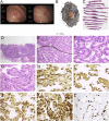
Results: The patients' ages ranged from 42 to 79 years, with a median age of 60. 17 were male and 20 were female. Morphologically, 19 OGAs, 16 GA-FGs, and two GA-FGMs were identified. Histopathological similarities exist between OGA, GA-FG, and GA-FGM. The tumors demonstrated well-formed glands, expanding with dense growth patterns comprising pale, blue-grey columnar cells with mild nuclear atypia. These cells resembled fundic gland cells. None of the OGA invaded the submucosal layer. The normal gastric pit epithelium covered the entire surface of the OGA and GA-FG, but the dysplasia pit epithelium covered the GA-FGM. Non-atrophic gastritis was observed in more than half of the background mucosa. All cases were diffusely positive for MUC6 and pepsinogen-I on immunohistochemistry. H+/K+-ATPase staining was negative or showed a scattered pattern in most cases. MUC5AC was expressed on the surface of GA-FGMs. p53 was focally expressed and the Ki67 index was low (1%-20%). Compared with OGA, GA-FG and GA-FGM were more prominent in the macroscopic view (p < 0.05) and had larger sizes (p < 0.0001). Additionally, GA-FG and GA-FGM exhibited higher Ki67 indices than OGA (p < 0.0001). Specimens with Ki-67 proliferation indices >2.5% and size >4.5 mm are more likely to be diagnosed with GA-FG and GA-FGM than OGA.
Conclusion: GEN-FGMLs are group of well-differentiated gastric tumors with favourable biological behaviours, low cellular atypia, and low proliferation. Immunohistochemistry is critical for confirming diagnosis. Compared with OGA, GA-FG and GA-FGM have larger sizes and higher Ki67 proliferation indices, indicating that they play a critical role in the identification of GEN-FGML. Pathologists and endoscopists should be cautious to prevent misdiagnosis and overtreatment, especially in biopsy specimens.
Keywords: gastric adenocarcinoma of fundic-gland mucosa type; gastric adenocarcinoma of fundic-gland type; gastric cancer; low-grade differentiated gastric tumors; oxyntic gland adenoma.
Copyright © 2024 Li, Zheng, Zhong, Yu, Zhang, Chen and Chen.
Conflict of interest statement
The authors declare that the research was conducted in the absence of any commercial or financial relationships that could be construed as a potential conflict of interest.
Figures


Similar articles
-
Gastric epithelial neoplasm of fundic-gland mucosa lineage: proposal for a new classification in association with gastric adenocarcinoma of fundic-gland type.J Gastroenterol. 2021 Sep;56(9):814-828. doi: 10.1007/s00535-021-01813-z. Epub 2021 Jul 15. J Gastroenterol. 2021. PMID: 34268625 Free PMC article.
-
Gastric adenocarcinoma of fundic gland mucosa type localized in the submucosa: A case report.Medicine (Baltimore). 2018 Sep;97(37):e12341. doi: 10.1097/MD.0000000000012341. Medicine (Baltimore). 2018. PMID: 30212986 Free PMC article.
-
[Clinical pathological characteristics of 4 cases of gastric gland-derived tumors].Zhonghua Zhong Liu Za Zhi. 2021 Jul 23;43(7):781-786. doi: 10.3760/cma.j.cn112152-20191202-00776. Zhonghua Zhong Liu Za Zhi. 2021. PMID: 34289573 Chinese.
-
Gastric adenocarcinoma of fundic gland type with signet-ring cell carcinoma component: A case report and review of the literature.World J Gastroenterol. 2018 Jul 14;24(26):2915-2920. doi: 10.3748/wjg.v24.i26.2915. World J Gastroenterol. 2018. PMID: 30018486 Free PMC article. Review.
-
Gastric adenocarcinoma of the fundic gland (chief cell-predominant type): A review of endoscopic and clinicopathological features.World J Gastroenterol. 2016 Dec 28;22(48):10523-10531. doi: 10.3748/wjg.v22.i48.10523. World J Gastroenterol. 2016. PMID: 28082804 Free PMC article. Review.
Cited by
-
An emerging entity of gastric adenocarcinoma: clinicopathological features and differential diagnosis of gastric adenocarcinoma of fundic-gland type in 25 retrospective cases.Virchows Arch. 2025 Mar 18. doi: 10.1007/s00428-025-04075-9. Online ahead of print. Virchows Arch. 2025. PMID: 40100386
-
Contract to kill: GNAS mutation.Mol Cancer. 2025 Mar 7;24(1):70. doi: 10.1186/s12943-025-02247-4. Mol Cancer. 2025. PMID: 40050874 Free PMC article. Review.
References
-
- Ueyama H, Yao T, Akazawa Y, Hayashi T, Kurahara K, Oshiro Y, et al. Gastric epithelial neoplasm of fundic-gland mucosa lineage: proposal for a new classification in association with gastric adenocarcinoma of fundic-gland type. J Gastroenterol (2021) 56(9):814–28. 10.1007/s00535-021-01813-z - DOI - PMC - PubMed
-
- Tanabe H, Iwashita A, Ikeda K. Histopathological characteristics of gastric adenocarcinoma of fundic gland type. StomachIntest (2015) 50:1521–31.
MeSH terms
Substances
LinkOut - more resources
Full Text Sources
Medical
Research Materials
Miscellaneous

