

Contemporary status of diagnostic endoluminal ultrasound and optical coherence tomography in the ureter
- PMID: 38873352
- PMCID: PMC11168776
- DOI: 10.1002/bco2.352
Contemporary status of diagnostic endoluminal ultrasound and optical coherence tomography in the ureter
Erratum in
-
Erratum.BJUI Compass. 2024 Dec 30;5(12):1324-1329. doi: 10.1002/bco2.482. eCollection 2024 Dec. BJUI Compass. 2024. PMID: 39744071 Free PMC article.
Abstract
Objective: To evaluate via a review of published literature, the efficacy of endoluminal ultrasound (ELUS) and optical coherence tomography (OCT) in the following ureteric diseases: urolithiasis, upper tract urothelial carcinoma, stricture disease and pelvic-ureteric junction obstruction (PUJO).
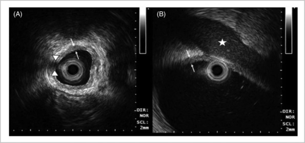
Patients and methods: Ureteric high-frequency ELUS provides 360° imaging, to a depth of 20 mm, and has been demonstrated to assess ureteric stricture length, degree of fibrosis and aetiology. OCT produces high-quality images with a penetration depth of 2 mm. ELUS has proven to be useful at the time of endopyelotomy for PUJO as it can identify crossing vessels, some not detectable on CT angiography, allowing the urologist to avoid these when making their incision. Ureteric ELUS may be utilised for submucosal ureteric stones as they are highly visible. Endoluminal ultrasound may be deployed in the case of known sub-mucosal urolithiasis when the ureter appears stone-free. It may help identify sub-mucosal stones or stones within diverticulum.
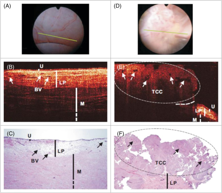
Results: Endoluminal ultrasound has been analysed for its use in determining muscle-invasive urothelial carcinoma of the ureter. The PPV for ≥pT2 was only 16.7% in one study of six patients with MIBC and 76.2% in 21 patients with <pT2 disease. Analysis of OCT for staging UTUC during ureteroscopy and biopsy demonstrated sensitivity for tumour invasion of 100% and specificity of 92%, 83% of lesion staging matched with histological analysis. Imaging analysis did not match histology in three patients with large exophytic tumours that exceeded the OCT depth penetration. Due to its superficial penetration, OCT cannot reliably stage large tumours.
Conclusions: Ureteric ELUS has been reported to be a useful tool in endopyelotomy, urolithiasis and stricture disease. The staging of ureteric urothelial carcinoma remains unsatisfactory with current imaging techniques and biopsy methods, and, based on the current literature, ELUS does not appear to have a strong enough PPV to determine muscle invasion. Ureteric OCT may be a useful tool in the future staging of upper tract urothelial carcinoma, particularly in differentiating the stage of small tumours. Further studies are needed in this area.
Keywords: endoluminal ultrasound; optical coherence tomography; ureter.
© 2024 The Authors. BJUI Compass published by John Wiley & Sons Ltd on behalf of BJU International Company.
Conflict of interest statement
None.
Figures
References
-
- Bus MT, Bruin DM, Kamphuis GM, Reijke TM, de la Rosette JJ. Beyond endoscopy‐ultrasound, optical coherence tomography and confocal laser endomicroscopy. In: Upper urinary tract urothelial carcinoma Springer; 2015. p. 121–132.
Publication types
LinkOut - more resources
Full Text Sources