. 2024 Feb 20;12(6):3893-3909.
doi: 10.1002/fsn3.4049. eCollection 2024 Jun.

Dihydromyricetin regulates KEAP1-Nrf2 pathways to enhance the survival of ischemic flap

Affiliations

Dihydromyricetin regulates KEAP1-Nrf2 pathways to enhance the survival of ischemic flap

Xianyao Tao et al. Food Sci Nutr. .

Abstract

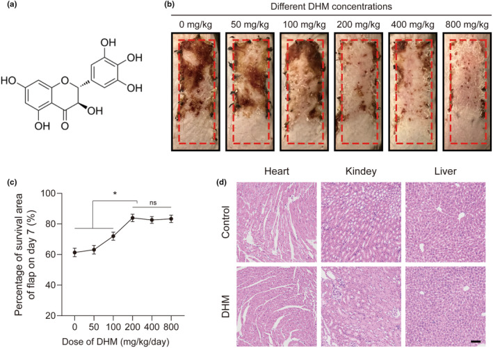
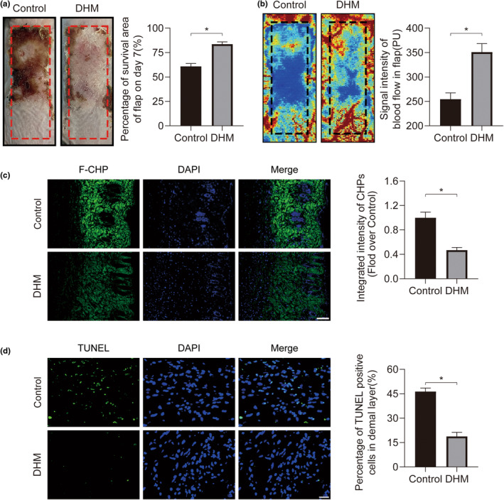
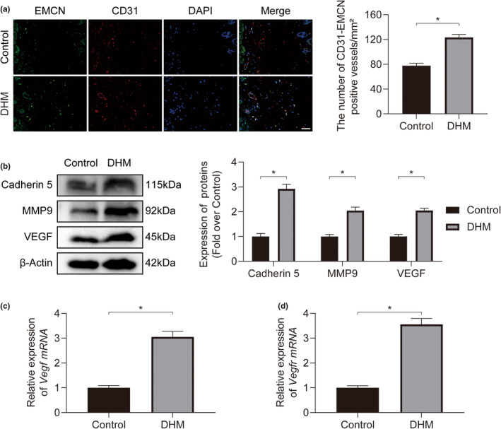
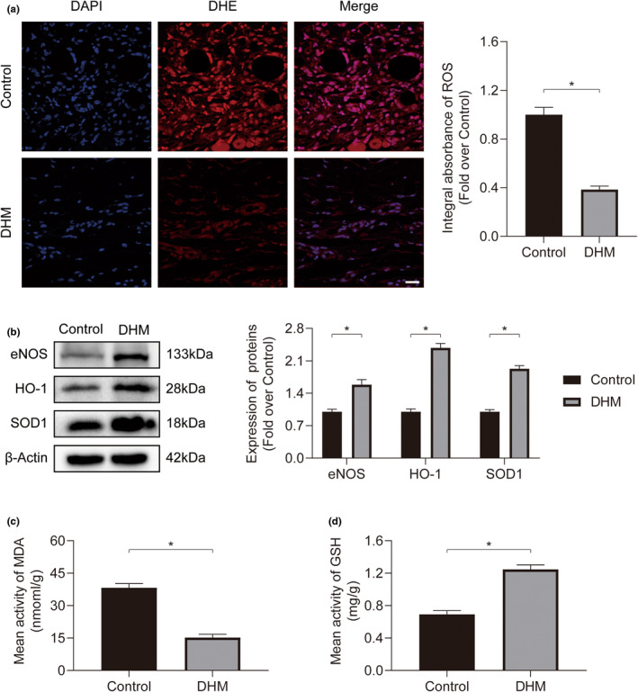
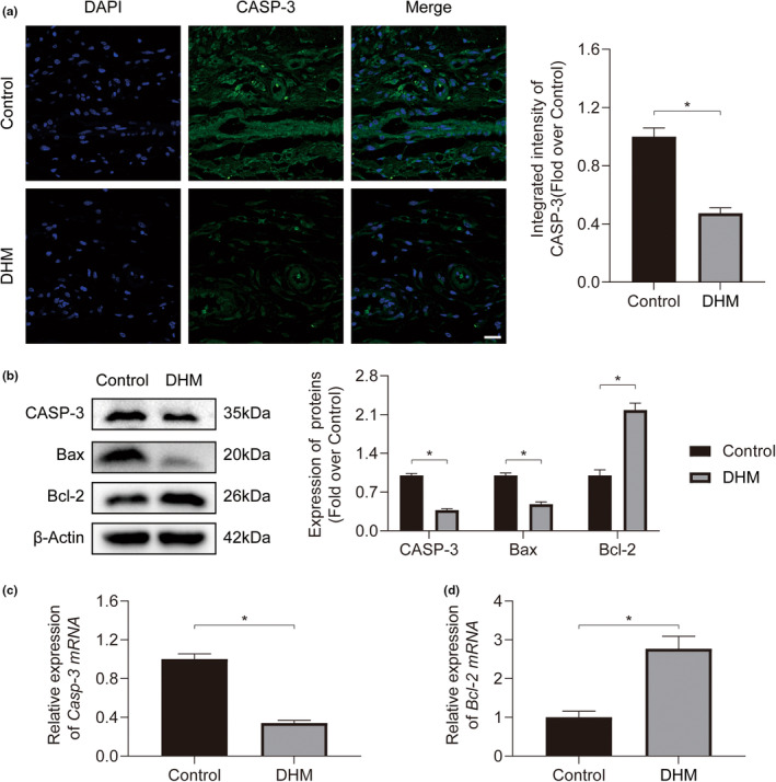
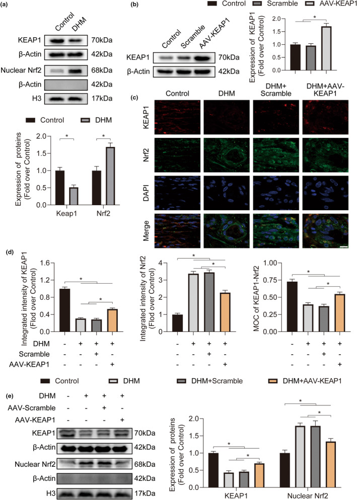
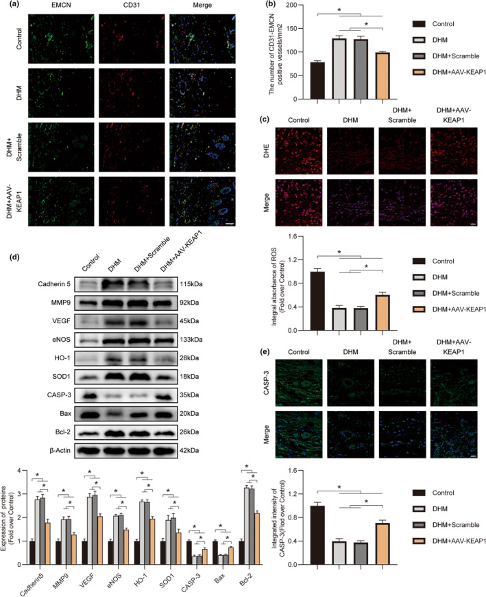
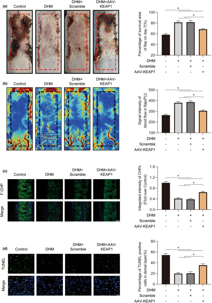
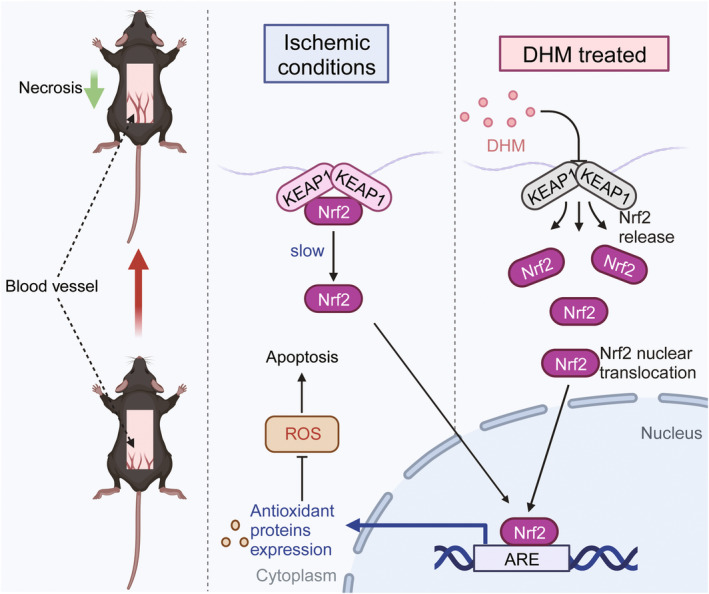
In clinical flap practice, there are a lot of studies being done on how to promote the survival of distal random flap necrosis in the hypoxic and ischemic state. As a traditional Chinese medicine, dihydromyricetin (DHM) is crucial in preventing oxidative stress and apoptosis in a number of disorders. In this work, we examined the impact of DHM on the ability to survive of ischemia flaps and looked into its fundamental mechanism. Our results showed that DHM significantly increased the ischemic flaps' survival area, encouraged angiogenesis and blood flow, reduced oxidative stress and apoptosis, and stimulated KEAP1-Nrf2 (Kelch-like ECH-associated protein 1-nuclear factor erythroid 2-related factor) signaling pathways. Adeno-associated virus (AAV) upregulation of KEAP1 expression also negated the favorable effects of DHM on flap survival. By activating KEAP1-Nrf2 signaling pathways, DHM therapy promotes angiogenesis while reducing oxidative stress and apoptosis.

Keywords: angiogenesis; apoptosis; dihydromyricetin; ischemic flap; oxidative stress.


Conflict of interest statement

No potential conflict of interest was reported by the author(s).

Figures

FIGURE 1
FIGURE 1
Optimal DHM concentration selection in ischemic flaps. (a) Dihydromyricetin (DHM) chemical structure. (b) Digital images of ischemic flaps after DHM treatment at different concentrations captured on POD7. (c) The dose–response curve showing the optimum dose of DHM (200 mg/kg/day) (n = 5). (d) Representative H&E staining of heart, liver, and kidney of mice in the Control group and the DHM group. 50 μm scale bars.
FIGURE 2
FIGURE 2
Dihydromyricetin (DHM) enhances the viability of ischemic flaps. (a) Digital photograph of the ischemic flap captured on POD7. Quantification of the percentage of viable flap area on POD7 for both experimental groups (n = 5). (b) Visual representation of the subcutaneous blood flow network on POD7. Quantification of blood flow signal intensity in the ischemic flaps for both groups on POD7 (n = 5). (c) Damaged collagen in ischemia flaps was found using F‐CHP staining on POD7. 100 μm scale bars. Comparison of the two groups' F‐CHP intensities (n = 5). (d) Terminal deoxynucleotidyl transferase dUTP nick end labeling (TUNEL) staining for the identification of dead cells in the ischemic flap on POD7. 50 μm scale bars. Calculation of the proportion of TUNEL‐positive cells in ischemic flaps for both groups (n = 5). Error bars are SEM. The band density was normalized and the loading control was β‐actin. Significance: *p < .05, substantially distinct as stated; two‐tailed, unpaired t‐test.
FIGURE 3
FIGURE 3
Dihydromyricetin (DHM) promotes angiogenesis in ischemic flaps. (a) Immunofluorescence (IF) staining of CD31 and EMCN in ischemic flaps on POD7. Scale bars: 50 μm. Quantification of CD31/EMCN‐positive blood vessel density between the two groups (n = 5). (b) Expression of Cadherin 5, MMP9, and VEGF proteins in ischemic flaps for both groups on POD7. Measurement of the expression of proteins associated with angiogenesis in both groups (n = 5). (c) Comparative analysis of relative Vegf mRNA expression in ischemic flaps for both groups on POD7 (n = 5). (d) Comparative analysis of relative Vegfr mRNA expression in ischemic flaps for both groups on POD7 (n = 5). Error bars are SEM. The band density was normalized and the loading control was β‐actin. Significance: *p < .05, substantially distinct as stated; two‐tailed, unpaired t‐test.
FIGURE 4
FIGURE 4
DHM suppresses oxidative stress in ischemic flaps. (a) Ischemic flap frozen sections from both experimental groups on POD7 were subjected to DHE staining. Scale bars: 50 μm. DHE intensity measurements for both groups (n = 5). (b) Expression of eNOS, HO‐1, and SOD1 proteins in ischemic flaps for both groups on POD7. Quantification of oxidative stress‐related protein expression for both groups (n = 5). (c) Measurement of MDA content in ischemic flaps for both groups on POD7 (n = 5). (d) Measurement of GSH content in ischemic flaps for both groups on POD7 (n = 5). Error bars are SEM. The band density was normalized and the loading control was β‐actin. Significance: *p < .05, substantially distinct as stated; two‐tailed, unpaired t‐test.
FIGURE 5
FIGURE 5
Dihydromyricetin (DHM) mitigates apoptosis in ischemic flaps. (a) Immunofluorescence (IF) staining of CASP‐3 in ischemic flaps on postoperative day 7 (POD7). Scale bars: 20 μm. Quantification of CASP‐3 intensity for both groups (n = 5). (b) Expression for CASP‐3, Bax, and Bcl‐2 proteins within ischemic flaps for both groups on POD7. Quantification of apoptosis‐related protein expression for both groups (n = 5). (c) Comparative analysis of relative Casp‐3 mRNA expression in ischemic flaps for both groups on POD7 (n = 5). (d) Comparative analysis of relative Bcl‐2 mRNA expression in ischemic flaps for both groups on POD7 (n = 5). Error bars are SEM. The band density was normalized and the loading control was β‐actin. Significance: *p < .05, substantially distinct as stated; two‐tailed, unpaired t‐test.
FIGURE 6
FIGURE 6
Dihydromyricetin (DHM) activates Nrf2 by suppressing KEAP1 within ischemic flaps. (a) Expression of KEAP1 and nuclear Nrf2 proteins in ischemic flaps for both groups on POD7. Quantification of KEAP1 and nuclear Nrf2 expression between the two groups is presented below (n = 5). (b) Kelch‐like ECH‐associated protein 1 (KEAP1) expression in ischemic flaps for all three groups on POD7. The quantified expression of KEAP1‐Nrf2 among these three groups is displayed to the right (n = 5). (c) Immunofluorescence (IF) staining for KEAP1 and Nrf2 within ischemic flaps on POD7. Scale bars: 10 μm. (d) Quantification for incorporated intensity in KEAP1 (left), nuclear Nrf2 (middle), and MOCs (Mander's overlap coefficients) of KEAP1‐Nrf2 (right) in ischemic flaps across all four groups (n = 5). (e) Expression of KEAP1 and nuclear Nrf2 proteins in ischemic flaps among all four groups on POD7. Quantification of KEAP1 and nuclear Nrf2 expression among all four groups. (n = 5). Error bars are SEM. β‐actin and H3 had been used as the loading control and for band density normalization. Significance: *p < .05, substantially distinct as indicated; two‐tailed, unpaired t‐test. ANOVA with LSD post hoc testing (categories with equivalent variances) or Dunnett's T3 technique (categories with different variances).
FIGURE 7
FIGURE 7
Molecular docking and dynamic simulation between DHM and Keap1‐Nrf2. (a) Kelch‐like ECH‐associated protein 1 (KEAP1) in complex with DHM at the center of the protein. (b) Three‐dimensional (3D) binding model of intermolecular interactions between KEAP1‐Nrf2 and DHM. (c) The space‐filling model of intermolecular interactions between KEAP1‐Nrf2 and DHM.
FIGURE 8
FIGURE 8
Dihydromyricetin (DHM) alters the KEAP1‐Nrf2 signaling network to promote angiogenesis while reducing oxidative damage and death. (a) Immunofluorescence (IF) staining of CD31 and EMCN in ischemic flaps on POD7. Scale bars: 50 μm. (b) Quantification of CD31/EMCN‐positive blood vessel density across all four groups (n = 5). (c) Frozen sections of ischemic flaps from all four groups on POD7 were subjected to DHE staining. Scale bars: 50 μm. Quantified DHE intensity across all four groups is presented below (n = 5). (d) Expression of angiogenesis‐, oxidative stress‐, and apoptosis‐related proteins in ischemic flaps among all four groups on POD7. Quantification of expression levels across all four groups is presented below (n = 5). (e) Immunofluorescence (IF) staining of CASP‐3 in ischemic flaps on POD7. Scale bars: 20 μm. Quantified CASP‐3 intensity across all four groups is presented below (n = 5). Error bars are SEM. The band density was normalized and the loading control was β‐actin. Significance: *p < .05, substantially distinct as specified; Dunnett's T3 technique or ANOVA with LSD post hoc testing (which are equivalent variances for the categories).
FIGURE 9
FIGURE 9
Dihydromyricetin (DHM) enhances the viability of ischemic flaps through the KEAP1‐Nrf2 signaling pathway. (a) Digital photograph of the ischemic flap on POD7. Calculating the proportion of flap area that survived in each of the four groups on POD7 (n = 5). (b) Visual representation of the subcutaneous blood flow network on POD7. Quantification of blood flow signal intensity in ischemic flaps across all five groups on POD7 (n = 5). (c) Damaged collagen in ischemia flaps was found using F‐CHP staining on POD7. 100 μm scale bars. Measurement of F‐CHP intensity in each of the four groups (n = 5). (d) Terminal deoxynucleotidyl transferase dUTP nick end labeling (TUNEL) staining for the identification of dead cells in the ischemic flap on POD7. 50 μm scale bars. The measurement of the proportion of TUNEL‐positive cells in ischemic flaps in all four groups (n = 5). Error bars are SEM. Significance: *p < .05, significantly different as indicated; Dunnett's T3 technique or ANOVA with LSD post hoc analysis (which are equivalent variances for the categories).
FIGURE 10
FIGURE 10
Schematic diagram of the mechanism of DHM promoting ischemic flap survival by inhibiting KEAP1 to upregulate Nrf2 and angiogenesis, while reducing oxidative stress and apoptosis.

Similar articles

Cited by

References

    1. Akhavani, M. A. , Sivakumar, B. , Paleolog, E. M. , & Kang, N. (2008). Angiogenesis and plastic surgery. Journal of Plastic, Reconstructive & Aesthetic Surgery, 61(12), 1425–1437. - PubMed
    1. Attinger, C. E. , & Colen, L. B. (2000). The role of microsurgical free flaps in foot and ankle surgery. Clinics in Podiatric Medicine and Surgery, 17(4), 649–680. - PubMed
    1. Bali, U. , Aydemir, I. , Keçeci, Y. , Yoleri, L. , & Tuğlu, M. İ. (2021). Effects of oxidative stress and apoptosis on vascularity and viability of perforator flaps. Biotechnic & Histochemistry, 96(7), 526–535. - PubMed
    1. Bardallo, R. G. , Panisello‐Roselló, A. , Sanchez‐Nuno, S. , Alva, N. , Roselló‐Catafau, J. , & Carbonell, T. (2022). Nrf2 and oxidative stress in liver ischemia/reperfusion injury. The FEBS Journal, 289(18), 5463–5479. - PubMed
    1. Brinkman, J. N. , Derks, L. H. , Klimek, M. , & Mureau, M. A. (2013). Perioperative fluid management and use of vasoactive and antithrombotic agents in free flap surgery: A literature review and clinical recommendations. Journal of Reconstructive Microsurgery, 29(6), 357–366. - PubMed