Review
. 2024 Nov;57(11):e13688.
doi: 10.1111/cpr.13688. Epub 2024 Jun 14.

Regulated vascular smooth muscle cell death in vascular diseases

Affiliations
Review

Regulated vascular smooth muscle cell death in vascular diseases

Zheng Yin et al. Cell Prolif. 2024 Nov.

Abstract

Regulated cell death (RCD) is a complex process that involves several cell types and plays a crucial role in vascular diseases. Vascular smooth muscle cells (VSMCs) are the predominant elements of the medial layer of blood vessels, and their regulated death contributes to the pathogenesis of vascular diseases. The types of regulated VSMC death include apoptosis, necroptosis, pyroptosis, ferroptosis, parthanatos, and autophagy-dependent cell death (ADCD). In this review, we summarize the current evidence of regulated VSMC death pathways in major vascular diseases, such as atherosclerosis, vascular calcification, aortic aneurysm and dissection, hypertension, pulmonary arterial hypertension, neointimal hyperplasia, and inherited vascular diseases. All forms of RCD constitute a single, coordinated cell death system in which one pathway can compensate for another during disease progression. Pharmacologically targeting RCD pathways has potential for slowing and reversing disease progression, but challenges remain. A better understanding of the role of regulated VSMC death in vascular diseases and the underlying mechanisms may lead to novel pharmacological developments and help clinicians address the residual cardiovascular risk in patients with cardiovascular diseases.


Conflict of interest statement

The authors declare no competing interests.

Figures

FIGURE 1
FIGURE 1
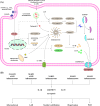
Types of regulated VSMC cell death in vascular diseases. ADCD, autophagy‐dependent cell death.
FIGURE 2
FIGURE 2
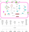
Schematic description of the signalling pathway of apoptosis. (A) The intrinsic pathway is triggered by diverse stimuli, such as DNA damage, hypoxia, oxidative stress, activated oncogenes, and irradiation. These stimuli promote the levels of transcription and post‐translation of BH3‐only proteins. BH3‐only proteins can directly or indirectly antagonize the pro‐survival protein BCL‐2, thereby unleashing BCL‐2‐associated X protein (BAX) and BCL‐2 antagonist/killer 1 (BAK) that mediate mitochondrial outer membrane permeabilization (MOMP). Subsequently, mitochondrial pro‐apoptotic factors, including cytochrome c and SMAC/HTRA2, are released into the cytoplasm. Cytochrome c can bind to apoptotic peptidase activating factor 1 (Apaf‐1), which induces a conformational change in Apaf‐1 that allows it to recruit pro‐caspase‐9, leading to the assembly of a complex termed apoptosome. In this complex, pro‐caspase‐9 is cleaved to form the active caspase‐9 protease, which then activates downstream effector caspase‐3/7. The X‐linked inhibitor of apoptosis (XIAP) inhibits the activation of pro‐caspase‐9 and the function of caspase‐3/7. To allow apoptosis to proceed, SMAC/HTRA2 in the cytosol can bind to XIAP, freeing these caspases. The extrinsic pathway is stimulated by the binding of death ligands to their cognate death receptors (e.g., FasL to Fas). Then, activated death receptors recruit adaptor proteins (e.g., FADD), allowing pro‐caspase 8 recruitment into the death‐inducing signalling complex (DISC). Caspase‐8 proteolytically activates executioner caspases for the downstream of apoptosis. Caspase‐8 also converts the BH3‐only protein BID into its pro‐apoptotic form, tBID, which triggers BAK/BAX‐mediated MOMP and subsequent apoptosis through the intrinsic pathway. Intriguingly, unlike other cell types, VSMCs exhibit resistance to Fas‐induced apoptosis due to the sequestration of the death receptor Fas/TNFR1 in the Golgi apparatus. However, various specific stimuli sensitize VSMCs to Fas‐mediated apoptosis by transporting Fas from the Golgi apparatus to the cell surface. (B) VSMC apoptosis in both intrinsic and extrinsic pathways has been well studied in various vascular diseases, with almost all players identified during apoptosis. VSMC apoptosis promotes atherosclerosis, AAD, and vascular senescence, while preventing intima hyperplasia, hypertension, and PAH. In turn, vascular senescence exhibit resistance to apoptosis.
FIGURE 3
FIGURE 3
Schematic description of the signalling pathway of necroptosis. (A) Necroptosis is triggered by the binding of death ligands to death receptors (e.g., TNF to TNFR1), allowing the recruitment of TRADD, receptor‐interacting protein kinase 1 (RIPK1), TNFR‐associated factor‐2/5 (TRAF2/5), and cellular inhibitor of apoptosis‐1/2 (cIAP1/2) for complex I formation. Subsequently, cIAP‐1/2 promotes RIPK1 ubiquitination via K63 ubiquitin chains, leading to the recruitment of the linear ubiquitin chain assembly complex (LUBAC). LUBAC generates M1 ubiquitin chains, which are added to RIPK1. Subsequently, M1‐ and K63‐ubiquitin chains serve as scaffolds for the recruitment of the nuclear factor‐κB (NF‐κB) essential modulator (NEMO)‐IκB kinase (IKK) complex and TGF‐β‐activated kinase 1 (TAK1)/TAK1‐binding protein 2/3 (TAB2/3), leading to NF‐κB and mitogen‐activated protein kinase (MAPK) activation. The destabilization of complex I leads to the formation of a second cytosolic complex IIa, which consists of TRADD, FADD and caspase‐8. Under conditions such as TNF stimulation following the loss or inhibition of IAP, a third cytosolic complex IIb, composed of RIPK1, RIPK3, FADD, and caspase‐8, is formed. Activated caspase‐8 within complex II triggers caspase‐3/7 activation, leading to RIPK1‐dependent apoptosis. However, in the absence of caspase‐8 activation, RIPK1 is activated, initiating the necroptosis pathway instead of RIPK1‐dependent apoptosis. This process occurs through autophosphorylation and transphosphorylation of RIPK1 and RIPK3, which recruit mixed lineage kinase domain‐like pseudokinase (MLKL) to induce its phosphorylation and oligomerization. Oligomerized MLKL is then transports to the plasma membrane via the Golgi‐microtubule‐actin machinery, leading to plasma membrane permeabilization, cytokine release, and eventually necroptosis. (B) VSMC necroptosis has been rarely studied in vascular diseases. RIPK1, RIPK3, and MLKL have been identified in VSMC necroptosis during AAD. The relationship between VSMC necroptosis and atherosclerosis is still unclear.
FIGURE 4
FIGURE 4
Schematic description of the signalling pathway of pyroptosis. (A) The NOD‐like receptor protein 3 (NLRP3) inflammasome is composed of a sensor NLRP3, an adapter apoptosis‐associated speck‐like protein containing a CARD (ASC), and a effector pro‐caspase‐1. Inflammasome assembly is mediated through the interaction between a pyrin domain (PYD) in ASC and an N‐terminal PYD in NLRP3, as well as the binding of the CARD in ASC to pro‐caspase‐1. This requires two steps: priming and activation. During the priming step, extracellular pathogen‐associated molecular patterns (PAMPs) and damage‐associated molecular patterns (DAMPs) are recognized by PRRs, which upregulates the expression of NF‐κB‐dependent target genes, including NLRP3, ASC, pro‐IL‐1β, pro‐IL‐18, pro‐caspase‐1, and pro‐caspase‐11. The second step, activation, involves various pathways such as potassium efflux, mitochondrial ROS generation, and lysosomal destabilization. After the inflammasome assembly, pro‐caspase‐1 (p45) is activated and hydrolysed into mature cleaved caspase‐1 (p10/p20 tetramer). On the one hand, activated caspase‐1 recognizes and cleaves precursors of IL‐1β and IL‐18 into their mature forms. On the other hand, activated caspase‐1 also cleaves the caustic executor protein GSDMD at the Asp275 site, forming the 22 kDa C‐terminus (GSDMD‐C) and 31 kDa N‐terminus (GSDMD‐N). GSDMD‐N oligomerizes and forms nonselective plasma membrane pores, which promotes the release of inflammatory cytokines, cell swelling, and finally, pyroptosis. In the non‐canonical pyroptosis pathway, the upstream sensory complex caspase‐11 and its human orthologs caspase‐4/5 can directly sense intracellular lipopolysaccharide (LPS) without the need for inflammasomes. Activated caspase‐11 and caspase‐4/5 result in GSDMD cleavage and pyroptosis. (B) VSMC pyroptosis has been identified to promote the development of various vascular diseases, with essential players, such as NLRP3 inflammasome, GSDMD‐N, IL‐1β, and IL‐18. AIM2 inflammation in VSMCs has only been identified during atherosclerosis and AAD.
FIGURE 5
FIGURE 5
Schematic description of the signalling pathway of ferroptosis. (A) The canonical ferroptosis‐suppressing pathway involves the uptake of cystine via the cystine‐glutamate antiporter (system xc‐), which results in glutathione (GSH) biosynthesis. Using GSH as a cofactor, the glutathione peroxidase 4 (GPX4) scavenges the harmful by‐products of iron‐dependent lipid peroxidation, thereby protecting the cell membrane against damage. Ferroptosis is also regulated by the iron metabolism pathway that involves iron uptake by transferrin (TF) and transferrin receptor 1 (TFR1), reduction by the metalloreductase STEAP3, transport by divalent metal transporter 1 (DMT1), storage with ferritin and heme, utilization for Fenton reaction, and export by ferroportin. Ferritin degradation by nuclear receptor coactivator 4 (NCOA4)‐mediated ferritinophagy and heme degradation by heme oxygenase‐1 (HO‐1) increase the labile iron pool (LIP), which sensitizes cells to ferroptosis via Fenton reaction. In addition, fatty‐acid CoA ligase 4 (LACS4), lysophospholipid acyltransferase 3 (LPLAT3), lipoxygenases (LOXs), and NADPH oxidases (NOXs) are involved in the lipid metabolic pathway for lipid peroxidation and ferroptosis. To prevent excessive ferroptosis, phospholipid peroxidation is also inhibited by the ferroptosis suppressor protein 1 (FSP1)‐coenzyme Q10 (CoQ10) system. Exogenously applied ferroptosis inhibitors include radical trapping antioxidants, such as ferrostatin‐1 (Fer‐1) and liproxstatin‐1 (Lip‐1), and iron chelators, such as deferoxamine. Ferroptosis is triggered by the disruption of cystine supply via erastin‐mediated system xc‐ inhibition, or the deficiency of GPX4 activity by RAS‐selective lethal 3 (RSL3). GSSG, glutathione disulphide. (B) Players in VSMC ferroptosis have been identified in different vascular diseases. VSMC ferroptosis promotes the development of atherosclerosis, AAD, neointima, vascular calcification, hypertension, and vascular senescence. In PAH, the role of VSMC ferroptosis has been controversial. ALOX15, arachidonate‐15‐lipoxygenase; COX2, cyclooxygenase 2; IRP2, iron regulatory protein 2.
FIGURE 6
FIGURE 6
Schematic description of the signalling pathway of parthanatos. (A) Various factors such as oxidative stress, inflammation, ischemia, hypoxia, hypoglycaemia, ionizing radiation, and alkylating agents can cause DNA fragmentation, which in turn activates PARP‐1. When DNA damage is mild, PARP‐1 is activated and recruits DNA damage repair proteins to facilitate the repair process. However, in cases of severe DNA damage, PARP‐1 becomes overactivated, leading to the synthesis of long‐chained and branched poly (ADP‐ribose) (PAR) polymers. These PAR polymers accumulate and are then released from the nucleus into the mitochondria, where they bind to apoptosis‐inducing factor (AIF). Subsequently, AIF is released from the mitochondria and translocated to the cytoplasm. In the cytoplasm, AIF forms a complex with macrophage migration inhibitory factor (MIF), a cytokine with nuclease activity. Subsequently, this complex translocates to the nucleus, triggering large‐scale DNA fragmentation, chromatin condensation, and cell death. (B) VSMC parthanatos has been rarely studied in vascular diseases. The best studied player, PARP‐1, has been identified to plays a role in different vascular diseases, which can provide clues for vascular parthanatos.
FIGURE 7
FIGURE 7
Schematic description of the signalling pathway of autophagy‐dependent cell death (ADCD). (A) The autophagy pathway includes initiation, nucleation, elongation, autophagosome maturation, autophagosome‐lysosome fusion, and autolysosome degradation. ADCD is dependent on autophagy machinery. ADCD is believed to be triggered by autophagy hyperactivation, for example, in response to treatment with natural compounds and specific cancer drugs. An increase in the number of autophagosomes and autolysosomes promotes autophagic flux, which leads to the excessive degradation of unselective cargo and cell death. Since ADCD is dependent on the autophagy machinery, genetically or chemically targeting essential autophagy genes and proteins inhibits the induction of ADCD. (B) Autophagy‐dependent VSMC death has been only identified in AAD, during which players ATG5, ATG7, SQSTM1, and BECN1 are required. 3‐Methyladenine (3‐MA) inhibits ADCD. Rapamycin promotes ADCD, while exert its anti‐inflammatory effects on AAD.
FIGURE 8
FIGURE 8
Interaction of RCD modalities. (A) Players in pyroptosis, apoptosis or necroptosis can be integrated to assemble different PANoptosome complexes, which leads to PANoptosis. (B) Caspase‐8 serves a pivotal role by promoting apoptosis and inhibiting necroptosis. Moreover, in the absence of caspase‐1 or GSDMD, caspase‐8 can trigger GSDMD‐independent pyroptosis, but its activity can be counteracted by GSDMD‐dependent pyroptosis. (C) Caspase‐3 induces apoptosis and GSDME‐dependent pyroptosis, with its activation mediated by gasdermin‐induced MOMP and subsequent cytochrome c release. (D) p53 functions as a crucial regulator in promoting apoptosis through modulation of pro‐apoptotic and anti‐apoptotic molecules, while also playing a role in promoting ferroptosis via the downregulation of SLC7A11.

Similar articles

Cited by

References

    1. Kerr JF, Wyllie AH, Currie AR. Apoptosis: a basic biological phenomenon with wide‐ranging implications in tissue kinetics. Br J Cancer. 1972;26(4):239‐257. doi:10.1038/bjc.1972.33 - DOI - PMC - PubMed
    1. Holler N, Zaru R, Micheau O, et al. Fas triggers an alternative, caspase‐8‐independent cell death pathway using the kinase RIP as effector molecule. Nat Immunol. 2000;1(6):489‐495. doi:10.1038/82732 - DOI - PubMed
    1. Wall VZ, Bornfeldt KE. Arterial smooth muscle. Arterioscler Thromb Vasc Biol. 2014;34(10):2175‐2179. doi:10.1161/atvbaha.114.304441 - DOI - PMC - PubMed
    1. Owens GK, Kumar MS, Wamhoff BR. Molecular regulation of vascular smooth muscle cell differentiation in development and disease. Physiol Rev. 2004;84(3):767‐801. doi:10.1152/physrev.00041.2003 - DOI - PubMed
    1. Xie C, Ritchie RP, Huang H, Zhang J, Chen YE. Smooth muscle cell differentiation in vitro: models and underlying molecular mechanisms. Arterioscler Thromb Vasc Biol. 2011;31(7):1485‐1494. doi:10.1161/atvbaha.110.221101 - DOI - PMC - PubMed