. 2024 Nov;20(11):2444-2459.
doi: 10.1080/15548627.2024.2367923. Epub 2024 Jul 3.

The autophagy protein RUBCNL/PACER represses RIPK1 kinase-dependent apoptosis and necroptosis

Affiliations

The autophagy protein RUBCNL/PACER represses RIPK1 kinase-dependent apoptosis and necroptosis

Diego Rojas-Rivera et al. Autophagy. 2024 Nov.

Abstract

Mesenchymal stem cells (MSCs) are used in cell therapy; nonetheless, their application is limited by their poor survival after transplantation in a proinflammatory microenvironment. Macroautophagy/autophagy activation in MSCs constitutes a stress adaptation pathway, promoting cellular homeostasis. Our proteomics data indicate that RUBCNL/PACER (RUN and cysteine rich domain containing beclin 1 interacting protein like), a positive regulator of autophagy, is also involved in cell death. Hence, we screened MSC survival upon various cell death stimuli under loss or gain of function of RUBCNL. MSCs were protected from TNF (tumor necrosis factor)-induced regulated cell death when RUBCNL was expressed. TNF promotes inflammation by inducing RIPK1 kinase-dependent apoptosis or necroptosis. We determine that MSCs succumb to RIPK1 kinase-dependent apoptosis upon TNF sensing and necroptosis when caspases are inactivated. We show that RUBCNL is a negative regulator of both RIPK1-dependent apoptosis and necroptosis. Furthermore, RUBCNL mutants that lose the ability to regulate autophagy, retain their function in negatively regulating cell death. We also found that RUBCNL forms a complex with RIPK1, which disassembles in response to TNF. In line with this finding, RUBCNL expression limits assembly of RIPK1-TNFRSF1A/TNFR1 complex I, suggesting that complex formation between RUBCNL and RIPK1 represses TNF signaling. These results provide new insights into the crosstalk between the RIPK1-mediated cell death and autophagy machineries and suggest that RUBCNL, due to its functional duality in autophagy and apoptosis/necroptosis, could be targeted to improve the therapeutic efficacy of MSCs. Abbreviations: BAF: bafilomycin A1; CASP3: caspase 3; Caspases: cysteine-aspartic proteases; cCASP3: cleaved CASP3; CQ: chloroquine; CHX: cycloheximide; cPARP: cleaved poly (ADP-ribose) polymerase; DEPs: differential expressed proteins; ETO: etoposide; MEF: mouse embryonic fibroblast; MLKL: mixed lineage kinase domain-like; MSC: mesenchymal stem cell; MTORC1: mechanistic target of rapamycin kinase complex 1; Nec1s: necrostatin 1s; NFKB/NF-kB: nuclear factor of kappa light polypeptide gene enhancer in B cells; PLA: proximity ligation assay; RCD: regulated cell death; RIPK1: receptor (TNFRSF)-interacting serine-threonine kinase 1; RIPK3: receptor-interacting serine-threonine kinase 3; RUBCNL/PACER: RUN and cysteine rich domain containing beclin 1 interacting protein like; siCtrl: small interfering RNA nonsense; siRNA: small interfering RNA; TdT: terminal deoxynucleotidyl transferase; Tm: tunicamycin; TNF: tumor necrosis factor; TNFRSF1A/TNFR1: tumor necrosis factor receptor superfamily, member 1a.

Keywords: Cell death; KIAA0226L; TNF; TNFR1; mesenchymal stem cells; necrostatin 1s.


Conflict of interest statement

No potential conflict of interest was reported by the author(s).

Figures

Figure 1.
Figure 1.
Proteomics and bioinformatic analysis suggest a role for RUBCNL in cell death. (A-G) Proteomic and bioinformatic analyses of MSCs treated with siCtrl or siRubcnl. The experiment was performed in triplicates and detected by mass spectrometry. Global changes in protein levels are displayed in (A). Differentially up- or downregulated proteins in the siRubcnl condition versus siCtrl are shown in a volcano plot (B). 174 proteins were consistently detected to be differentially expressed passing the p-value 0.05. Of those, 88 were upregulated and 86 were downregulated (C). GO term (D) and Pathway (E) analysis showed that a significant number of proteins associated to the process of apoptosis, which were identified in (F) by overlapping analyses performed in (C-E), as well as by displaying levels of differentially detected proteins in (G). (H-I) The effect of RUBCNL loss of function on different cell death modalities was interrogated using different stimuli. MSCs were stably transduced with lentiviral expression vectors for shRubcnl or shCtrl. Cells described were treated with indicated compounds (50 μM H2O2, 10 μM Tm, 10 μg/ml Tg, 10 μM ML162, 5 μM ETO, and 10 ng/mL TNF) and cell death was measured in function of time by SG-positivity. The results are presented as mean±S.E.M. of three independent experiments. Statistical significance was determined by two-way ANOVA. Significance between samples is indicated as follows: n.s., p > 0.05; ****, p ≤ 0.0001.
Figure 2.
Figure 2.
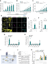
TNF induces RIPK1-dependent apoptosis in MSCs. (A) WT MSCs were pre-treated or not for 20 min with 10 μM Nec-1s, and then MSCs were exposed to indicated concentrations of TNF by 21 h, and cell death was measured by SytoxGreen (SG) positivity. (B-C) WT MSCs were pre-treated or not for 20 min with 10 μM Nec1s, and then cells were exposed to 10 ng/ml TNF, and TNF-mediated cell death (B) and caspase activity (C) were measured in function of time, respectively, by SG-positivity and DEVD-AMC fluorescence. (D-F) WT MSCs were pre-treated or not for 20 min with 10 μM Nec1s, and then stimulated with 10 ng/ml TNF for 48 h. Then, cPARP+ and TUNEL+ cells were observed by IF and confocal microscopy. (D) Representative images show: DAPI in cyan, and TUNEL+ in magenta, cPARP+ in green, and F-actins in yellow. Scale bar: 20 µm. (E) Quantification of cPARP+ cells (%). (F) Quantification of TUNEL+ cells (%). (G and H) WT MSCs were pre-treated for 20 min with 10 μM Nec1, 10 μM Nec1s, 10 μM GSK963, 1 μM GSK157 and 10 GSK840, and then cells were exposed to 10 ng/ml TNF for 21 h, and TNF-mediated cell death (G) and CASP3 activity (H) were measured, respectively, by SG positivity and DEVD-AMC fluorescence. (I) Model of signaling following TNFRSF1A activation. The TNF signaling pathway begins with the activation of its transmembrane receptor TNFRSF1A. (I.i) RIPK1 has a scaffold protein role in the TNFR1-NFKB pathway, positively impacting cell survival. (I.ii) RIPK1 enzymatic activity in the presence of CASP8 activity induces apoptosis. (I.iii) RIPK1 enzymatic activity positively regulates necroptosis with RIPK3 and MLKL. (J) WT MSCs were transfected with siRnaNAs targeting Ripk1, Ripk3, Mlkl mRNA, or the control (siCtrl). After 48 h, total protein extracts were generated and probed as indicated. (K) Cells described in (J) were pre-treated for 20 min with 50 μM zVAD or DMSO (control), and then stimulated with 10 ng/ml TNF for 21 h. TNF-mediated cell death was measured by SG-positivity. The results are presented as mean±S.E.M. of three or four independent experiments. Statistical significance was determined by two-way ANOVA. Significance between samples is indicated as follows: n.s., p > 0.05; ***, p ≤ 0.001; ****, p ≤ 0.0001.
Figure 3.
Figure 3.
RUBCNL deficiency sensitizes to apoptosis and necroptosis. (A) MSCs were transfected with siRnas targeting Rubcnl mRNA (siRubcnl) or the siRNA control (siCtrl). After 48 h, total protein extracts were generated and probed as indicated. (B) MSCs described in (A) were treated with 10 ng/ml TNF by 21 h, and cell morphology was visualized by phase contrast microscopy. (C-D) Cells were pre-treated for 20 min with 10 μM Nec1s, and then cells were exposed to 10 ng/ml TNF. TNF-mediated cell death (C) and CASP3 activity (D) were measured in function of time, respectively, by SG positivity and DEVD-AMC fluorescence. (E-F) Cells were pre-treated for 20 min with 50 μM zVAD presence or absence of Nec1s and stimulated with 10 ng/ml TNF, and cell death was measured in function of time, by SG-positivity fluorescence, and protein extracts were generated and probed as indicated. (G) MEFs were transfected with siRNAs targeting Rubcnl mRNA (siRubcnl) or the siRNA control (siCtrl). After 48 h, total cells were exposed to 10 ng/ml TNF+zVAD and cell death was measured in function of time, by SG. The results are presented as mean±S.E.M. of four independent experiments. Statistical significance was determined by two-way ANOVA. Significance between samples is indicated as follows: ****, p ≤ 0.0001.
Figure 4.
Figure 4.
RUBCNL overexpression protects from TNF-induced cell death in MSCs. (A) MSCs were stably transduced with lentiviral expression vectors for RUBCNL-Flag or empty vector (Mock) (see also Figure S2B). Total protein extracts were generated and probed as indicated. (B) Cells described in (A), were pre-treated for 20 min with 10 μM Nec1s, and then cells were exposed to 10 ng/ml TNF and cell death was measured by SG-positivity in function of the time. (C-D) Cells described in (A) were pre-treated with MAP3K7-inh. (C) or zVAD (D), in presence or absence of Nec1s and stimulated with 10 ng/ml TNF, and cell death was measured in function of time by SG-positive fluorescence. (E) L929 cells were transfected with expression vectors for RUBCNL-V5 or empty vector (Mock) by 48 h. Total protein extracts were generated and probed as indicated. (F) Cells described in (E) were pre-treated for 20 min with 10 μM Nec1s and 50 μM zVAD, and then cells were exposed to 10 ng/ml TNF and cell death was measured by SG-positivity in function of the time. The results are presented as mean±S.E.M. of three or four independent experiments. Statistical significance was determined by two-way ANOVA. Significance between samples is indicated as follows: n.s., p > 0.05; ****, p ≤ 0.0001.
Figure 5.
Figure 5.
Autophagy counteracts TNF-induced cell death, but it is dispensable for RIPK1-dependent apoptosis and necroptosis repression by RUBCNL upon TNFR1 activation. (A) MSCs stably transduced with lentiviral expression vectors for RUBCNL-Flag or empty vector (Mock) (Fig. S2B) were transfected with siRnas targeting Atg5 mRNA (siAtg5) or the control (siCtrl). Total protein extracts were generated and probed as indicated. (B and C) Cells described in (A), were treated with TNF (B) or TNF+zVAD (C). Cell death was measured by SG-positivity in function of time. (D-F) WT MSCs were transfected with siCtrl (2×) (40 nM), siCtrl (20 nM) + siAtg5 (20 nM), siCtrl (20 nM) + siRubcnl (20 nM), and siAtg5 (20 nM) + siRubcnl (20 nM). (D) Efficient knockdown of Rubcnl and Atg5 was assessed at 48 h by qPCR. Foldchange of Rubcnl or Atg5 mRNA levels was calculated using Actin mRNA levels as a reference. Cells were additionally treated with TNF (E) or TNF+zVAD (F) and cell death was measured at indicated times. (G) Illustration of autophagy competent or incompetent V5-tagged RUBCNL mutants. (H and I) Cells described in (Fig. S5C-F) were stimulated with TNF (H) or TNF+zVAD (I), and cell death rate was measured by SG-positivity on the time. The results are presented as mean±S.E.M. of three or four independent experiments. Statistical significance was determined by one-way ANOVA or two-way ANOVA. Significance between samples is indicated as follows: n.s., p > 0.05; **, p ≤ 0.01; ***, p ≤ 0.001; ****, p ≤ 0.0001.
Figure 6.
Figure 6.
Treatment with TNF reduces the interaction between RUBCNL and RIPK1 in MSCs. (A) MSC treated with shCtrl or shRubcnl (Fig. S1B) were stimulated with FLAG-HsTNF. Complex I was immunoprecipitated, and TNFRSF1A-bound RIPK1 was analyzed by immunoblotting. (B) Levels of RIPK1 in complex with TNFRSF1A were quantified in 3 independent experiments. (C and D) Detection of an interaction between RUBCNL and RIPK1 by PLA in vitro. Representative images of MSCs were transfected with Mock or V5-tagged RUBCNL and then treated with TNF for 120 minutes. Complex formation between RUBCNL-V5 and endogenous RIPK1 was observed by confocal microscopy. (C) Representative images are shown for PLA test. DAPI is depicted in cyan, and the RUBCNL-RIPK1 complex in magenta. Scale bar 20 µm. (D) Quantification of the amount of RUBCNL-RIPK1 complex spots per nucleus radius in each sample. Complex formation was observed under non-treated conditions or TNF treatment for 10, 30, 60, and 120 min. (E) HEK293T cells were transfected with empty vector (Mock) or a vector for RUBCNL-V5 and treated or not with TNF (10 ng for 120 min). Forty-eight h post-transfection, a co-immunoprecipitation was performed using the V5 tag on RUBCNL. RIPK1 and V5-tagged RUBCNL were detected by western blot. Representative blots of 3 independent experiments are shown. (F) Model of a possible mode of action of RUBCNL on RIPK1-dependent cell death signaling. Statistical significance was determined by one-way ANOVA. Significance between samples is indicated as follows: n.s., p > 0.05; ****, p ≤ 0.0001.

References

    1. Liu X, Chen H, Chen H, et al. Angiopoietin-1 preconditioning enhances survival and functional recovery of mesenchymal stem cell transplantation. J Zhejiang Univ Sci B [Internet]. 2012. [cited 2018 May 21];13(8):616–623. Available from: http://www.ncbi.nlm.nih.gov/pubmed/22843181 - PMC - PubMed
    1. Li L, Chen X, Wang WE, et al. How to improve the survival of transplanted mesenchymal stem cell in ischemic heart? STEM Cells Int [Internet]. 2016. [cited 2018 May 21];2016:9682757. Available from: http://www.hindawi.com/journals/sci/2016/9682757/ - PMC - PubMed
    1. Eggenhofer E, Benseler V, Kroemer A, et al. Mesenchymal stem cells are short-lived and do not migrate beyond the lungs after intravenous infusion. Front Immunol [Internet]. 2012. [cited 2018 May 21];3:297. Available from 10.3389/fimmu.2012.00297 - DOI - PMC - PubMed
    1. Prockop DJ. Repair of tissues by adult stem/progenitor cells (MSCs): controversies, myths, and changing paradigms. Mol Ther [Internet]. 2009. [cited 2022 Jan 11];17(6):939–946. Available from: https://pubmed.ncbi.nlm.nih.gov/19337235/ - PMC - PubMed
    1. Noronha NC, Mizukami A, Caliári-Oliveira C, et al. Priming approaches to improve the efficacy of mesenchymal stromal cell-based therapies. Stem Cell Res Ther [Internet]. 2019. [cited 2019 May 2];10(1):131. Available from 10.1186/s13287-019-1224-y - DOI - PMC - PubMed

Publication types

MeSH terms

Substances