. 2024 Sep;23(9):e14244.
doi: 10.1111/acel.14244. Epub 2024 Jun 14.

TAF15 downregulation contributes to the benefits of physical training on dendritic spines and working memory in aged mice

Affiliations

TAF15 downregulation contributes to the benefits of physical training on dendritic spines and working memory in aged mice

Yun He et al. Aging Cell. 2024 Sep.

Abstract

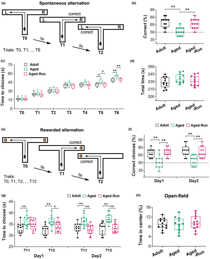
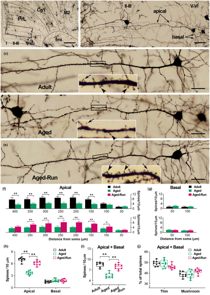
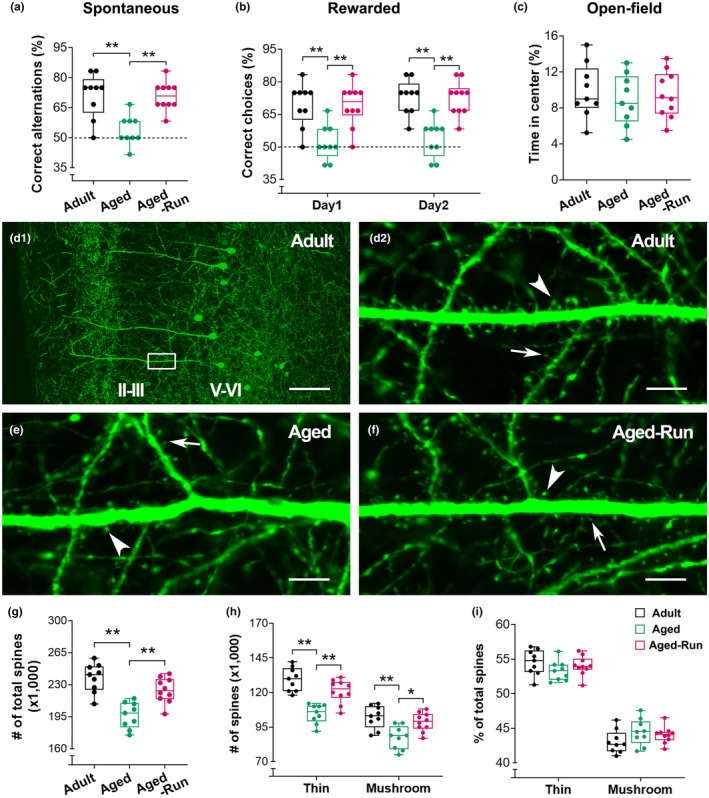
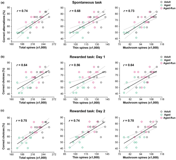
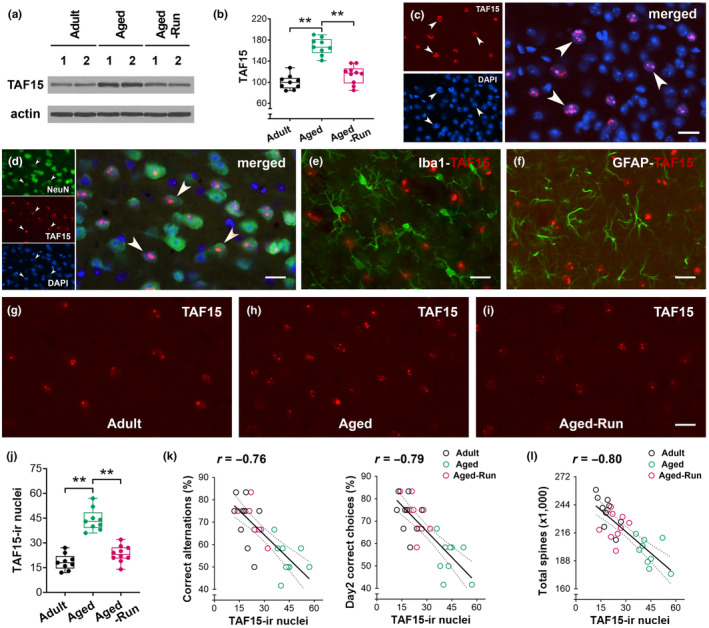
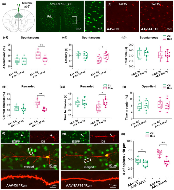
Moderate physical training has been shown to hinder age-related memory decline. While the benefits of physical training on hippocampal memory function are well-documented, little is known about its impact on working memory, which is linked to the prelimbic cortex (PrL), one major subdivision of the prefrontal cortex. Here, we examined the effects of physical training on spatial working memory in a well-established animal model of physical training, starting at 16 months of age and continuing for 5 months (running wheel 1 h/day and 5 days/week). This training strategy improved spatial working memory in aged mice (22-month-old), which was accompanied by an increased spine density and a lower TAF15 expression in the PrL. Specifically, physical training affected both thin and mushroom-type spines on PrL pyramidal cells, and prevented age-related loss of spines on selective segments of apical dendritic branches. Correlation analysis revealed that increased TAF15-expression was detrimental to the dendritic spines. However, physical training downregulated TAF15 expression in the PrL, preserving the dendritic spines on PrL pyramidal cells and improving working memory in trained aged mice. When TAF15 was overexpressed in the PrL via a viral approach, the benefits of physical training on the dendritic spines and working memory were abolished. These data suggest that physical training at a moderate pace might downregulate TAF15 expression in the PrL, which favors the dendritic spines on PrL pyramidal cells, thereby improving spatial working memory.

Keywords: TAF15; aging; physical running; prefrontal cortex; spine; working memory.


Conflict of interest statement

The authors have no conflict of interest to declare.

Figures

FIGURE 1
FIGURE 1
Physical training prevents age‐related decline of spatial working memory. (a–d) Working memory was tested using a T‐maze‐based spontaneous alternation task that contains a sample trial (T0) and six test trials (T1‐T6). A correct alternation was recorded when a mouse visited a goal arm different from its preceding trial, for example mouse visited right arm in T1 compared to the left in T0 (a). The percentage of correct alternations over six trials was used to represent working memory. Both adults and aged runners had better memory performance than aged controls (F 2,33 = 12.81; **p < 0.01; n = 12) (b). Choice latency (c) of mice among the three groups displayed an increased trend across the testing trials (F 2,33 = 18.50, p < 0.0001). The aged mice exhibited an increased latency at T5 and T6 to make choices compared to the adults (*p < 0.05, **p < 0.01, respectively). However, mice that were trained in running wheels for 5 months (Aged‐Run) demonstrated a lower decision‐making latency versus aged controls (*p < 0.05, **p < 0.01). No difference was found in the total time to complete the trials in the test (F 2,33 = 1.86, p = 0.17) (d). (e–g) Improved working memory was evident in a win‐shift rewarded task in the runners. The task contains a sample trial (T0) and 12 test trials (T1‐T12) with an intertrial interval of 5 s (e). In the sample trial, both goal arms were baited with food pellets. During the test trials, rewarded food was provided only in the arm that was not visited in the previous trial. Entries into baited arms were recorded as correct choices. The aged runners displayed a higher percentage of correct choices versus aged controls (Day 1: F 2,33 = 14.17, Day 2: F 2,33 = 10.90; **p < 0.01) (f). The adults also performed better than the aged controls on both trials (post hoc, **p < 0.01) (f). The aged control mice spent a long time to make a choice compared to the runners and adults (Day 1: F 2,33 = 10.44, Day 2: F 2,33 = 13.18; *p < 0.05, **p < 0.01) (g). (h) In an open‐field test, no difference was observed in the time spent at the center among the three groups (F 2,33 = 0.28, p = 0.76).
FIGURE 2
FIGURE 2
Physical training protects against age‐related loss of spines on PrL pyramidal cells. (a, b) A representative image to show the PrL and adjacent areas in an aged mouse (22‐month‐old). The layers of the PrL are indicated by I, II–III, and V–VI. Boxed area in (a) was magnified in (b) to present the apical and basal dendrites of a PrL pyramidal cell in layers V–VI. Cg1: cingulate cortex; M2: secondary motor cortex; fmi: forceps minor of the corpus callosum. (c–e) Representative PrL pyramidal cells in layers V–VI from an adult, aged control, and aged runner. Boxed segments were amplified to show thin (arrows) and mushroom‐type (arrowheads) spines. Loss of spines on apical dendrites was apparent in the aged control compared to the runner as well as the adult. The PrL pyramidal cell in (d) was derived from image (b). (f, g) On the apical dendrites, age‐related spine loss was observed on segments at 150–400 μm from the soma (Aged vs. Adult), but to a lesser extent in mice that underwent 5 months of physical training (Aged‐Run vs. Adult) (F 2,23 = 94.64; *p < 0.05, **p < 0.01; n = 8 or 9) (f). Training prevented spine loss on the apical dendritic segments (Aged‐Run vs. Aged) (*p < 0.05; **p < 0.01) (f). No difference was observed on the basal dendrites (F 2,23 = 1.52; p > 0.05) (G). Two‐way RM ANOVA. Error bars in (f) and (g) represent the standard error of the mean (Mean ± SEM). (h–j) Physical training protected against age‐related spine loss on the apical dendrites (F 2,23 = 45.52; **p < 0.01) (h). Aged mice had a lower average spine density compared to the runners and adults (F 2,23 = 45.61; **p < 0.01) (i). The percentage of thin and mushroom‐type spines on both apical and basal dendrites was not affected (j). Scale bars = 250 μm (a), 50 μm (b), and 25 μm (c–e).
FIGURE 3
FIGURE 3
Improved working memory performance and reserved spines on PrL pyramidal cells in a cohort of Thy1‐YFPH aged runners. (a) The mice trained for 5 months (Aged‐Run; n = 10) displayed a higher percentage of correct alternations in a spontaneous task versus aged controls (22‐month‐old; n = 9), while the later had a lower percentage of correct alternations compared to adult controls (16‐month‐old; n = 9) (F 2,25  = 12.29; **p < 0.01). (b) Improved memory performance in the aged runners was evident in a 2‐day win‐shift rewarded task. The runners had a higher percentage of correct choices versus aged controls (two‐way RM ANOVA, main effect of running, F 2,25 = 20.16; **p < 0.01). (c) In an open‐field test, no difference was observed in the time spent at the center among the three groups (F 2,25 = 0.29, p = 0.75). (d) Representative confocal images from an adult mouse to clarify the layers of the PrL. The cell bodies of Thy1‐expressing pyramidal cells were located in layers V–VI. The dendritic segments covered with numerous spines were mainly observed in layers II–III, where the spines were counted. The boxed area in (d1) was magnified in (d2) to show the dendritic branches with thin spines (arrows) and mushroom‐type spines (arrowheads). (e, f) Representative images from an aged runner and an aged control. Spine loss is apparent in the control. (g–i) Stereological quantitative analysis. Runners had an increased number of spines versus age‐matched controls (F 2,25 = 18.53; **p < 0.01) (g). Training for 5 months protected against age‐related loss of both thin and mushroom‐type spines in the PrL (F 2,25 = 18.02; *p < 0.05, **p < 0.01) (h). No difference was observed in the percentage of either thin or mushroom‐type spines (F 2,25 = 0.99, p = 0.39) (i). Counts of GFP‐labeled spines in a volume of 6 × 105 μm3. Scale bars = 100 μm (d1) and 7 μm (d2, e, f).
FIGURE 4
FIGURE 4
Correlation between YFP‐labeled dendritic spines in the PrL and spatial working memory measured via two T‐maze based tasks. (a) Positive correlations were observed between the correct alternations in a spontaneous alternation task and the numbers of total spines in the PrL (Pearson r = 0.74). When dendritic spines were classified as thin or mushroom‐type and analyzed separately, positive correlations were also detected (Pearson r = 0.68 and 0.73, respectively). (b, c) In a 2‐day rewarded win‐shift task, the correct choices positively correlated with the number of total, thin, and mushroom‐type spines (Day 1: r = 0.64, 0.56 and 0.64, respectively; Day 2: r = 0.75, 0.74, and 0.70, respectively; p < 0.01), uncovering the critical role of dendritic spines in spatial working memory performance.
FIGURE 5
FIGURE 5
Physical running downregulates TAF15 expression in the PrL of aged mice. (a, b) A decreased TAF15 expression was identified in aged runners (F 2,25 = 46.80; **p < 0.01; n = 9 or 10). The protein levels of TAF15 were normalized to respective Actin levels and expressed as a percentage of adult controls. (c) TAF15 was primarily detected in DAPI‐counterstained nuclei. The co‐localization was pointed by arrowheads. (d–f) TAF15 was detected in NeuN‐labeled neurons (arrowheads), but not Iba1‐labeled microglial cells and GFAP‐labeled astrocytes. (g–i) Representative confocal images from an adult, aged control, and aged runner to show TAF15‐ir nuclei in layers V–VI. (j) A decreased number of TAF15‐ir nuclei in the aged runners versus aged controls (F 2,25 = 53.68; **p < 0.01). (k) Negative correlations were observed between the numbers of TAF15‐ir nuclei (per 5 × 104 μm2) and the spontaneous alternations (left, r = −0.76, p < 0.01) as well as rewarded correct choices (right, r = −0.79, p < 0.01). (l) A negative correlation between TAF15‐ir nuclei and total dendritic spines (r = −0.80, p < 0.01). Scale bars = 20 μm (c, d–i).
FIGURE 6
FIGURE 6
Overexpression of TAF15 abolishes the effects of physical training on working memory and dendritic spines. (a, b) AAV‐TAF15‐EGFP or control AAV‐EGFP was bilaterally injected into the PrL of 16‐month‐old mice, followed by physical training and memory tests, which lasted 20 and 3 weeks, respectively. Robust EGFP‐expressing cells were observed in the PrL. IL, infralimbic cortex; fmi, forceps minor of the corpus callosum. (c) In a spontaneous T‐maze test, runners with AAV‐TAF15 had similar correct alternation (c1) and choice latency (c2) compared to controls (F 1,32 = 11.03, 4.93; Fisher's post hoc, p > 0.05; n = 9). Improved memory was only observed in runners with control virus (effect of AAV‐TAF15: F 1,32 = 5.40; effect of running: F 1,32 = 11.03; **p < 0.01). No difference in the time to complete the testing (AAV effect: F 1,32 = 0.24; running effect: F 1,32 = 1.03; post hoc p > 0.05) (c3). (d) In a rewarded task, the AAV‐TAF15 runners displayed a comparable percentage of correct choices versus their sedentary controls (F 1,32 = 10.28; post hoc, p > 0.05) (d1). They also spent equal time to make a choice at the last test trial (T12) (F 1,32 = 4.28; p > 0.05) (d2). However, the runners with control AAV had a good memory performance (*p < 0.05; **p < 0.01). (e) No difference in the time spent at the center in an open‐field test. (f–h) TAF15‐EGFP expressing PrL pyramidal cells were labeled with DiI (red) to show their dendritic spines. Representative images were from a runner with control AAV (f) and a runner with AAV‐TAF15 (g). Spine density in the runners with AAV‐TAF15 was lower than those with control AAV (AAV effect: F 1,24 = 32.73; running effect: F 1,24 = 19.22; post hoc, **p < 0.01; n = 7). The control mice with AAV‐TAF15 also had a lower spine density versus those with control AAV (*p < 0.05) (h). The benefit of training on spines was only observed in the runners with control AAV (**p < 0.01, not marked in the graph).

Similar articles

References

    1. Ahlbeck, J. , Song, L. , Chini, M. , Bitzenhofer, S. H. , & Hanganu‐Opatz, I. L. (2018). Glutamatergic drive along the septo‐temporal axis of hippocampus boosts prelimbic oscillations in the neonatal mouse. eLife, 7, e33158. 10.7554/eLife.33158 - DOI - PMC - PubMed
    1. Aoki, N. , Higashi, S. , Kawakami, I. , Kobayashi, Z. , Hosokawa, M. , Katsuse, O. , Togo, T. , Hirayasu, Y. , & Akiyama, H. (2012). Localization of fused in sarcoma (FUS) protein to the post‐synaptic density in the brain. Acta Neuropathologica, 124(3), 383–394. 10.1007/s00401-012-0984-6 - DOI - PubMed
    1. Benoit, L. J. , Holt, E. S. , Teboul, E. , Taliaferro, J. P. , Kellendonk, C. , & Canetta, S. (2020). Medial prefrontal lesions impair performance in an operant delayed nonmatch to sample working memory task. Behavioral Neuroscience, 134(3), 187–197. 10.1037/bne0000357 - DOI - PMC - PubMed
    1. Bloss, E. B. , Janssen, W. G. , Ohm, D. T. , Yuk, F. J. , Wadsworth, S. , Saardi, K. M. , McEwen, B. S. , & Morrison, J. H. (2011). Evidence for reduced experience‐dependent dendritic spine plasticity in the aging prefrontal cortex. The Journal of Neuroscience, 31(21), 7831–7839. 10.1523/JNEUROSCI.0839-11.2011 - DOI - PMC - PubMed
    1. Bonilla‐Quintana, M. , Wörgötter, F. , Tetzlaff, C. , & Fauth, M. (2020). Modeling the shape of synaptic spines by their Actin dynamics. Frontiers in Synaptic Neuroscience, 12, 9. 10.3389/fnsyn.2020.00009 - DOI - PMC - PubMed

Substances

LinkOut - more resources