Review
. 2024 Aug;11(30):e2401069.
doi: 10.1002/advs.202401069. Epub 2024 Jun 14.

Extracellular Vesicle Preparation and Analysis: A State-of-the-Art Review

Affiliations
Review

Extracellular Vesicle Preparation and Analysis: A State-of-the-Art Review

Zesheng Wang et al. Adv Sci (Weinh). 2024 Aug.

Abstract

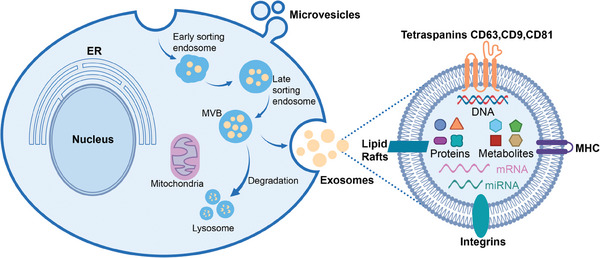
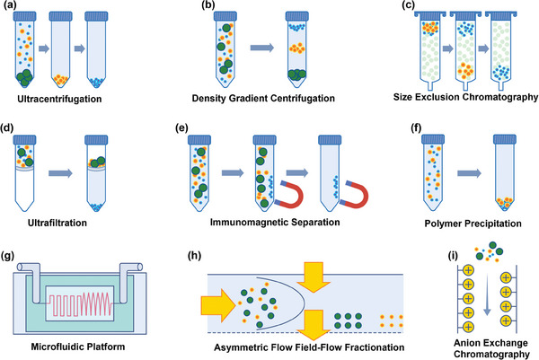
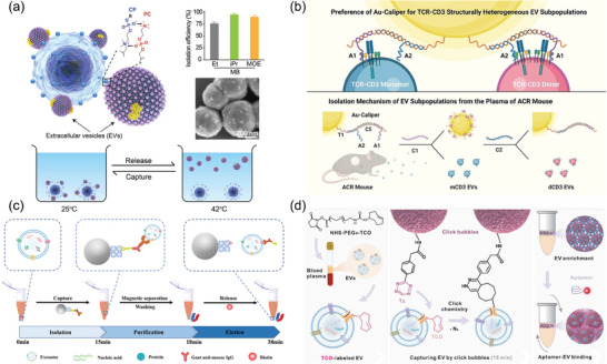
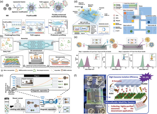
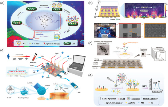
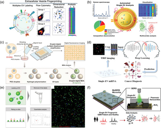
In recent decades, research on Extracellular Vesicles (EVs) has gained prominence in the life sciences due to their critical roles in both health and disease states, offering promising applications in disease diagnosis, drug delivery, and therapy. However, their inherent heterogeneity and complex origins pose significant challenges to their preparation, analysis, and subsequent clinical application. This review is structured to provide an overview of the biogenesis, composition, and various sources of EVs, thereby laying the groundwork for a detailed discussion of contemporary techniques for their preparation and analysis. Particular focus is given to state-of-the-art technologies that employ both microfluidic and non-microfluidic platforms for EV processing. Furthermore, this discourse extends into innovative approaches that incorporate artificial intelligence and cutting-edge electrochemical sensors, with a particular emphasis on single EV analysis. This review proposes current challenges and outlines prospective avenues for future research. The objective is to motivate researchers to innovate and expand methods for the preparation and analysis of EVs, fully unlocking their biomedical potential.

Keywords: artificial intelligence; disease diagnosis; extracellular vesicles; single EV.


Conflict of interest statement

The authors declare no conflict of interest.

Figures

Figure 1
Figure 1
An overview of the biogenesis pathways for two primary types of EVs (exosomes and microvesicles), along with a structural schematic of exosomes. ER: endoplasmic reticulum; MVB: multivesicular bodies; MHC: major histocompatibility complex I/II.
Figure 2
Figure 2
Illustration of commonly used techniques for EV preparation. These techniques include a) ultracentrifugation, which leverages high‐speed centrifugal forces; b) density gradient centrifugation, which separates EVs based on their buoyancy; c) size‐exclusion chromatography, for isolating EVs by size; d) ultrafiltration, a method that uses membrane filters for size‐based separation; e) immunomagnetic separation, which targets specific EV markers with magnetic beads; f) polymer precipitation, where polymers are used to precipitate EVs out of solution; g) microfluidics platforms, offering precise manipulation of fluids to isolate EVs; h) asymmetric flow field‐flow fractionation, which separates particles based on their hydrodynamic size in a flow; i) anion exchange chromatography, which isolates EVs based on charge differences.
Figure 3
Figure 3
Latest reported affinity‐based methods for EV preparation include: a) The mechanism for adhesion of EVs to a magnetic bead coated with choline phosphate, utilizing the high‐affinity interaction between phosphatidylcholine and choline phosphate, followed by subsequent release after capture. Reproduced with permission.[ 150 ] Copyright 2023, American Association for the Advancement of Science. b) Isolation mechanism for structurally heterogeneous TCR‐CD3 EV subpopulations. Reproduced with permission.[ 152 ] Copyright 2023, Wiley‐VCH, GmbH. c) The exosome isolation process using the SIMI system. Reproduced with permission.[ 153 ] Copyright 2023, American Chemical Society. d) EVs captured via a bioorthogonal click chemistry reaction. Reproduced with permission.[ 155 ] Copyright 2023, Wiley‐VCH, GmbH.
Figure 4
Figure 4
Cutting‐edge microfluidic techniques for EV preparation. a) Fabrication of FluidFaceMB and EV capture. Reproduced with permission.[ 165 ] Copyright 2023, Wiley‐VCH, GmbH. b) Herringbone‐patterned microfluidic device for capturing HER2‐positive cancer exosomes in urine. Reproduced with permission.[ 166 ] Copyright 2024, Elsevier. c) An immuno‐magnetophoresis‐based microfluidic chip for EV isolation. Reproduced with permission.[ 167 ] Copyright 2023, Elsevier. d) EV isolation using a digital microfluidic platform. Reproduced with permission.[ 168 ] Copyright 2024, Elsevier. e) Immuno‐inertial microfluidics for EV isolation. Reproduced with permission.[ 169 ] Copyright 2023, Elsevier. f) A microfluidic device using metallic arrays for EV isolation. Reproduced with permission.[ 170 ] Copyright 2023, Elsevier.
Figure 5
Figure 5
Nanomaterials, including nanoparticles and nanowires, serve as innovative tools for the efficient isolation of EVs. a) 3D porous sponge chip for EV isolation. Reproduced with permission.[ 171 ] Copyright 2023, Wiley‐VCH, GmbH. b) SiO2 microsphere‐coated 3D hierarchical porous chip for the enrichment of EVs. Reproduced with permission.[ 172 ] Copyright 2023, Wiley‐VCH, GmbH. c) ExoSIC chip for the continuous isolation of EVs. Reproduced with permission.[ 173 ] Copyright 2023, Royal Society of Chemistry. d) ZnO nanowires for EV isolation. Reproduced with permission.[ 176 ] Copyright 2023, American Chemical Society. e) DNA‐based hydrogel for EV isolation. Reproduced with permission.[ 177 ] Copyright 2023, National Academy of Sciences.
Figure 6
Figure 6
Various methods for EV analysis include: NTA: Nanoparticle Tracking Analysis; AFM: Atomic Force Microscopy; TRPS: Tunable Resistive Pulse Sensing; SERS: Surface‐Enhanced Raman Scattering; SEM: Scanning Electron Microscopy; TEM: Transmission Electron Microscopy; DLS: Dynamic Light Scattering; MS: Mass Spectrometry; PCR: Polymerase Chain Reaction; FC: Flow Cytometry; WB: Western Blotting; SPR: Surface Plasmon Resonance; ELISA: Enzyme‐Linked Immunosorbent Assay; TIRF: Total Internal Reflection Fluorescence.
Figure 7
Figure 7
Labeled SERS for analysis of EVs. a) SERS profiling of three biomarkers on plasma‐derived exosomes for the diagnosis of osteosarcoma. Reproduced with permission.[ 257 ] Copyright 2022, Elsevier. b) Structure and working principle of the on‐chip SERS‐based EV analysis platform. Reproduced with permission.[ 258 ] Copyright 2023, Elsevier. c) A paper‐based SERS‐vertical flow biosensor for multiplexed quantitative profiling of EV proteins. Reproduced with permission.[ 259 ] Copyright 2023, American Chemical Society. d) The “sandwich” SERS immunoassay for small EVs. Reproduced with permission.[ 260 ] Copyright 2023, Royal Society of Chemistry.
Figure 8
Figure 8
Recent progress in AI‐assisted SERS for the analysis of EVs. a) SERS dataset with deep‐learning algorithm for the analysis of plasma exosomal proteins. Reproduced with permission.[ 265 ] Copyright 2023, American Chemical Society. b) Label‐free SERS combined with machine learning for EV detection. Reproduced with permission.[ 266 ] Copyright 2023, American Chemical Society. c) Cellular origin identified by AI‐assisted SERS of EV analysis. Reproduced with permission.[ 267 ] Copyright 2023, Wiley‐VCH, GmbH. d) Multi‐cancer simultaneous diagnosis utilizing AI‐SERS in EV analysis. Reproduced with permission.[ 268 ] Copyright 2023, Springer Nature. e) Diagnosing major depressive disorder (MDD) in vitro through the detection of plasma exosomes via SERS and analysis using AI. Reproduced with permission.[ 269 ] Copyright 2023, American Chemical Society.
Figure 9
Figure 9
Recent advances in electrochemical sensors for EV analysis. a) Electrochemiluminescence for EV detection utilizing a glycosyl‐imprinted polymer membrane and an aptamer for dual recognition. Reproduced with permission.[ 287 ] Copyright 2024, American Chemical Society. b) Enhanced electrokinetic, label‐free plasmonic sensing for the detection of tumor‐derived EVs. Reproduced with permission.[ 288 ] Copyright 2023, Elsevier. c) Detection of urinary EVs using an electrochemical immunoassay in a microtiter plate. Reproduced with permission.[ 289 ] Copyright 2023, American Chemical Society. d) The filter‐electrochemical microfluidic chip for EV detection. Reproduced with permission.[ 290 ] Copyright 2023, Elsevier. e) An electrochemical strategy for EV detection assisted by Au nanoparticles (NPs). Reproduced with permission.[ 291 ] Copyright 2023, Elsevier.
Figure 10
Figure 10
Innovative techniques for single‐EV research. a) Single‐vesicle flow cytometry with multiple parameters for the analysis of EV heterogeneity. Reproduced with permission.[ 302 ] Copyright 2024, American Chemical Society. b) Single EV analysis using Single Particle Automated Raman Trapping Analysis platform (SPARTA). Reproduced with permission.[ 303 ] Copyright 2021, American Chemical Society. c) Hydrogel droplet digital amplification for profiling DNA cargos in single EV. Reproduced with permission.[ 304 ] Copyright 2024, American Chemical Society. d) A method to identify multi‐miRNA in single EV through the integration of Total Internal Reflection Fluorescence imaging and deep learning analysis. Reproduced with permission.[ 305 ] Copyright 2024, American Chemical Society. e) Multiplexed protein measurements in single EV through fluorescence imaging. Reproduced with permission.[ 306 ] Copyright 2022, American Association for the Advancement of Science. f) A microfluidic surface embedded with arrayed nanocavity microchips enables SERS detection of single EV from glioblastoma. Reproduced with permission.[ 307 ] Copyright 2023, American Chemical Society.

Similar articles

Cited by

References

    1. a) Pan B.‐T., Johnstone R. M., Cell 1983, 33, 967; - PubMed
    2. b) Harding C., Heuser J. M., Stahl P. D., J. Cell Biol. 1983, 97, 329. - PMC - PubMed
    1. Trams E. G., Lauter C. J., Salem N. Jr., Heine U., Biochimica et Biophysica Acta (BBA) – Biomembranes 1981, 645, 63. - PubMed
    1. Johnstone R. M., Adam M., Hammond J. R., Orr L., Turbide C., J. Biol. Chem. 1987, 262, 9412. - PubMed
    1. Thery C., Witwer K. W., Aikawa E., Alcaraz M. J., Anderson J. D., Andriantsitohaina R., Antoniou A., Arab T., Archer F., Atkin‐Smith G. K., Ayre D. C., Bach J. M., Bachurski D., Baharvand H., Balaj L., Baldacchino S., Bauer N. N., Baxter A. A., Bebawy M., Beckham C., Bedina Zavec A., Benmoussa A., Berardi A. C., Bergese P., Bielska E., J Extracell Vesicles 2018, 7, 1535750. - PMC - PubMed
    1. Gyorgy B., Szabo T. G., Pasztoi M., Pal Z., Misjak P., Aradi B., Laszlo V., Pallinger E., Pap E., Kittel A., Nagy G., Falus A., Buzas E. I., Cell. Mol. Life Sci. 2011, 68, 2667. - PMC - PubMed

MeSH terms

LinkOut - more resources