. 2024 Aug 21;39(8):1132-1146.
doi: 10.1093/jbmr/zjae093.

diABZI and poly(I:C) inhibit osteoclastic bone resorption by inducing IRF7 and IFIT3

Affiliations

diABZI and poly(I:C) inhibit osteoclastic bone resorption by inducing IRF7 and IFIT3

Yingkang Huang et al. J Bone Miner Res. .

Abstract

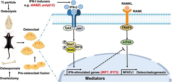
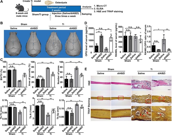
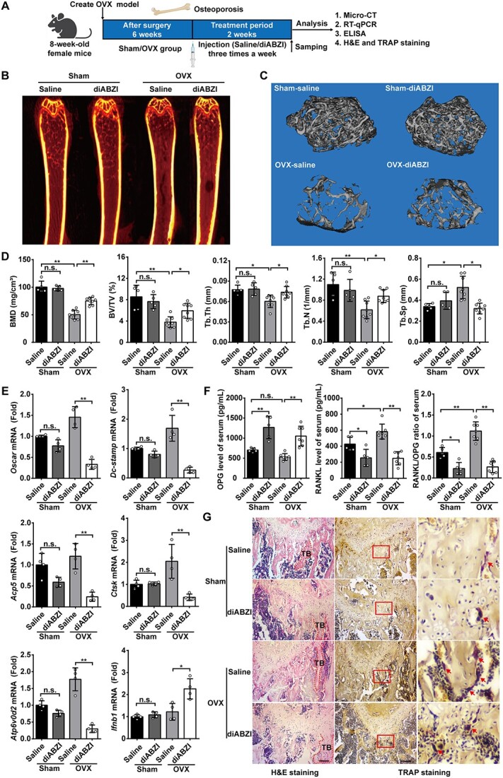
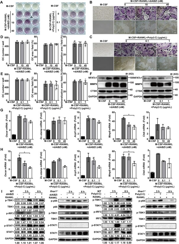
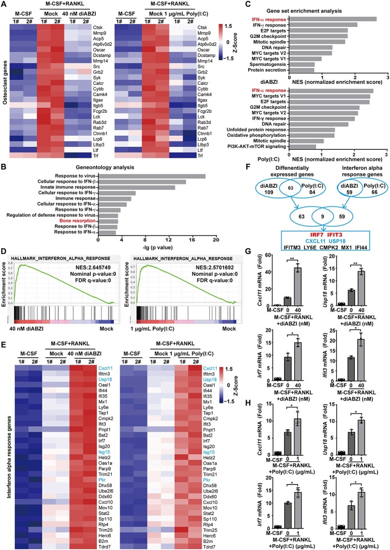
Type I interferons (IFN-I) are pleiotropic factors endowed with multiple activities that play important roles in innate and adaptive immunity. Although many studies indicate that IFN-I inducers exert favorable effects on broad-spectrum antivirus, immunomodulation, and anti-tumor activities by inducing endogenous IFN-I and IFN-stimulated genes, their function in bone homeostasis still needs further exploration. Here, our study demonstrates 2 distinct IFN-I inducers, diABZI and poly(I:C), as potential therapeutics to alleviate osteolysis and osteoporosis. First, IFN-I inducers suppress the genes that control osteoclast (OC) differentiation and activity in vitro. Moreover, diABZI alleviates bone loss in Ti particle-induced osteolysis and ovariectomized -induced osteoporosis in vivo by inhibiting OC differentiation and function. In addition, the inhibitory effects of IFN-I inducers on OC differentiation are not observed in macrophages derived from Ifnar1-/-mice, which indicate that the suppressive effect of IFN-I inducers on OC is IFNAR-dependent. Mechanistically, RNAi-mediated silencing of IRF7 and IFIT3 in OC precursors impairs the suppressive effect of the IFN-I inducers on OC differentiation. Taken together, these results demonstrate that IFN-I inducers play a protective role in bone turnover by limiting osteoclastogenesis and bone resorption through the induction of OC-specific mediators via the IFN-I signaling pathway.

Keywords: IFIT3; IFN-I inducer; IRF7; ISG mediator; bone resorption; osteoclast; osteolysis; osteoporosis.

Plain language summary

OCs are responsible for bone resorption, and their excessive differentiation and enhanced activity will lead to bone resorption diseases such as osteoporosis and osteolysis. Here, our study demonstrates 2 distinct IFN-I inducers, diABZI and poly(I:C), as potential therapeutics to alleviate osteolysis and osteoporosis. IFN-I inducers suppress OC differentiation, and particularly diABZI alleviates bone loss in osteolysis and osteoporosis mouse models. Taken together, IFN-I inducers play a protective role in bone turnover by limiting osteoclastogenesis and bone resorption through the induction of OC-specific mediators via the IFN-I signaling pathway. Our in-depth and comprehensive discovery of the IFN-I inducer would provide new insight into OC biology and therapeutic targets for osteoclastic bone resorption diseases.


Conflict of interest statement

All authors state that they have no conflicts of interest.

Figures

Graphical Abstract
Graphical Abstract
Figure 1
Figure 1
IFN-I inducers inhibit OC differentiation and function in vitro. (A) The general view of TRAP staining of BMMs after 6 d osteoclastogenic induction in the presence of diABZI and poly(I:C). (B, C) Representative images of TRAP staining of BMMs after 6 d osteoclastogenic induction and the demineralization area after 6 d osteoclastogenic induction in the presence of diABZI and poly(I:C). Scale bar: 100 μm. (D–G) Quantification of number and area of TRAP-positive cells after 6 d osteoclastogenic induction in the presence of diABZI and poly(I:C). TRAP-positive cells with 3 or more nuclei were counted as OC. (H, I) Quantification of the demineralization area after 6 d osteoclastogenic induction in the presence of diABZI and poly(I:C). (J, K) Relative mRNA expression of OC-specific genes in BMMs including Oscar, Dc-stamp, Acp5, Mmp9, and Ctsk in BMMs after 3 d osteoclastogenic induction in the presence of diABZI and poly(I:C). Gapdh as an internal control gene. (L, M) Protein expression of OC-specific proteins in BMMs including NFATc1, MMP9, and CTSK in BMMs after 3 d osteoclastogenic induction in the presence of diABZI and poly(I:C). Data of (A–C) are representative images from 3 independent experiments. Data of (D–K) are shown as the mean ± SD from at least 3 independent experiments. Data of (L) and (M) are shown as representative of 3 independent experiments. *p<.05, **p<.01 by one-way ANOVA.
Figure 2
Figure 2
diABZI alleviates Ti particle-induced osteolysis in vivo. (A) Schematic diagram of the titanium particle-induced osteolysis model. (B) Representative micro-CT images of murine calvaria 3D reconstruction figure. (C) Quantitative parameters of morphological bone alterations, including BV/TV, area of porosity, number of pores, Tb.Th, Tb.N, and Tb.Sp (Sham, n=3; Ti, n=7). (D) RANKL and OPG serum concentrations and RANKL/OPG ratio (Sham, n=3; Ti, n=7). (E) Representative images of histological staining with HE and TRAP staining. Arrows indicate OC. Scale bar: 100 μm. Data of (B) and (E) are representative images of one representative experiment from 3 independent experiments. Data of (C) and (D) are shown as the mean ± SD of one representative experiment from 3 independent experiments. *p<.05, **p<.01, n.s.: not significant by one-way ANOVA.
Figure 3
Figure 3
diABZI alleviates OVX-induced osteoporosis in vivo. (A) Schematic diagram of the OVX-induced osteoporosis model. (B, C) Representative micro-CT images of murine femur calvaria showing 2D fault and 3D reconstruction figure (D) Quantitative parameters of morphological bone alterations, including BMD, BV/TV, Tb.Th, Tb.N, and Tb.Sp (Sham, n=5; OVX, n=8). (E) Relative mRNA expression of OC-specific genes, including Oscar, Dc-stamp, Acp5, Ctsk, Atp6v0d2, and Ifnb1 in bone tissues (n=4). Gapdh as an internal control gene. (F) RANKL and OPG serum concentrations and RANKL/OPG ratio (Sham, n=5; OVX, n=8). (G) Representative images of histological staining with HE and TRAP staining. Scale bar: 100 μm. TB: trabecular bone. Arrows indicate OC. Data of (B), (C), and (G) are representative images of one representative experiment from 3 independent experiments. Data of (D–F) are shown as the mean ± SD of one representative experiment from 3 independent experiments. *p<.05, **p<.01, n.s.: not significant by one-way ANOVA.
Figure 4
Figure 4
IFNAR is required for the suppressive effect of IFN-I inducers on osteoclastogenesis. (A) The general view of TRAP staining in Ifnar1−/− BMMs after 6 d osteoclastogenic induction in the presence of diABZI and poly(I:C). (B, C) Representative images of TRAP staining after 6 d osteoclastogenic induction and the demineralization area after 6 d osteoclastogenic induction in Ifnar1−/− BMMs in the presence of diABZI and poly(I:C). (D, E) Quantification of number and area of TRAP-positive cells, and the demineralization area in the presence of diABZI and poly(I:C). TRAP-positive cells with 3 or more nuclei were counted as OC. (F) Protein expression of OC-specific proteins in Ifnar1−/− BMMs including NFATc1, MMP9, and CTSK after 3 d osteoclastogenic induction in the presence of diABZI and poly(I:C). (G, H) Relative mRNA expression of OC-specific genes in Ifnar1−/− BMMs including Oscar, Dc-stamp, Acp5, Mmp9, and Ctsk after 3 d osteoclastogenic induction in the presence of diABZI and poly(I:C). Gapdh as an internal control gene. (I) Protein expression of IFN-I signaling in WT BMMs including p-TBK1, TBK1, p-IRF3, IRF3, p-STAT1, and STAT1 after 3 h and 6 h osteoclastogenic induction in the presence of diABZI and poly(I:C). Protein expression of IFN-I signaling in Ifnar1−/− BMMs including p-p65, p65, p-TBK1, TBK1, p-STAT1, and STAT1 after 3 h and 6 h osteoclastogenic induction in the presence of diABZI and poly(I:C). Scale bar: 100 μm. Data of (A–C) are representative images from 3 independent experiments. Data of (D–E) and (G–H) are shown as the mean ± SD from at least 3 independent experiments. Data of (F) and (I) are shown as representative of 3 independent experiments. *p<.05 by one-way ANOVA.
Figure 5
Figure 5
IRF7 and IFIT3 are common mediators during IFN-I inducers inhibiting osteoclastogenesis. (A) Heatmap of OC marker genes in BMMs after 3 d osteoclastogenic induction in the presence of diABZI and poly(I:C) (B) Geneontology analysis of RANKL-inducible genes regulated by diABZI and poly(I:C). (C, D) GSEA of RANKL-inducible genes regulated by diABZI and poly(I:C) ranked by NES of Hallmark. (E) Heatmap of the enriched interferon alpha response genes in BMMs after 3 d osteoclastogenic induction in the presence of diABZI and poly(I:C). (F) Left Venn diagram showing the overlap of the DEGs between diABZI and poly(I:C); right Venn diagram showing the overlap of the interferon alpha response genes between diABZI and poly(I:C); finally overlap the DEGs and interferon alpha response genes. (G, H) The mRNA expression of the possible OC-related ISGs including Cxcl11, Usp18, Irf7, and Ifit3 during RANKL-induced osteoclastogenesis in the presence of diABZI and poly(I:C). Gapdh as an internal control gene. Data of (G) and (H) are shown as the mean ± SD from 3 independent experiments. *p<.05, **p<.01 by one-way ANOVA.
Figure 6
Figure 6
Induction of IRF7 and IFIT3 mediates the suppressive effect of IFN-I inducers on OC differentiation and function. (A, B) Efficiency of silencing of IRF7 and IFIT3 and kinetics of IRF7 and IFIT3 mRNA expression during osteoclastogenic induction at indicated time points. (C, D) The general view of TRAP staining of BMMs after 6 d osteoclastogenic induction in the presence of si-IRF7 and si-IFIT3 with diABZI and poly(I:C). (E, F) Representative images of TRAP staining of BMMs after 6 d osteoclastogenic induction in the presence of si-IRF7 and si-IFIT3 with diABZI and poly(I:C). (G, H) Quantification of number and area of TRAP-positive cells after 6 d osteoclastogenic induction in the presence of si-IRF7 and si-IFIT3 with diABZI and poly(I:C). TRAP-positive cells with 3 or more nuclei were counted as OC (I, J) Relative mRNA expression of OC-specific genes including Oscar, Dc-stamp, Acp5, and Mmp9 in BMMs after 3 d osteoclastogenic induction in the presence of si-IRF7 and si-IFIT3 with diABZI and poly(I:C). Gapdh as an internal control gene. Scale bar: 100 μm. Data of (A) and (B) and (G–J) are shown as the mean ± SD from at least 3 independent experiments. Data of (C–F) are representative images from 3 independent experiments. *p<.05, **p<.01 by one-way ANOVA.

Similar articles

Cited by

References

    1. Du T, Yan Z, Zhu S, et al. QKI deficiency leads to osteoporosis by promoting RANKL-induced osteoclastogenesis and disrupting bone metabolism. Cell Death Dis. 2020;11(5):330. 10.1038/s41419-020-2548-3 - DOI - PMC - PubMed
    1. Boyle WJ, Simonet WS, Lacey DL. Osteoclast differentiation and activation. Nature. 2003;423(6937):337–342. 10.1038/nature01658 - DOI - PubMed
    1. Wang R, Yang Y, Zhang Z, et al. Major vault protein (MVP) suppresses aging- and estrogen deficiency-related bone loss through Fas-mediated apoptosis in osteoclasts. Cell Death Dis. 2023;14(9):604. 10.1038/s41419-023-05928-4 - DOI - PMC - PubMed
    1. Adam S, Simon N, Steffen U, et al. JAK inhibition increases bone mass in steady-state conditions and ameliorates pathological bone loss by stimulating osteoblast function. Sci Transl Med. 2020;12(530):eaay4447. 10.1126/scitranslmed.aay4447 - DOI - PubMed
    1. Teitelbaum SL. Bone resorption by osteoclasts. Science (New York, NY). 2000;289(5484):1504–1508. 10.1126/science.289.5484.1504 - DOI - PubMed

MeSH terms