. 2024 Jul 16;62(7):e0004224.
doi: 10.1128/jcm.00042-24. Epub 2024 Jun 14.

Generation of recombinant viruses directly from clinical specimens of COVID-19 patients

Affiliations

Generation of recombinant viruses directly from clinical specimens of COVID-19 patients

Hirotaka Yamamoto et al. J Clin Microbiol. .

Abstract

Rapid characterization of the causative agent(s) during a disease outbreak can aid in the implementation of effective control measures. However, isolation of the agent(s) from crude clinical samples can be challenging and time-consuming, hindering the establishment of countermeasures. In the present study, we used saliva specimens collected for the diagnosis of SARS-CoV-2-a good example of a practical target-and attempted to characterize the virus within the specimens without virus isolation. Thirty-four saliva samples from coronavirus disease 2019 patients were used to extract RNA and synthesize DNA amplicons by PCR. New primer sets were designed to generate DNA amplicons of the full-length spike (S) gene for subsequent use in a circular polymerase extension reaction (CPER), a simple method for deriving recombinant viral genomes. According to the S sequence, four clinical specimens were classified as BA. 1, BA.2, BA.5, and XBB.1 and were used for the de novo generation of recombinant viruses carrying the entire S gene. Additionally, chimeric viruses carrying the gene encoding GFP were generated to evaluate viral propagation using a plate reader. We successfully used the RNA purified directly from clinical saliva samples to generate chimeric viruses carrying the entire S gene by our updated CPER method. The chimeric viruses exhibited robust replication in cell cultures with similar properties. Using the recombinant GFP viruses, we also successfully characterized the efficacy of the licensed antiviral AZD7442. Our proof-of-concept demonstrates the novel utility of CPER to allow rapid characterization of viruses from clinical specimens.

Importance: Characterization of the causative agent(s) for infectious diseases helps in implementing effective control measurements, especially in outbreaks. However, the isolation of the agent(s) from clinical specimens is often challenging and time-consuming. In this study, saliva samples from coronavirus disease 2019 patients were directly subjected to purifying viral RNA, synthesizing DNA amplicons for sequencing, and generating recombinant viruses. Utilizing an updated circular polymerase extension reaction method, we successfully generated chimeric SARS-CoV-2 viruses with sufficient in vitro replication capacity and antigenicity. Thus, the recombinant viruses generated in this study were applicable for evaluating the antivirals. Collectively, our developed method facilitates rapid characterization of specimens circulating in hosts, aiding in the establishment of control measurements. Additionally, this approach offers an advanced strategy for controlling other (re-)emerging viral infectious diseases.

Keywords: SARS-CoV-2; clinical specimens; reporter assay; reverse genetics.


Conflict of interest statement

The authors declare no conflict of interest.

Figures
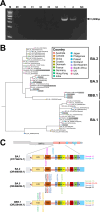
Fig 1
Fig 1
Viral characterization of SARS-CoV-2 spike (S) obtained from clinical specimens. (A) Gel electrophoresis analysis of S gene amplicons generated by PCR from the saliva of four groups with different Ct values. M, 1 kb DNA ladder. 1, 2, and 29–34: sample ID. Arrow indicates base pair (bp) of the amplified spike protein gene. (B) The phylogenetic tree of SARS-CoV-2 clinical specimens used in this study. The tree was analyzed using BEAST software (26). The 28 clinical specimens are highlighted. The color of the branch indicates the country where the viruses were found. (C) A schematic representation of the SARS-CoV-2 viral genome. The mutations observed in S within the clinical samples are indicated by arrows and color-coded by individual samples. GenBank accession numbers of the reference strains are OY724674.1 for BA.1, OR496569.1 for BA.2, OR496284.1 for BA.5, and OR239444.1 for XBB.1.
Fig 2
Fig 2
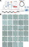
Generation of recombinant viruses from clinical specimens using the updated CPER method. (A) A schematic overview of the updated CPER method for generating chimeric SARS-CoV-2 from 28 clinical saliva specimens. A fragment (shown in red) covering the full-length S gene was amplified with a new primer set (F8 Fw and Full-length S protein F8 Rv, Table 2). This fragment along with the eight fragments covering the remaining SARS-CoV-2 genome were assembled with a UTR linker fragment by CPER. The resulting CPER products were transfected into the susceptible cell line HEK293-3P6C33. Illustration was created with BioRender.com. (B) The cytopathic effects were observed in the VeroE6/TMPRSS2 cells upon inoculation with the 28 chimeric viruses generated by the updated CPER method. Numbers above the photos (1–28) indicate sample ID. Bars indicate 200 μm.
Fig 3
Fig 3
Characterization of the chimeric viruses carrying the S of clinical specimens. (A) The four selected chimeric viruses carrying the S of BA.1 (ID: 9), BA.2 (ID: 14), BA.5 (ID: 1), XBB.1.5 (ID: 25), and the B.1.1 (WT) were inoculated into the VeroE6/TMPRSS2 cells, and the morphological changes upon infection were observed serially at the indicated time points. Bars indicate 200 µm. (B) The growth kinetics of WT and the recombinant chimeric viruses in vitro. VeroE6/TMPRSS2 cells were infected with either WT or the recombinant chimeric viruses (MOI = 0.001). Infectious titers in the culture supernatants were determined at the indicated time points. Assays were performed independently in duplicate. Statistical analyses between each virus across time points were conducted by one-way ANOVA. (C) The virus yields of WT and chimeric viruses in HEK293-3P6C33 cells. HEK293-3P6C33 cells were infected with the viruses (MOI = 0.01). Supernatants were collected at 48 hpi, and the infectious titer was determined. Statistical analyses between each virus across time points were conducted by one-way ANOVA.
Fig 4
Fig 4
Neutralizing activity of AZD4772 against the chimeric viruses carrying the S of clinical specimens. (A) Captured differential interference contrast images (top) and the GFP fluorescence (bottom) of VeroE6/TMPRSS2 cells 12–36 h post-infection with the chimeric GFP viruses. Bars indicate 200 µm. (B) Schematic of the neutralization assay. Created with BioRender.com. (C) Neutralizing antibody titers of the monoclonal antibody combination AZD4772 against the respective chimeric GFP viruses. Statistically significant differences between each virus across time points were determined by the two-tailed Student’s t-test. A dashed line indicates the limitation of detection. ND, not detected. (D) Neutralizing antibody titers of the monoclonal antibody combination AZD4772 against the respective clinical isolates. Statistically significant differences between each virus across time points were determined by the two-tailed Student’s t-test. A dashed line indicates the limitation of detection. ND, not detected.

Similar articles

References

    1. Morens DM, Folkers GK, Fauci AS. 2004. The challenge of emerging and re-emerging infectious diseases. Nature 430:242–249. doi:10.1038/nature02759 - DOI - PMC - PubMed
    1. Organization WH . 2023. WHO Coronavirus (COVID-19) dashboard. Available from: https://covid19.who.int. Retrieved 14 Nov 2023.
    1. Venter JC, Glass JI, Hutchison CA, Vashee S. 2022. Synthetic chromosomes, genomes, viruses, and cells. Cell 185:2708–2724. doi:10.1016/j.cell.2022.06.046 - DOI - PMC - PubMed
    1. Torii S, Ono C, Suzuki R, Morioka Y, Anzai I, Fauzyah Y, Maeda Y, Kamitani W, Fukuhara T, Matsuura Y. 2021. Establishment of a reverse genetics system for SARS-CoV-2 using circular polymerase extension reaction. Cell Rep 35:109014. doi:10.1016/j.celrep.2021.109014 - DOI - PMC - PubMed
    1. Motozono C, Toyoda M, Zahradnik J, Saito A, Nasser H, Tan TS, Ngare I, Kimura I, Uriu K, Kosugi Y, et al. . 2021. SARS-CoV-2 spike L452R variant evades cellular immunity and increases infectivity. Cell Host Microbe 29:1124–1136. doi:10.1016/j.chom.2021.06.006 - DOI - PMC - PubMed

LinkOut - more resources