. 2024 Jul 17;15(7):e0079524.
doi: 10.1128/mbio.00795-24. Epub 2024 Jun 14.

Targeted eradication of EBV-positive cancer cells by CRISPR/dCas9-mediated EBV reactivation in combination with ganciclovir

Affiliations

Targeted eradication of EBV-positive cancer cells by CRISPR/dCas9-mediated EBV reactivation in combination with ganciclovir

Febri Gunawan Sugiokto et al. mBio. .

Abstract

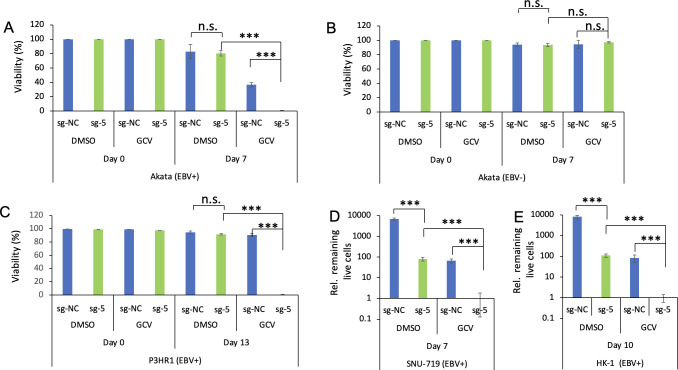
Epstein-Barr virus (EBV) is a ubiquitous human tumor virus that establishes lifelong, persistent infections in B cells. The presence of EBV in cancer cells presents an opportunity to target these cells by reactivating the virus from latency. In this study, we developed a novel approach for EBV reactivation termed clustered regularly interspaced short palindromic repeats (CRISPR)/dCas9-mediated EBV reactivation (CMER) strategy. Using modified CRISPR-associated protein 9 (dCas9) fused with VP64, we designed 10 single guide RNAs (sgRNAs) to target and activate the EBV immediate-early gene promoter. In Akata Burkitt lymphoma cells, 9 out of 10 CMER sgRNAs effectively reactivated EBV. Among these, CMER sgRNA-5 triggered robust reactivation across various cell types, including lymphoma, gastric cancer, and nasopharyngeal carcinoma cells. Importantly, the combination of CMER and ganciclovir selectively eliminated EBV-positive cells, regardless of their cell origin. These findings indicate that targeted virus reactivation by CMER, combined with nucleoside analog therapy, holds promise for EBV-associated cancer treatment.

Importance: This study explores a novel strategy called clustered regularly interspaced short palindromic repeats (CRISPR)/dCas9-mediated Epstein-Barr virus (EBV) reactivation (CMER) to reactivate the Epstein-Barr virus in cancer cells. EBV is associated with various cancers, and reactivating EBV from latency offers a potential therapeutic strategy. We utilized an enzymatically inactive CRISPR-associated protein 9 (dCas9) fused with VP64 and designed 10 single guide RNAs to target the EBV immediate-early gene promoter. Nine of these sgRNAs effectively reactivated EBV in Burkitt lymphoma cells, with CMER sgRNA-5 demonstrating strong reactivation across different cancer cell types. Combining CMER with ganciclovir selectively eliminated EBV-positive cells, showing promise for EBV-associated cancer treatment.

Keywords: CMER; CRISPR/dCas9 activation; EBV; ganciclovir; gastric cancer; latency; lymphoma; nasopharyngeal carcinoma; reactivation; targeted therapy.


Conflict of interest statement

The application for a patent related to this study has been filed by the University of Pittsburgh and Virginia Commonwealth University. R.L. and F.G.S. are inventors on this provisional patent application.

Figures

Fig 1
Fig 1
The design of sgRNA targeting EBV ZTA promoter. (A) Schematic representation of CRISPR/dCas9-VP64 targeting EBV ZTA promoter. The relative positions of sgRNA targeting sites were labeled as indicated. sgRNA-1 and sgRNA-5 (sg-1 and sg-5) target the sense strand, while the remaining sgRNAs target the anti-sense strand. (B) Sequence alignment of the sgRNA targeting sequences from 10 different EBV strains. Polymorphisms are highlighted in yellow. There are no polymorphisms in the protospacer adjacent motif sequences of the 10 sgRNAs.
Fig 2
Fig 2
CMER promotes EBV reactivation in Akata (EBV+) Burkitt lymphoma cells. (A) Akata (EBV+) cells were used to create cell lines using lentivirus carrying dCas9-VP64 with control (sg-NC) and 10 ZTA promoter-targeting sgRNAs. The cells were uninduced (0 hour) or induced using anti-IgG for 24 hours. The expression levels of ZTA and BGLF4 were monitored by Western blot. β-Actin blot was included as loading control. (B) The relative extracellular EBV copy numbers were measured using quantitative polymerase chain reaction as described in Materials and Methods. The value of lane 1 was set as 1. Results from three biological replicates are presented. Error bars indicate the standard deviation (mean ± SD, **P < 0.01 and ***P < 0.001).
Fig 3
Fig 3
CMER triggers EBV reactivation in P3HR1 Burkitt lymphoma cells. (A) EBV-positive P3HR1 cells were used to create cell lines carrying dCas9-VP64 with control (sg-NC) and two ZTA promoter-targeting sgRNAs, sg-1 and sg-5. The cells were uninduced (0 hour) or induced using 12-O-tetradecanoylphorbol-13-acetate (TPA) and sodium butyrate (TPA/NaBu) for 24 and 48 hours. The expression levels of ZTA and BGLF4 were monitored by Western blot. β-Actin blot was included as loading control. (B) The relative intracellular EBV DNA copy numbers were measured using quantitative polymerase chain reaction (qPCR) as described in Materials and Methods. (C) The relative extracellular virion-derived DNA copy numbers were measured using qPCR as described in Materials and Methods. The value of lane 1 was set as 1. Results from three biological replicates are presented. Error bars indicate the standard deviation (mean ± SD, *P < 0.05; **P < 0.01; and ***P < 0.001).
Fig 4
Fig 4
CMER triggers EBV reactivation in SNU-719 gastric cancer cells. (A) Lentiviruses carrying dCas9-VP64 with control (sg-NC), sg-1, or sg-5 sgRNAs were used to transduce EBV-positive SNU-719 cells. The relative EBV copy numbers secreted to the medium (72 hours post-lentiviral transduction) were measured using quantitative polymerase chain reaction (qPCR) as described in Materials and Methods. The cells were subsequently transferred to the T25 flask for cell line establishment. (B) SNU-719 cells carrying dCas9-VP64 with control (sg-NC), sg-1, or sg-5 sgRNAs were either uninduced (0 hour) or induced using TPA and sodium butyrate (TPA/NaBu) for 24 and 48 hours. The expression levels of ZTA and BGLF4 were monitored by Western blot. β-Actin blot was included as loading control. (C) The relative intracellular EBV DNA copy numbers were measured using qPCR as described in Materials and Methods. (D) The relative extracellular virion-derived DNA copy numbers were measured using qPCR as described in Materials and Methods. The value of lane 1 was set as 1. Results from three biological replicates are presented. Error bars indicate the standard deviation (mean ± SD, *P < 0.05 and **P < 0.01).
Fig 5
Fig 5
CMER triggers EBV reactivation in HK-1 (EBV+) nasopharyngeal carcinoma cells. (A) Lentiviruses carrying dCas9-VP64 with control (sg-NC), sg-1, or sg-5 sgRNAs were used to transduce HK-1 (EBV+) cells. The relative EBV copy numbers secreted to the medium (72 hours post-lentiviral transduction) were measured using quantitative polymerase chain reaction (qPCR) as described in Materials and Methods. The cells were subsequently transferred to the T25 flask for cell line establishment. (B) HK-1 (EBV+) cells carrying dCas9-VP64 with control (sg-NC), sg-1, or sg-5 sgRNAs were either uninduced (0 hour) or induced using TPA and sodium butyrate (TPA/NaBu) for 24 and 48 hours. The expression levels of ZTA and BGLF4 were monitored by Western blot. β-Actin blot was included as loading control. (C) The relative intracellular EBV DNA copy numbers were measured using qPCR as described in Materials and Methods. (D) The relative extracellular virion-derived DNA copy numbers were measured using qPCR as described in Materials and Methods. The value of lane 1 was set as 1. Results from three biological replicates are presented. Error bars indicate the standard deviation (mean ± SD, *P < 0.05; **P < 0.01; and ***P < 0.001).
Fig 6
Fig 6
CMER and GCV treatment selectively kills EBV-infected cells. (A) Akata (EBV+) cells were transduced with lentiviruses containing dCas9-VP64 with sg-NC or sg-5. The cells were grown under puromycin selection together with DMSO or GCV for 7 days. The cell viability (live to total cells ratio) was measured as described in Materials and Methods. (B) Akata (EBV−) cells were transduced with lentiviruses containing dCas9-VP64 with sg-NC or sg-5. The cells were grown under puromycin selection together with DMSO or GCV for 7 days. The cell viability (live to total cells ratio) was measured as described in Materials and Methods. (C) P3HR1 (EBV+) cells were transduced with lentiviruses containing dCas9-VP64 with sg-NC or sg-5. The cells were grown under puromycin selection together with DMSO or GCV for 13 days. The cell viability was measured as described in Materials and Methods. (D) SNU-719 (EBV+) cells were transduced with lentiviruses containing dCas9-VP64 with sg-NC or sg-5. The cells were grown under puromycin selection together with DMSO or GCV for 7 days. The relative live cell numbers were counted. The number of sg-5-expressing cells treated with GCV was set as 1. (E) HK-1 (EBV+) cells were transduced with lentiviruses containing dCas9-VP64 with sg-NC or sg-5. The cells were grown under puromycin selection together with DMSO or GCV for 10 days. The relative live cell numbers were counted. The number of sg-5-expressing cells treated with GCV was set as 1. Results from three biological replicates are presented. Error bars indicate the standard deviation (mean ± SD, ***P < 0.001). n.s., no significance.
Fig 7
Fig 7
Delivery of CRISRP/dCas9-VP64 by transient transfection triggers EBV reactivation and subsequent cell death induced by GCV. (A) SNU-719 (EBV+) cells were transfected with pAC152-dual-dCas9VP64-sg-NC and pAC152-dual-dCas9VP64-sg-5 for 48 hours. The expression levels of ZTA, RTA, and BGLF4 were monitored by Western blot. β-Actin blot was included as loading controls. (B) The relative extracellular virion-derived DNA copy numbers were measured using quantitative polymerase chain reaction as described in Materials and Methods. The value of lane 1 was set as 1. (C) The transfected cells (corresponidng to lane 2 and lane 5 conditions in panel A) were treated with DMSO or GCV for 7 days. The relative live cell numbers were counted. The number of sg-5-expressing cells treated with GCV was set as 1. Results from three biological replicates are presented. Error bars indicate the standard deviation (mean ± SD, *P < 0.05; **P < 0.01; and ***P < 0.001). n.s., no significance.
Fig 8
Fig 8
CMER reactivates EBV with 100% efficiency. Akata (EBV+) cells (A–D) and SNU-719 (EBV+) cells (E–H) carrying CRISPR/dCAS9-VP64-sgNC or sg-5 were blocked with 3% bovine serum albumin in PBS at room temperature for 1 hour and then incubated with anti-ZTA (A, B, E, and F) or anti-gp350/250 (C, D, G, and H) antibodies. Subsequently, the Alexa Fluor 488-labeled goat anti-mouse IgG antibody was added to the cells. Cell nuclei were stained with 4′,6-diamidino-2-phenylindole (DAPI) and visualized using a Nikon AXR microscope.
Fig 9
Fig 9
CMER triggers the expression of both lytic and latent genes. Akata (EBV+) cells (A–C) and SNU-719 (EBV+) cells (D–F) carrying CRISPR/dCAS9-VP64-sgNC or sg-5 were lysed to extract protein and RNA. (A and D) The expression levels of ZTA, RTA, p18, and EBNA1 were detected by Western blot. β-Actin blot was included as a loading control. (B and E) The mRNA levels of lytic genes (ZTA, RTA, BGLF4, BALF5, BMRF1, and BLLF1) were measured using RT-qPCR as described in Materials and Methods. (C and F) The mRNA levels of latent genes (EBNA1, EBNA3A, EBNA3B, and LMP1) were measured using RT-qPCR as described in Materials and Methods. Results from three biological replicates are presented. Error bars indicate the standard deviation (mean ± SD, *P < 0.05; **P < 0.01; and ***P < 0.001).
Fig 10
Fig 10
Model summarizing CMER in promoting cell death by EBV reactivation and GCV-mediated DNA synthesis inhibition. Lentiviral delivery or transfection of CRISPR/dCas9-VP64 and ZTA promoter targeting sgRNA (Zp-sg-5) promotes the expression of EBV ZTA and the downstream viral protein kinase (BGLF4). Strong lytic replication and the phosphorylation of nucleoside analog GCV by BGLF4 lead to the eradication of EBV-infected cancer cells.

Similar articles

Cited by

References

    1. Young LS, Rickinson AB. 2004. Epstein-Barr virus: 40 years on. Nat Rev Cancer 4:757–768. doi:10.1038/nrc1452 - DOI - PubMed
    1. Young LS, Yap LF, Murray PG. 2016. Epstein-Barr virus: more than 50 years old and still providing surprises. Nat Rev Cancer 16:789–802. doi:10.1038/nrc.2016.92 - DOI - PubMed
    1. Messick TE, Smith GR, Soldan SS, McDonnell ME, Deakyne JS, Malecka KA, Tolvinski L, van den Heuvel APJ, Gu B-W, Cassel JA, Tran DH, Wassermann BR, Zhang Y, Velvadapu V, Zartler ER, Busson P, Reitz AB, Lieberman PM. 2019. Structure-based design of small-molecule inhibitors of EBNA1 DNA binding blocks Epstein-Barr virus latent infection and tumor growth. Sci Transl Med 11:eaau5612. doi:10.1126/scitranslmed.aau5612 - DOI - PMC - PubMed
    1. Meng Q, Hagemeier SR, Fingeroth JD, Gershburg E, Pagano JS, Kenney SC. 2010. The Epstein-Barr virus (EBV)-encoded protein kinase, EBV-PK, but not the thymidine kinase (EBV-TK), is required for ganciclovir and acyclovir inhibition of lytic viral production. J Virol 84:4534–4542. doi:10.1128/JVI.02487-09 - DOI - PMC - PubMed
    1. Westphal EM, Mauser A, Swenson J, Davis MG, Talarico CL, Kenney SC. 1999. Induction of lytic Epstein-Barr virus (EBV) infection in EBV-associated malignancies using adenovirus vectors in vitro and in vivo. Cancer Res 59:1485–1491. - PubMed

MeSH terms

LinkOut - more resources