. 2024 Jun 13;16(12):10321-10347.
doi: 10.18632/aging.205931. Epub 2024 Jun 13.

Identification of VEGFs-related gene signature for predicting microangiogenesis and hepatocellular carcinoma prognosis

Affiliations

Identification of VEGFs-related gene signature for predicting microangiogenesis and hepatocellular carcinoma prognosis

Shengpan Jiang et al. Aging (Albany NY). .

Abstract

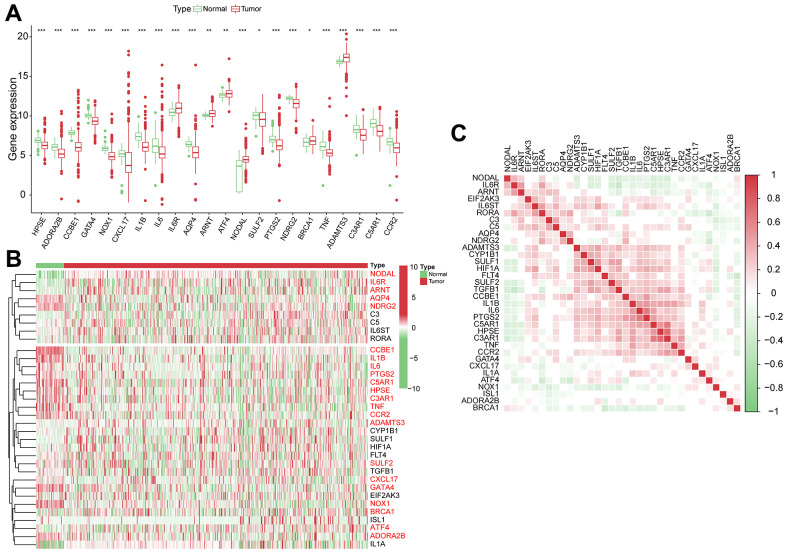
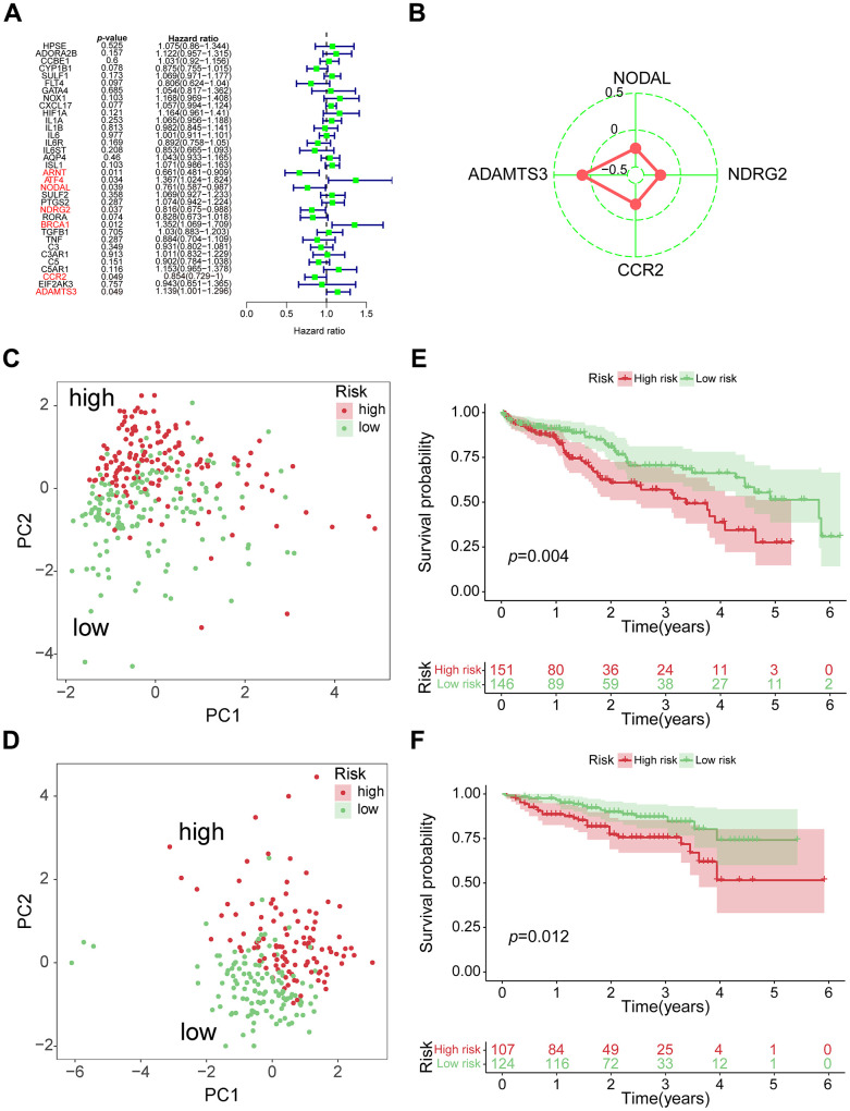
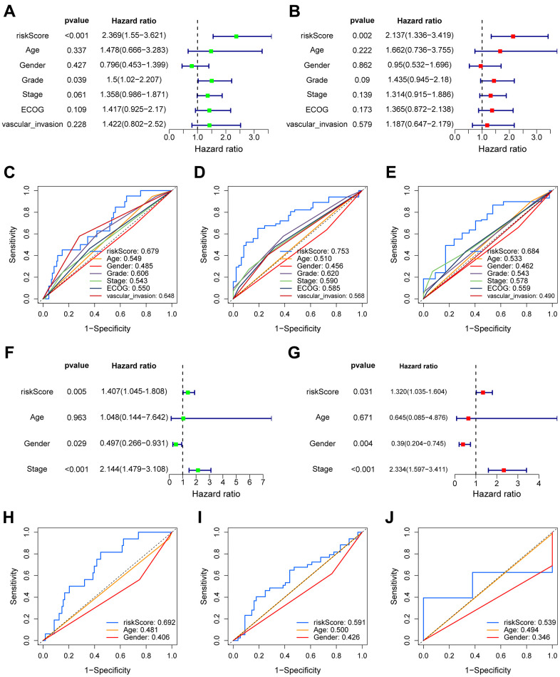
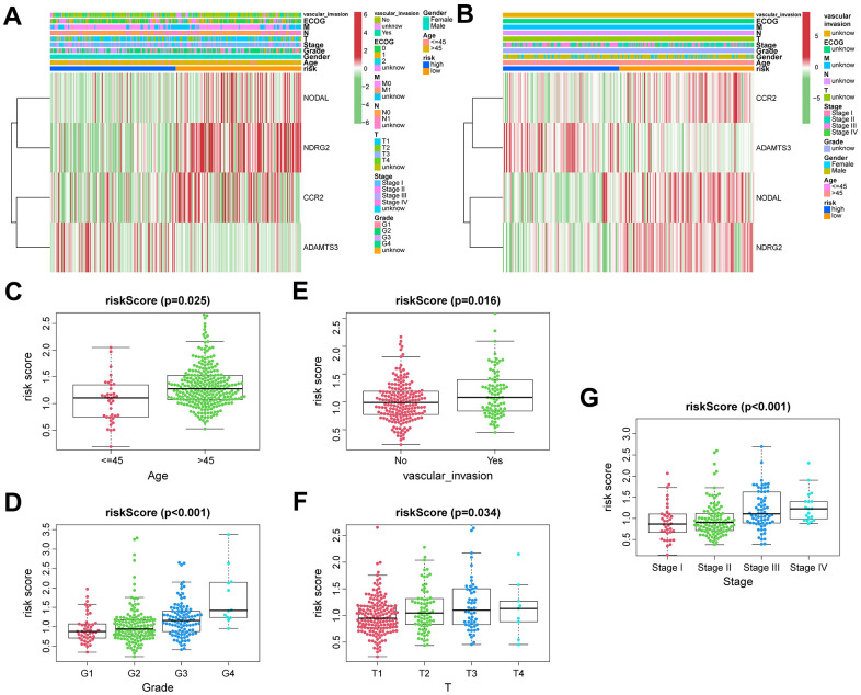
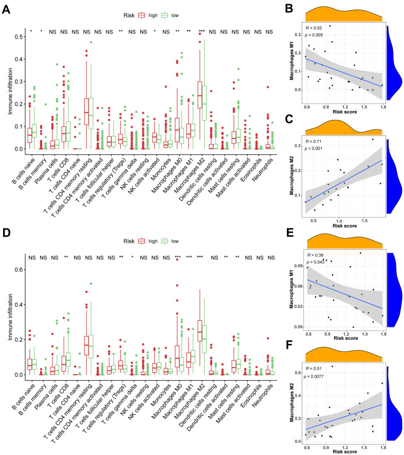
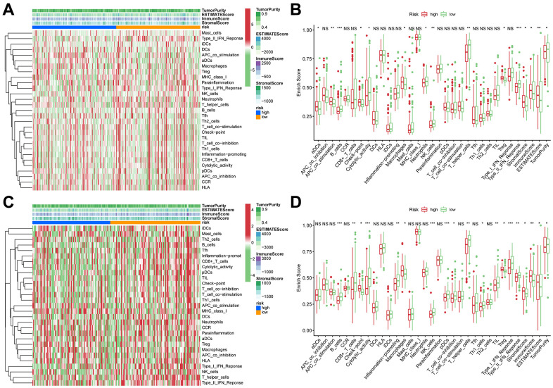
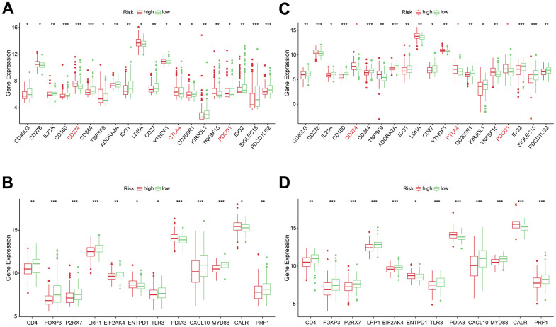
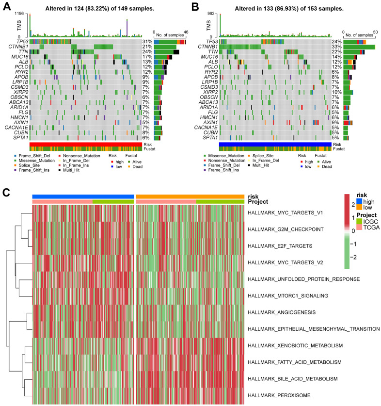
Microangiogenesis is an important prognostic factor in various cancers, including hepatocellular carcinoma (HCC). The Vascular Endothelial Growth Factor (VEGF) has been shown to contribute to tumor angiogenesis. Recently, several studies have investigated the regulation of VEGF production by a single gene, with few researchers exploring all genes that affect VEGF production. In this study, we comprehensively analyzed all genes affecting VEGF production in HCC and developed a risk model and gene-based risk score based on VEGF production. Moreover, the model's predictive capacity on prognosis of HCCs was verified using training and validation datasets. The developed model showed good prediction of the overall survival rate. Patients with a higher risk score experienced poor outcomes compared to those with a lower risk score. Furthermore, we identified the immunological causes of the poor prognosis of patients with high-risk scores comparing with those with low-risk scores.

Keywords: VEGF; gene signature; hepatocellular carcinoma; immune cell; microangiogenesis.


Conflict of interest statement

CONFLICTS OF INTEREST: The authors declare that they have no conflicts of interest.

Figures

Figure 1
Figure 1
The genomic characterization of VPRGs. (A) Boxplot for differentially expressed VPRGs. (B) Heatmap for differentially expressed VPRGs; genes with red color are significantly differently expressed between normal liver tissues and HCC tissues. (C) Correlation plot for VPRGs; red and green squares indicate positive and inverse correlation respectively. ***p < 0.001, **p < 0.001, *p < 0.05.
Figure 2
Figure 2
Construction of 4-genes VPRS. (A) Forest plot for the survival analysis of HCC patients with a univariate Cox model after adjustment for VPRGs; red color represents p < 0.05. (B) Radar diagram of efficiency of the 4 genes in VPRS; the closer the red dot is to the outside, the greater the value it represents. (C) PCA of HCC samples in TCGA; dots in red and green represent samples in high-risk and low-risk groups respectively. (D) Overall survival analysis of risk score for HCC patients in TCGA. (E) PCA in ICGC-HCC. (F) Survival analysis in ICGC-HCC.
Figure 3
Figure 3
The prognostic value of VPRS. In the training set, forest plot on the left for the univariate Cox test (A) evaluating the correlation of the risk score and clinical factors with OS of patients, and forest plot on the right for the multivariate Cox analysis (B) identifying independent risk factors for the OS of patients. The ROC curve of risk score and clinical factors to predict 1- (C), 3- (D), and 5-year (E) OS. In the validation set, univariate (F) and multivariate (G) COX analysis of risk score and clinical factors. ROC curve of risk score compared with other clinical factors to predict 1- (H), 3- (I), and 5-year (J) OS.
Figure 4
Figure 4
The correlation between clinicopathological factors and risk score. Heatmap of the correlations between clinicopathological characteristics of HCC and risk score in the TCGA (A) and ICGC (B) cohorts. Distribution of vascular endothelial growth factor production-related risk signature within HCC patients stratified by age, tumor grade, T classification, and vascular invasion in TCGA (CF) and tumor stage in ICGC (G) cohorts.
Figure 5
Figure 5
The association between TIICs and VPRS. (A) Differential analysis of 22 kinds of TIICs in the low and high-risk groups in training sets. (B, C) Spearman’s correlation analysis for risk score and M1 and M2 macrophages in TCGA cohorts, each dot plot represents a subject, and the correlation is fitted into a straight blue line. (D) Differential analysis of 22 kinds of TIICs in the low and high-risk groups invalidation sets. (E, F) Spearman’s correlation analysis for risk score and M1 and M2 macrophages in ICGC cohorts, each dot plot represents a subject, and the correlation is fitted into a straight blue line. R, rho; ***p < 0.001, **p < 0.001, *p < 0.05.
Figure 6
Figure 6
ssGSEA of immune hallmarks. (A) Heatmap of ssGSEA scores among high- and low-risk groups in training sets (green = negative, red = positive). (B) Boxplot of ssGSEA scores, immune score, stromal score, ESTIMATE score, and tumor purity among high- and low-risk groups in TCGA cohorts. (C) Heatmap of ssGSEA scores among high- and low-risk groups in validation sets (green = negative, red = positive). (D) Boxplot of ssGSEA scores, immune score, stromal score, ESTIMATE score, and tumor purity among high- and low-risk groups in ICGC cohorts. ***p < 0.001, **p < 0.001, *p < 0.05.
Figure 7
Figure 7
Correlation between risk subtypes and ICPs and ICD modulators. (A) Differential expression of ICP genes among the risk subtypes in TCGA cohorts. (B) Differential expression of ICD modulator genes among the risk subtypes in TCGA cohorts. (C) Differential expression of ICP genes among the risk subtypes in ICGC cohorts. (D) Differential expression of ICD modulator genes among the risk subtypes in ICGC cohorts.
Figure 8
Figure 8
Gene set variation analysis and correlation between mutation and risk subtype. (A) Top 20 highly mutated genes in HCC high-risk group. (B) Top 20 highly mutated genes in HCC low-risk group. (C) Heatmap for the contribution of GSVA scores of hallmarks in high- and low-risk groups. The red color represents up-regulated terms in the high-risk group, green color shows the down-regulated terms in the low-risk group.
Figure 9
Figure 9
Verification of the prognostic value and expression of hub genes of VPRS. Survival analysis of ADAMTS3, CCR2, NDRG2, and NODAL for patients in TCGA (AD) and ICGC (EH) cohorts. The protein expression level of ADAMTS3 (I), CCR2 (J), NDRG2 (K), and NODAL (L) in normal and HCC tissues based on the HPA database. (M) The relative mRNA expression levels of ADAMTS3, CCR2, NDRG2, and NODAL are compared between HCC and non-tumor tissues with real-time PCR results. ***p < 0.001, **p < 0.001, *p < 0.05.
Figure 10
Figure 10
Prognostic nomogram was established by combining risk score and tumor stage characters. (A) Nomogram for assessing the 1-, 3-, and 5-year OS for HCC patients in the TCGA dataset. ROC curves of 1-, 3-, and 5-year in the TCGA (B) and ICGC (C) datasets.

Similar articles

Cited by

References

    1. Wang Y, Song F, Zhang X, Yang C. Mitochondrial-Related Transcriptome Feature Correlates with Prognosis, Vascular Invasion, Tumor Microenvironment, and Treatment Response in Hepatocellular Carcinoma. Oxid Med Cell Longev. 2022; 2022:1592905. 10.1155/2022/1592905 - DOI - PMC - PubMed
    1. Akinyemiju T, Abera S, Ahmed M, Alam N, Alemayohu MA, Allen C, Al-Raddadi R, Alvis-Guzman N, Amoako Y, Artaman A, Ayele TA, Barac A, Bensenor I, et al. , and Global Burden of Disease Liver Cancer Collaboration. The Burden of Primary Liver Cancer and Underlying Etiologies From 1990 to 2015 at the Global, Regional, and National Level: Results From the Global Burden of Disease Study 2015. JAMA Oncol. 2017; 3:1683–91. 10.1001/jamaoncol.2017.3055 - DOI - PMC - PubMed
    1. Morse MA, Sun W, Kim R, He AR, Abada PB, Mynderse M, Finn RS. The Role of Angiogenesis in Hepatocellular Carcinoma. Clin Cancer Res. 2019; 25:912–20. 10.1158/1078-0432.CCR-18-1254 - DOI - PubMed
    1. Felmeden DC, Blann AD, Lip GY. Angiogenesis: basic pathophysiology and implications for disease. Eur Heart J. 2003; 24:586–603. 10.1016/s0195-668x(02)00635-8 - DOI - PubMed
    1. Karamysheva AF. Mechanisms of angiogenesis. Biochemistry (Mosc). 2008; 73:751–62. 10.1134/s0006297908070031 - DOI - PubMed

Publication types

MeSH terms

Substances