. 2024 Jul 22;65(4):482-490.
doi: 10.1093/jrr/rrae044.

Phase-separated super-enhancers confer an innate radioresistance on genomic DNA

Affiliations

Phase-separated super-enhancers confer an innate radioresistance on genomic DNA

Koki Matsumoto et al. J Radiat Res. .

Abstract

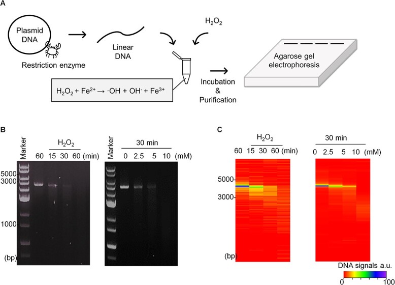
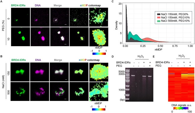
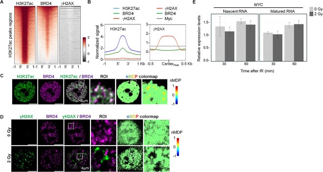
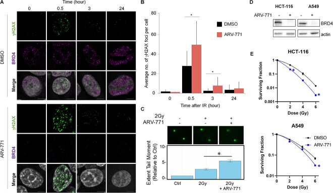
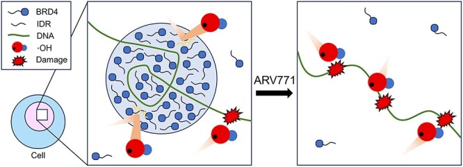
Recently, biomolecular condensates formed through liquid-liquid phase separation have been widely reported to regulate key intracellular processes involved in cell biology and pathogenesis. BRD4 is a nuclear protein instrumental to the establishment of phase-separated super-enhancers (SEs) to direct the transcription of important genes. We previously observed that protein droplets of BRD4 became hydrophobic as their size increase, implying an ability of SEs to limit the ionization of water molecules by irradiation. Here, we aim to establish if SEs confer radiation resistance in cancer cells. We established an in vitro DNA damage assay that measures the effect of radicals provoked by the Fenton reaction on DNA integrity. This revealed that DNA damage was markedly reduced when BRD4 underwent phase separation with DNA. Accordingly, co-focal imaging analyses revealed that SE foci and DNA damage foci are mutually exclusive in irradiated cells. Lastly, we observed that the radioresistance of cancer cells was significantly reduced when irradiation was combined with ARV-771, a BRD4 de-stabilizer. Our data revealed the existence of innately radioresistant genomic regions driven by phase separation in cancer cells. The disruption of these phase-separated components enfolding genomic DNA may represent a novel strategy to augment the effects of radiotherapy.

Keywords: brd4; phase separation; radiation damage; super-enhancer.


Conflict of interest statement

The authors declare no competing financial interest.

Figures

Fig. 1
Fig. 1
Establishment of in vitro DNA damage assay. (A) Scheme illustrating the preparation of linear DNA, ROS generation and quantification of DNA damage using electrophoresis. (B) Result of DNA damage represented in reaction time-dependent manner (left) and concentration-dependent manner (right). (C) Heatmap visualization of the extent of DNA amounts. Maximum signal of DNA (non-damaged DNA) was prepared as 100%.
Fig. 2
Fig. 2
Phase-separated BRD4-IDRs minimize the DNA damage in vitro. (A) BRD4-IDRs were diluted in the buffer to a final concentration of 5 μM in the presence of DNA (5 ng/μl) and indicated conditions. Colocalization in ROI was visualized using nMDP colormap. (B) BRD4-IDRs and DNA were diluted in the buffer to a final concentration of 5 μM and indicated conditions in the presence of 10% PEG. (C) The extent of colocalization was quantified using nMDP value and visualized by density plot. (D) Result of DNA damage represented in the presence of BRD4-IDRs with/without 10% PEG. (E) Heatmap visualization of the extent of DNA amounts. Maximum signal of DNA (non-damaged DNA) was prepared as 100%.
Fig. 3
Fig. 3
Genomic DNA occupied by BRD4 is resistant to DNA damage upon IR. (A) Heatmaps of ChIP-Seq signals at H3K27ac peak regions (±1 kb of peak region). (B) Line plots showing the distribution of indicated ChIP-Seq signals at H3K27ac (left; ±1 kb of peak region) and γH2AX (right; ±0.5 kb of peak centers). (C, D) IF imaging of H3K27ac and BRD4 in TE5 cells (C), γH2AX and BRD4 in HCT116 cells (D). Colocalization in ROI was visualized using nMDP colormap. (E) qRT-PCR analysis of the levels of MYC transcripts using cDNA prepared from non-IR and irradiated HCT-116 cells. A non-irradiated sample at 0 min was used as a reference point. The Intronic primer set was used to detect nascent RNA (left), and an exonic primer set was used to detect matured RNA. Data show mean ± SEM from three independent experiments (n = 3).
Fig. 4
Fig. 4
Pharmacological degradation of BRD4 works as radiation sensitizer. (A) Representative images of BRD4 and IR-induced γH2AX in the cells with either 2 Gy IR alone or a combination of IR and BRD4 degradation. (B) Quantification of γH2AX foci (30 cells) based on a single experiment. Similar results were obtained in two independent experiments. Data show mean ± SD. (C) Fluorescence microscopy images of neutral comet assay in cells upon 2 Gy pretreated for 12 h with DMSO or ARV-771 (n = 3). The bar graph shows mean ± SEM. Significance was assessed using a Student’s t-test (*P < 0.05). (D) Western blotting analysis of BRD4 upon ARV-771 treatment in HCT116 and A549 cell lines. (E) Survival fraction of HCT116 and A549 cell lines upon IR alone and IR combined with ARV-771.
Fig. 5
Fig. 5
A model whereby phase-separated SEs prevent DNA damage from radiation or ROS.

References

    1. Hyman AA, Weber CA, Julicher F. Liquid-liquid phase separation in biology. Annu Rev Cell Dev Biol 2014;30:39–58. 10.1146/annurev-cellbio-100913-013325. - DOI - PubMed
    1. Boija A, Klein IA, Young RA. Biomolecular condensates and cancer. Cancer Cell 2021;39:174–92. 10.1016/j.ccell.2020.12.003. - DOI - PMC - PubMed
    1. Cheng X, Case LB. Phase separation in chemical and mechanical signal transduction. Curr Opin Cell Biol 2023;85:102243. 10.1016/j.ceb.2023.102243. - DOI - PMC - PubMed
    1. Laghmach R, Di Pierro M, Potoyan D. A liquid state perspective on dynamics of chromatin compartments. Front Mol Biosci 2021;8:781981. 10.3389/fmolb.2021.781981. - DOI - PMC - PubMed
    1. Boija A, Klein IA, Sabari BR, et al. Transcription factors activate genes through the phase-separation capacity of their activation domains. Cell 2018;175:1842–1855.e16. 10.1016/j.cell.2018.10.042. - DOI - PMC - PubMed

LinkOut - more resources