. 2024 Aug;11(30):e2400242.
doi: 10.1002/advs.202400242. Epub 2024 Jun 14.

Sensory Nerve Regulation via H3K27 Demethylation Revealed in Akermanite Composite Microspheres Repairing Maxillofacial Bone Defect

Affiliations

Sensory Nerve Regulation via H3K27 Demethylation Revealed in Akermanite Composite Microspheres Repairing Maxillofacial Bone Defect

Kaijun Gu et al. Adv Sci (Weinh). 2024 Aug.

Abstract

Maxillofacial bone defects exhibit intricate anatomy and irregular morphology, presenting challenges for effective treatment. This study aimed to address these challenges by developing an injectable bioactive composite microsphere, termed D-P-Ak (polydopamine-PLGA-akermanite), designed to fit within the defect site while minimizing injury. The D-P-Ak microspheres biodegraded gradually, releasing calcium, magnesium, and silicon ions, which, notably, not only directly stimulated the osteogenic differentiation of bone marrow mesenchymal stem cells (BMSCs) but also activated sensory nerve cells to secrete calcitonin gene-related peptide (CGRP), a key factor in bone repair. Moreover, the released CGRP enhanced the osteogenic differentiation of BMSCs through epigenetic methylation modification. Specifically, inhibition of EZH2 and enhancement of KDM6A reduced the trimethylation level of histone 3 at lysine 27 (H3K27), thereby activating the transcription of osteogenic genes such as Runx2 and Osx. The efficacy of the bioactive microspheres in bone repair is validated in a rat mandibular defect model, demonstrating that peripheral nerve response facilitates bone regeneration through epigenetic modification. These findings illuminated a novel strategy for constructing neuroactive osteo-inductive biomaterials with potential for further clinical applications.

Keywords: bioactive ions; bone regeneration; composite microspheres; methylation modification; sensory nerve.


Conflict of interest statement

The authors declare no conflict of interest.

Figures
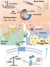
Figure 1
Figure 1
Schematic Diagram of Injectable D‐PLGA‐Ak Composite Microspheres for Maxillofacial Bone Repair. The jigsaw‐like background symbolizes the synergistic effect of osteogenesis promotion by bioactive ions and sensory nerve cells. Uniform‐sized composite microspheres were synthesized using microfluidics, followed by polydopamine coating, and loaded into a syringe for injection into the defect site. As the D‐PLGA‐Ak particles biodegraded, they released bioactive ions crucial for osteo‐differentiation of mesenchymal stem cells. Simultaneously, trigeminal sensory nerve cells responded to these ions and secreted CGRP, initiating the demethylation of histone3 lysine 27, which promotes the transcription of Runx2 and Osx genes.
Figure 2
Figure 2
Preparation, Morphological Characteristics and Properties of Microspheres. A) Preparation and coating process of the (D‐)PLGA‐Ak microspheres. B) (a) The stereoscope observation and (b,c) the SEM surface observation of PLGA/P10Ak/P20Ak/P30Ak series microspheres. Mapping of microspheres shows the distribution of Mg/Ca/Si. Scale bar is 200 µm. C) (a) The stereoscope observation and (b,c) the SEM surface observation of D‐PLGA/D‐P10Ak/D‐P20Ak/D‐P30Ak series microspheres with PDA coating. Scale bar is 200 µm. D) Element mapping and analysis of P10Ak/P20Ak/P30Ak series microspheres by energy dispersive spectroscopy analysis. E) Average size of PLGA/P10Ak/P20Ak/P30Ak composite microspheres. 50 microspheres were measured in every group. *P < 0.05. F) pH changes of P‐Ak microspheres in PBS at 37 °C for 6 weeks. 4 mL of the solution were refreshed with fresh PBS every week. 3 parallel samples were tested at every time point. G) Accumulative ion release profiles of P‐Ak composite microspheres in 8 weeks. H) Injectability of the microsphere. (a) microspheres suspended in sheep plasma in gradient weight percentage. (b) microspheres were squeezed through a 1 mL syringe holding to the universal mechanical testing machine. (c) the stress‐time curves of microspheres extruded at a fixed rate of 2 mL min−1.
Figure 3
Figure 3
Effect of Microspheres on BMSCs in vitro. A) Biocompatibility of microspheres. (a) Images of Live/Dead analysis for BMSCs showing viability after co‐cultured indirectly with the microspheres’ conditional medium for 2 days. The conditional medium was 7‐day extraction of different microspheres immersing in ɑ‐MEM complete medium respectively. Red staining indicates dead cells and green staining indicates live cells. (b,c) count and proportion of living cells. 3 duplicate samples were set for each group. CTRL, BMSCs cultured with only complete ɑ‐MEM. *P < 0.05, Scale bar is 200 µm. B) CCK‐8 assay of BMSCs indirect co‐cultured with microspheres for 1, 3, 5, and 7 days. CTRL, BMSCs cultured with only complete ɑ‐MEM. 3 duplicate samples were set for each group. *P < 0.05. C) Cell attachment assay of BMSCs implanted on microspheres for 24 hours with (a) calcein staining. (b) sketch map of cell spreading on different surfaces. Scale bar is 200 µm. D) Transwell assay of microspheres. (a) schematic representation of BMSCs migration experiment for 48 hours. (b,c) Images and analysis of migrating cells at the lower surface of the membrane with crystal violet stain. CTRL, both chambers were filled with only complete ɑ‐MEM. 3 visions of view were selected for measurements in each group. Scale bar is 200 µm. *P < 0.05. E) (a) ALP staining images on day 7 and (b) quantitative analysis of ALP activity, the results were normalized with the total protein concentration. CTRL, BMSCs cultured with only complete ɑ‐MEM. 3 duplicate samples were set for each group. Scale bar is 2 mm. *P < 0.05. F) Osteoblastic genes expressions of BMSCs indirectly co‐cultured with microspheres after 4 days. CTRL, BMSCs cultured with only complete ɑ‐MEM. 3 duplicates were tested for each group. The qRT‐PCR results were expressed as the mean ± error. Statistical analysis was performed by one‐way ANOVA. *P < 0.05.
Figure 4
Figure 4
In vivo Bone Repair Potential of D‐P‐Ak Microspheres. A) A schematic demonstration of rat mandibular defect. First column is 3D reconstructed micro‐CT images of mandibles after implantation for 4 weeks. CTRL, the incision was sutured without any filling in the defect. Following column displays histological evaluation of new bone formation revealed by Masson staining and Van Gesson staining. Scale bar is 600 µm and 1 mm respectively. Sequential fluorescent labeling images at 4, 10, 18 days after the operation. 4 duplicate sites were constructed in 4 independent rats for each group of microspheres. Scale bar is 100 µm. B) (a) Quantitative analysis of bone volume/total tissue volume (BV/TV), (b) number of trabecula (Tb. N) and (c) bone mineral density (BMD). (d) Quantitative evaluation of the area of stained bone. Three visions of view were selected for measurements in each group. *P < 0.05.
Figure 5
Figure 5
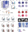
The Synergistic Effect of Trigeminal Sensory Nerve Cells on Osteogenesis. A) Heat map of different expressed genes between trigeminal nerve cells treated with complete F12 medium (CTRL) and D‐P20Ak conditional medium. Fold change data was logarithmically processed. #, genes related to receptors in nerve cells. ##, gene encoding endothelin. ###, genes correlated with neuro‐metabolism. *, genes regarding cytochrome P450 family and lipid metabolism. **, genes encoding central neurotransmitter. Three duplicates were tested for each group, P < 0.05. B) CGRP gene expression of mice trigeminal sensory nerve cells co‐cultured with microsphere extraction medium for three days. CTRL, BMSCs cultured with only complete F12 medium. Three duplicates were tested for each group. *P < 0.05. C) ELISA showing the CGRP concentration in the supernatant of the trigeminal neurons, which were co‐cultured with conditional medium for 3 days. CTRL, BMSCs cultured with only complete F12 medium. 3 duplicates were tested for each group, *P < 0.05. D) Schematic illustration of the experimental verification of synergistic effect and the relationship among “biomaterial, nerve cells and stem cells” triad. E) Osteoblastic genes expressions of BMSCs indirectly co‐cultured with microspheres and nerve cells for 4 days. The conditional medium is 7‐day extraction complete α‐MEM medium collected from trigeminal nerve culture dish. CTRL, BMSCs cultured with only complete ɑ‐MEM. 3 duplicates were tested for each group. The qRT‐PCR results were expressed as the mean ± error. Statistical analysis was performed by one‐way ANOVA. *P < 0.05. F) Osteoblastic genes expressions of BMSCs stimulated by recombinant CGRP protein in gradient concentration for 4 days. CTRL, BMSCs cultured with only complete ɑ‐MEM. 3 duplicates were tested for each group. The qRT‐PCR results were expressed as the mean ± error. Statistical analysis was performed by one‐way ANOVA. *P < 0.05. G) Microscopic images of ALP staining of CGRP‐treated and CGRP+BIBN‐4096‐treated BMSCs on day 4. CTRL, BMSCs cultured with only complete ɑ‐MEM. 3 parallel samples were set for each group. Scale bar is 5 mm and 100 µm respectively. H) Alizarin red staining assays for calcium deposition along with osteo‐inductive culture on day 14. CTRL, BMSCs cultured with only osteo‐inductive medium. 3 parallel samples were set for each group. Scale bar is 5 mm and 100 µm respectively. I) In vivo study of CGRP's role in bone reconstruction. The alveolar bone defects were established at the buccal side of the upper incisor of C57BL/6 mice. BIBN‐4096 were dissolved 750 µg mL−1 in DMSO and injected intraperitoneally every 3 days. CTRL, no injection was performed after the surgery. 4 duplicate sites were constructed in 4 independent mice for each group of treatment. (a) 3D reconstructed micro‐CT images of mouse maxillary 4 weeks after operation. (b) Histological evaluation of new alveolar bone formation revealed by Masson staining. (c) Quantitative analysis of BV/TV. Scale bar is 200 µm. 3 samples were scanned and measured in each group of treatment. *P < 0.05.
Figure 6
Figure 6
Epigenetic Mechanism of CGRP In Bone Regeneration. A) Western blotting analysis of the expression of methyltransferase of Histone3 in BMSCs treated with 1 nм CGRP or together with nonpeptide CGRP‐receptor antagonist BIBN‐4096 for 1 hour in vitro. Quantitative analysis of the expression of KDM6A, EZH2, H3K27me3 proteins were presented below. CTRL, BMSCs cultured with only complete ɑ‐MEM. 3 parallel samples were analyzed for each group of treatment. *P < 0.05. B) Microscopic images of (a) ALP staining of CGRP‐treated and CGRP+GSK‐J4‐treated BMSCs on day 4. CTRL, BMSCs cultured with only complete ɑ‐MEM. Scale bar is 5 mm. C) Osteoblastic genes expressions of BMSCs stimulated by recombinant CGRP or CGRP+GSK‐J4 for 4 days. CTRL, BMSCs cultured with only complete ɑ‐MEM. 3 duplicates were tested for each group. The qRT‐PCR results were expressed as the mean ± error. Statistical analysis was performed by one‐way ANOVA. *P < 0.05. D) Illustration of the epigenetic modulation initiated by CGRP on osteogenic gene transcription.

Similar articles

Cited by

References

    1. Checinska K., Checinski M., Cholewa‐Kowalska K., Sikora M., Chlubek D., Int. J. Mol. Sci. 2022, 23, 7473. - PMC - PubMed
    1. a) Costela‐Ruiz V. J., Melguizo‐Rodriguez L., Bellotti C., Illescas‐Montes R., Stanco D., Arciola C. R., Lucarelli E., Int. J. Mol. Sci. 2022, 23; - PMC - PubMed
    2. b) Kang Y., Xu C., Meng L., Dong X., Qi M., Jiang D., Bioact Mater. 2022, 18, 26; - PMC - PubMed
    3. c) He Y., Wang W., Lin S., Yang Y., Song L., Jing Y., Chen L., He Z., Li W., Xiong A., Yeung K. W. K., Zhao Q., Jiang Y., Li Z., Pei G., Zhang Z. Y., Bioact. Mater. 2022, 9, 491; - PMC - PubMed
    4. d) Silva M. M., Cyster L. A., Barry J. J., Yang X. B., Oreffo R. O., Grant D. M., Scotchford C. A., Howdle S. M., Shakesheff K. M., Rose F. R., Biomaterials 2006, 27, 5909. - PubMed
    1. a) Haidar Z. S., Hamdy R. C., Tabrizian M., Biotechnol. Lett. 2009, 31, 1817; - PubMed
    2. b) Kim D.‐S., Lee J.‐K., Kim Jun H., Lee J., Kim Dong S., An S., Park S.‐B., Kim T.‐H., Rim Jong S., Lee S., Han Dong K., Sci. Adv. 2021, 7, eabj1083; - PMC - PubMed
    3. c) Fang T., Yuan Z., Zhao Y., Li X., Zhai Y., Li J., Wang X., Rao N., Ge L., Cai Q., Chem. Eng. J. 2019, 370, 573.
    1. Cai Z., Jiang H., Lin T., Wang C., Ma J., Gao R., Jiang Y., Zhou X., Mater. Today Adv. 2022, 16, 100315.
    1. a) Cui J., Xia L., Lin K., Wang X., J. Mater. Chem. B 2021, 9, 9505; - PubMed
    2. b) Huang Y., Jin X., Zhang X., Sun H., Tu J., Tang T., Chang J., Dai K., Biomaterials 2009, 30, 5041. - PubMed

MeSH terms

LinkOut - more resources