. 2024 Jun 26;16(25):31922-31935.
doi: 10.1021/acsami.4c02575. Epub 2024 Jun 14.

Cellular and Molecular Insights into the Divergence of Neural Stem Cells on Matrigel and Poly-l-lysine Interfaces

Affiliations

Cellular and Molecular Insights into the Divergence of Neural Stem Cells on Matrigel and Poly-l-lysine Interfaces

Cuiping Wu et al. ACS Appl Mater Interfaces. .

Abstract

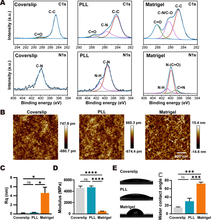
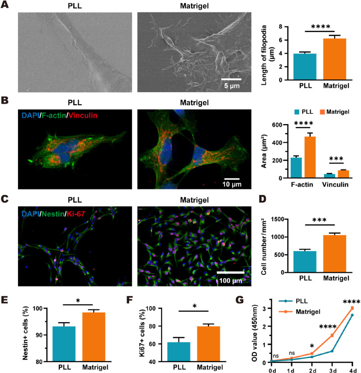
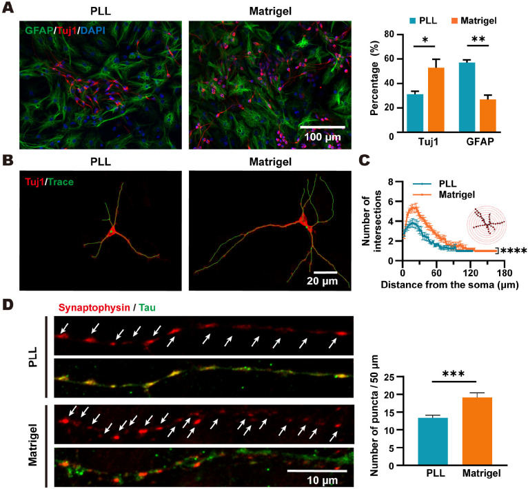
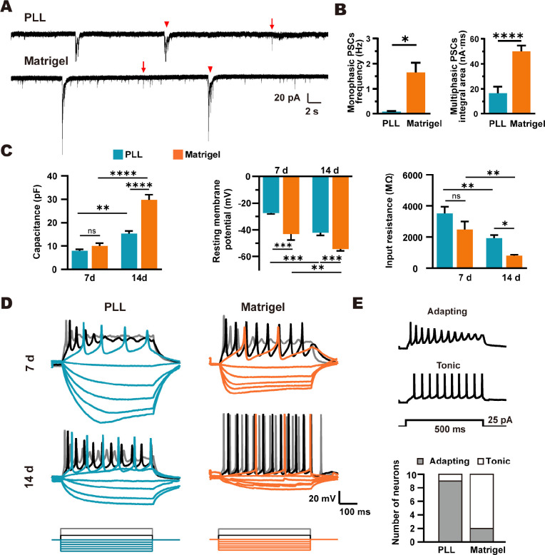
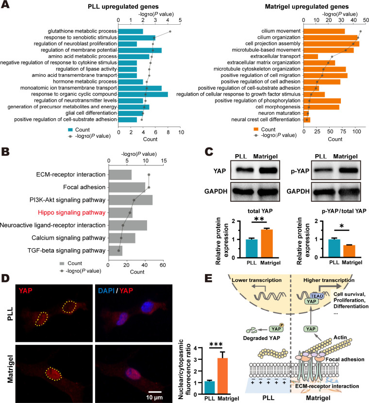
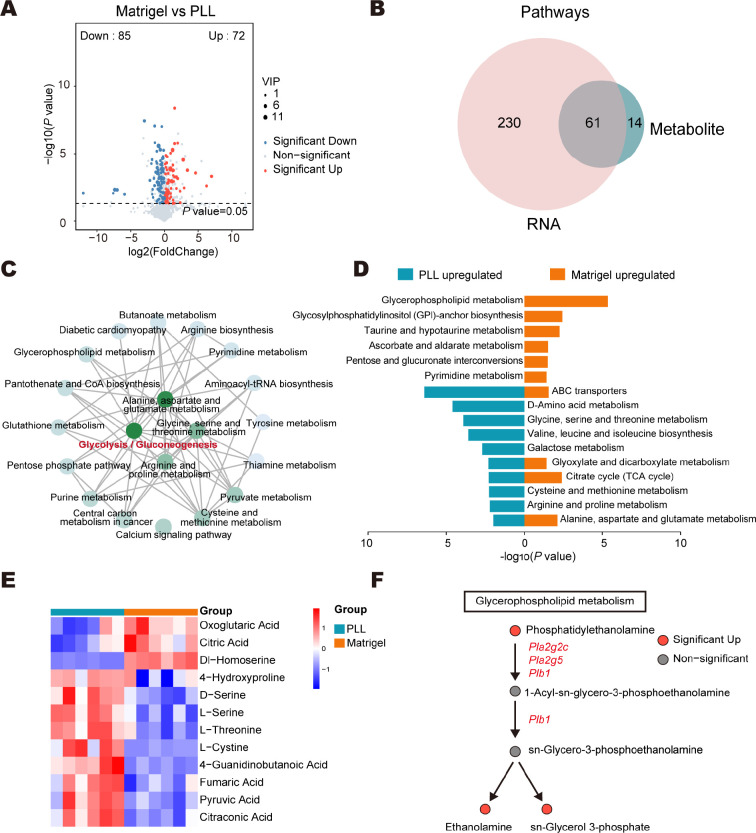
Poly-l-lysine (PLL) and Matrigel, both classical coating materials for culture substrates in neural stem cell (NSC) research, present distinct interfaces whose effect on NSC behavior at cellular and molecular levels remains ambiguous. Our investigation reveals intriguing disparities: although both PLL and Matrigel interfaces are hydrophilic and feature amine functional groups, Matrigel stands out with lower stiffness and higher roughness. Based on this diversity, Matrigel surpasses PLL, driving NSC adhesion, migration, and proliferation. Intriguingly, PLL promotes NSC differentiation into astrocytes, whereas Matrigel favors neural differentiation and the physiological maturation of neurons. At the molecular level, Matrigel showcases a wider upregulation of genes linked to NSC behavior. Specifically, it enhances ECM-receptor interaction, activates the YAP transcription factor, and heightens glycerophospholipid metabolism, steering NSC proliferation and neural differentiation. Conversely, PLL upregulates genes associated with glial cell differentiation and amino acid metabolism and elevates various amino acid levels, potentially linked to its support for astrocyte differentiation. These distinct transcriptional and metabolic activities jointly shape the divergent NSC behavior on these substrates. This study significantly advances our understanding of substrate regulation on NSC behavior, offering novel insights into optimizing and targeting the application of these surface coating materials in NSC research.

Keywords: Differentiation; Electrophysiological function; Matrigel; Metabolomics; Neural stem cell; Poly-l-lysine; YAP.


Conflict of interest statement

The authors declare no competing financial interest.

Figures

Figure 1
Figure 1
Characterization of biophysical and biochemical properties of PLL and Matrigel substrates. (A) High resolution of XPS C 1s and N 1s spectra of uncoated, PLL, and Matrigel-coated substrates. (B) AFM images of different substrate surfaces. (C) Rq of different substrate surfaces. n = 3. (D) Modulus of different substrates. n = 3. (E) Water contact angles of different substrate surfaces. n = 3. One-way ANOVA with Tukey’s multiple correction test. The data are presented as the mean ± s.e.m. *P < 0.05, ***P < 0.001, ****P < 0.0001, ns, not significant.
Figure 2
Figure 2
Adhesion, spreading, and proliferation of NSCs on Matrigel and PLL. (A) SEM images of NSCs. Bar graph shows the length of filopodia. More than 20 cells were measured. (B) Fluorescence images of NSCs with phalloidin (F-actin, green), vinculin (red), and cell nuclei (blue) after 2 days of proliferation. Bar graphs show the quantification of cell spreading area and focal adhesion area. n = 12 cells. (C–F) Fluorescence images of NSCs showing cell nuclei (blue), NSC marker nestin (green), and proliferation marker Ki-67 (red) after 2 days of proliferation. Bar graphs show cell number and percentage of nestin+ and Ki-67+ cells. n = 3. (G) Cell growth was assessed by CCK-8 assay, measured daily. n = 5. Two-tailed unpaired Student’s t test (A–F) or two-way ANOVA with Šídák’s multiple comparisons test (G). The data are presented as the mean ± s.e.m. *P < 0.05, ***P < 0.001, ****P < 0.0001, ns, not significant.
Figure 3
Figure 3
NSC differentiation and synaptic connection of NSC-derived neurons on PLL and Matrigel. (A) Representative fluorescence images of NSCs cultured on Matrigel and PLL after 7 days of differentiation with the neuron marker Tuj1 (red) and astrocyte marker GFAP (green). Nuclei stained with DAPI (blue). Bar graphs show the quantification of the percentage of Tuj1+ and GFAP+ cells. n = 3. (B) Representative neuron morphology reconstruction (green) based on Tuj1 (red) fluorescence images. (C) Sholl analysis of NSC-derived neurons. The number of neurites that intersect concentric circles at varying distances from the soma was counted (PLL, n = 16 neurons; Matrigel, n = 14 neurons). (D) Representative fluorescence images of the presynaptic marker synaptophysin (red) and the axonal marker Tau (green) after 14 days of differentiation. Bar graph shows number of synaptophysin puncta (indicated by white arrows) per 50 μm. n = 16 neurons. Two-tailed unpaired Student’s t test (A and D), two-way ANOVA test (C). All data are presented as mean ± s.e.m. *P < 0.05, **P < 0.01, ***P < 0.001, ****P < 0.0001, ns, not significant.
Figure 4
Figure 4
Physiological maturation of NSC-derived neurons on PLL and Matrigel. (A) Representative traces of spontaneous PSCs of NSC-derived neurons after 14 days of differentiation. Arrows indicate monophasic PSCs, and arrowheads indicate multiphasic PSCs. (B) Frequency of monophasic PSCs and integral area of multiphasic PSCs. Ten neurons were analyzed in each group. (C) Capacitance, RMP, and input resistance of NSCs-derived neurons after 7 and 14 days of differentiation. 7–12 cells were analyzed in each group. (D) Membrane voltage responses to hyperpolarizing (−25 pA to 0 pA in 5 pA increments) and depolarizing (10 and 25 pA) current steps. Cells were clamped at −60 mV membrane potential. (E) Representative traces of two firing patterns: adapting or tonic. Bar graph shows the number of neurons exhibiting adapting or tonic firing patterns after 14 days of differentiation. Two-tailed unpaired Student’s t test (B) or two-way ANOVA test with Tukey’s multiple comparisons test (C). Data are presented as mean ± s.e.m. *P < 0.05, **P < 0.01, ***P < 0.001,****P < 0.0001, ns, not significant.
Figure 5
Figure 5
The effect of different substrates on gene expression of NSC. (A) GO biological process enrichment analysis of upregulated DEGs in the PLL and Matrigel groups, respectively. (B) KEGG pathway enrichment analysis of all DEGs. (C) Western blot detection of total and phosphorylated (p)-YAP. n = 3. (D) Fluorescence images of YAP (red) and nucleus (stained with DAPI) and quantification of nuclear/cytoplasmic fluorescence ratio. The yellow dotted line indicates the nucleus. PLL, n = 58 cells; Matrigel, n = 63 cells. (E) Schematic diagram illustrating molecular regulation of NSCs by PLL and Matrigel substrates. Two-tailed unpaired Student’s t test. All data are presented as mean ± s.e.m. *P < 0.05, **P < 0.01, ***P < 0.001.
Figure 6
Figure 6
Metabolic profiles of NSC differentiation on PLL and Matrigel. (A) Volcano plot showing upregulated (red) and downregulated (blue) DEMs in Matrigel group compared to PLL. DEMs were identified with VIP values >1.0 and P values <0.05. (B) Venn gram of DEG and DEM-mapped KEGG pathways. (C) KGML network of the top 20 interaction pathways, with darker node colors indicating a higher number of interactions. (D) KEGG pathway enrichment analysis of upregulated metabolites in the PLL and Matrigel groups, respectively. (E) Heatmap of DEMs from the enriched amino acid metabolic pathways. (F) DEMs and DEGs in the glycerophospholipid metabolism pathway. Red indicates upregulated and gray indicates not significant.

Similar articles

Cited by

References

    1. Liu D. D.; He J. Q.; Sinha R.; Eastman A. E.; Toland A. M.; Morri M.; Neff N. F.; Vogel H.; Uchida N.; Weissman I. L. Purification and Characterization of Human Neural Stem and Progenitor Cells. Cell 2023, 186 (6), 1179–1194.e15. 10.1016/j.cell.2023.02.017. - DOI - PMC - PubMed
    1. Hayashi Y.; Lin H.-T.; Lee C.-C.; Tsai K.-J. Effects of Neural Stem Cell Transplantation in Alzheimer’s Disease Models. Journal of Biomedical Science 2020, 27 (1), 29.10.1186/s12929-020-0622-x. - DOI - PMC - PubMed
    1. Zhang B.; Yan W.; Zhu Y.; Yang W.; Le W.; Chen B.; Zhu R.; Cheng L. Nanomaterials in Neural-Stem-Cell-Mediated Regenerative Medicine: Imaging and Treatment of Neurological Diseases. Adv. Mater. 2018, 30 (17), e170569410.1002/adma.201705694. - DOI - PubMed
    1. Hu Y.; Chen Z.; Wang H.; Guo J.; Cai J.; Chen X.; Wei H.; Qi J.; Wang Q.; Liu H.; Zhao Y.; Chai R. Conductive Nerve Guidance Conduits Based on Morpho Butterfly Wings for Peripheral Nerve Repair. ACS Nano 2022, 16 (2), 1868–1879. 10.1021/acsnano.1c11627. - DOI - PubMed
    1. Marques-Almeida T.; Lanceros-Mendez S.; Ribeiro C. State of the Art and Current Challenges on Electroactive Biomaterials and Strategies for Neural Tissue Regeneration. Adv. Healthcare Mater. 2024, 13 (1), e230149410.1002/adhm.202301494. - DOI - PubMed