. 2024 Jun 14;51(1):734.
doi: 10.1007/s11033-024-09664-6.

Human umbilical cord-derived mesenchymal stem cells attenuate hepatic stellate cells activation and liver fibrosis

Affiliations

Human umbilical cord-derived mesenchymal stem cells attenuate hepatic stellate cells activation and liver fibrosis

Xiaoyu Shi et al. Mol Biol Rep. .

Abstract

Background: Liver cirrhosis, a prevalent chronic liver disease, is characterized by liver fibrosis as its central pathological process. Recent advancements highlight the clinical efficacy of umbilical cord mesenchymal stem cell (UC-MSC) therapy in the treatment of liver cirrhosis.

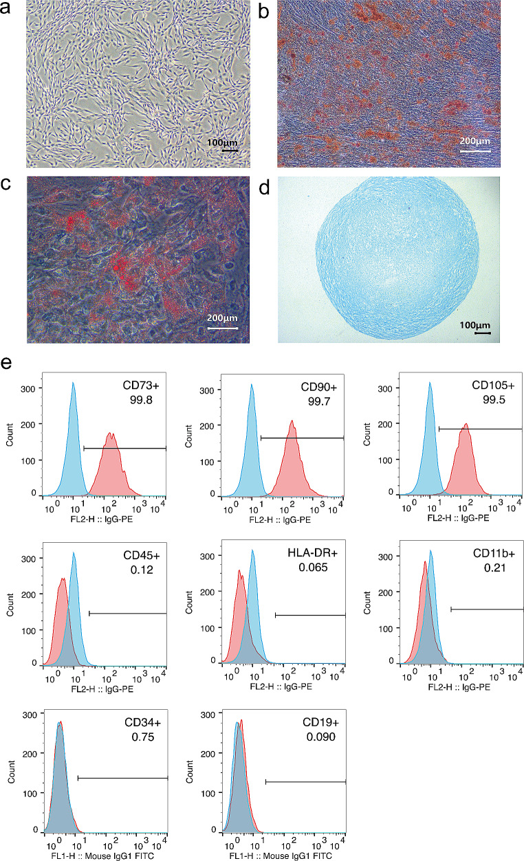
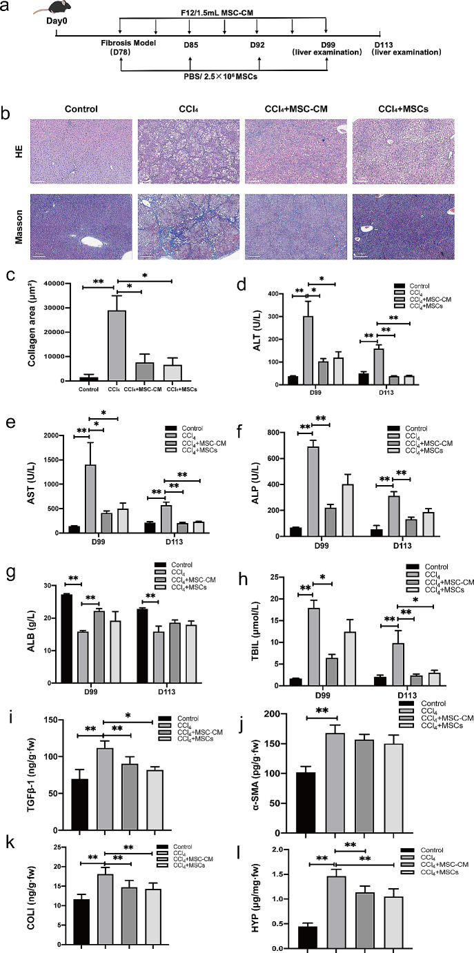
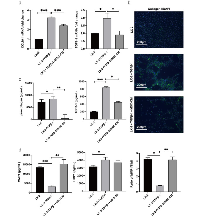
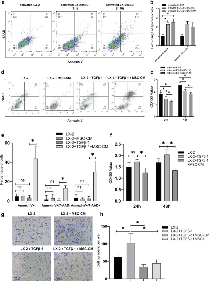
Methods and results: We investigated the pharmacodynamic effects of UC-MSCs and MSC conditional medium (MSC-CM) in vivo, utilizing a carbon tetrachloride (CCl4)-induced fibrotic rat model. Concurrently, we assessed the in vitro impact of MSCs and MSC-CM on various cellular process of hepatic stellate cells (HSCs), including proliferation, apoptosis, activation, immunomodulatory capabilities, and inflammatory factor secretion. Our results indicate that both MSCs and MSC-CM significantly ameliorate the pathological extent of fibrosis in animal tissues, reducing the collagen content, serum biochemical indices and fibrosis biomarkers. In vitro, MSC-CM significantly inhibited the activation of the HSC line LX-2. Notably, MSC-CM modulated the expression of type I procollagen and TGFβ-1 while increasing MMP1 expression. This modulation restored the MMP1/TIMP1 ratio imbalance and extracellular matrix deposition in TGFβ-1 induced fibrosis. Both MSCs and MSC-CM not only induced apoptosis in HSCs but also suppressed proliferation and inflammatory cytokine release from activated HSCs. Furthermore, MSCs and MSC-CM exerted a suppressive effect on total lymphocyte activation.

Conclusions: UC-MSCs and MSC-CM primarily modulate liver fibrosis severity by regulating HSC activation. This study provides both in vivo and in vitro pharmacodynamic evidence supporting the use of MSCs in liver fibrosis treatment.

Keywords: CCL4-induced liver fibrosis; Hepatic stellate cell activation; Immunomodulation; MSC conditional medium; UC-MSC.


Conflict of interest statement

The authors declare no competing interests.

Figures

Fig. 1
Fig. 1
Phenotypic characteristics of UC-MSCs. (a) Light microscopic visualization of UC-MSCs (Passage 5), revealing their morphological features. (b) Alizarin red staining highlighting osteogenic differentiation of UC-MSCs. (c) Oil red-O staining emphasizing adipogenic differentiation of UC-MSCs. (d) Alcian blue stain depicting chondrogenic differentiation of UC-MSCs. (e) Flow cytometric analysis revealing the characteristic phenotype markers of MSCs
Fig. 2
Fig. 2
MSC-CM and UC-MSCs profoundly alleviate liver fibrosis in rat models. (a) Schematic representation of the administration timeline for intravenous injection of either MSC-CM or MSCs. Rats were administered bi-weekly CCl4 injections, in conjunction with specialized feed, until a successful fibrosis model was established. Following this, the animals received bi-weekly MSC-CM injections or weekly MSCs injections via the tail vein. Hepatic tissue samples and blood serum were collected on Day 99 and 113, corresponding to 3 weeks and 5 weeks post intravenous administration, respectively. (b) Representative images of HE staining and Masson’s trichrome staining are shown for hepatic sections obtained from oil-treated rats (Control), CCl4-induced fibrotic livers treated with PBS (CCl4), fibrotic rats treated with MSC-CM (CCl4 + MSC-CM), and fibrotic rats treated with UC-MSCs (CCl4 + MSCs). (c-h) Quantitative analysis of the fold changes in liver collagen content (c) and serum levels of ALT (d), AST (e), ALP (f), ALB (g) and TBIL (h) among different experimental groups is presented. (i-l) Alterations in the expression of hepatic fibrosis markers, including TGFβ-1, α-SMA, COL I, and HYP, were analyzed in each experimental group. Statistical significance was denoted as *P < 0.05, **P < 0.01, and ***P < 0.001
Fig. 3
Fig. 3
MSC-CM attenuates LX-2 activation. (a) qRT-PCR analysis of mRNA expression levels for COL1A1 and TGFβ-1 in LX-2 cultured alone (designated as LX-2 control), LX-2 activated with 10 ng/mL TGFβ1 (denoted as LX-2 + TGFβ-1) and treated with MSC-CM following TGFβ-1 activation (labled as LX-2 + TGFβ-1 + MSC-CM). (b) Immunofluorescence micrographs were captured to visualized the cells, with green fluorescence denoting collagen expression and blue fluorescence for nuclear staining. (c) The protein concentrations of pre-collagen and TGFβ-1 assessed by ELISA. (d) Quantification of MMP1 and TIMP1 protein measured by ELISA. All values were presented as mean ± SD, obtained from triplicate experiments. Statistical significance was as follows: *P < 0.05, **P < 0.01, and ***P < 0.001
Fig. 4
Fig. 4
UC-MSCs modulate apoptosis and activation of activated HSCs. (a) Apoptosis in activated LX-2 cells with TGFβ-1 pre-treatment was significantly induced co-cultured with UC-MSCs for 48 h, as visualized by the fluorescent Annexin V/7-AAD assay. (b) Apoptosis induction was analyzed at varying target-effector ratios, with a 1:1 and 1:10 ratio of activated LX-2 cells to MSCs, revealing a proportional change in apoptotic cells. (c) Cell viability of activated LX-2 was evaluated using the CCK8 assay after co-culturing with varying concentrations of MSCs for 24 and 48 h. (d) A significant increase in apoptosis of activated LX-2 cells was observed 48 h post MSC-CM treatment, as demonstrated by fluorescent Annexin V/7-AAD assay. (e) A comparative analysis of apoptotic cell percentages was conducted among quiescent LX-2, quiescent LX-2 treated with MSC-CM, activated LX-2 and MSC-CM treated activated LX-2 cells. (f) Cell viability of LX-2, activated LX-2 with TGFβ-1 treatment and activated LX-2 with MSC-CM treatment was assessed over a period of 24 and 48 h. (g) A photograph depicting various HSCs groups, includingLX-2 alone, activated LX-2, activated LX-2 treated with MSC-CM, and MSC-cocultured activated LX-2). (h) Quantitative analysis of migrated cells across the distinct treatment groups. All experiments were performed in triplicate, and the data are presented as mean ± SD. *P < 0.05
Fig. 5
Fig. 5
Immunosuppressive impact of MSC-CM on both HSCs and PBMCs. (a) qRT-PCR was utilized to assess the mRNA levels of IL-6 and IL-8 under three experimental conditions: untreated LX-2 cells serving as a control (designated as LX-2), TGFβ-1 stimulated LX-2 cells (designated as LX-2 + TGFβ-1), and MSC-CM treated activated LX-2 cells (designated as LX-2 + TGFβ-1 + MSC-CM). (b) The protein concentrations of IL-6 and IL-8 of different groups were quantified by ELISA. (c) CFSE staining was employed to determine the proportion of progenitor cells within native PBMCs, PBMCs activated with 10 µg/mL PHA, and activated PBMCs treated with three distinct MSC-CMs. Protein quantifications are presented as mean ± SD, with each experiment replicated thrice. ∗P < 0.05 and ∗∗P < 0.01

Similar articles

Cited by

References

    1. Guo Y, Chen B, Chen LJ, Zhang CF, Xiang C (2016) Current status and future prospects of mesenchymal stem cell therapy for liver fibrosis. J Zhejiang Univ Sci B 17(11):831–841 - PMC - PubMed
    1. Cao Y, Ji C, Lu L (2020) Mesenchymal stem cell therapy for liver fibrosis/cirrhosis. Annals Translational Med 8(8):562 - PMC - PubMed
    1. Lee Y, Friedman SL (2010) Fibrosis in the liver: acute protection and chronic disease. Prog Mol Biol Transl Sci 97:151–200 - PubMed
    1. Tsuchida T, Friedman SL (2017) Mechanisms of hepatic stellate cell activation. Nat Rev Gastroenterol Hepatol 14(7):397–411 - PubMed
    1. Kadyk LC, Collins LR, Littman NJ, Millan MT (2015) Proceedings: moving toward cell-based therapies for liver disease. Stem Cells Transl Med 4(3):207–210 - PMC - PubMed

MeSH terms