. 2024 Jul 1;35(5):238-247.
doi: 10.1097/MBC.0000000000001308. Epub 2024 Jun 10.

Characterization of zebrafish coagulation cofactors Fviii and Fv mutants and modeling hemophilia A and factor V deficiency

Affiliations

Characterization of zebrafish coagulation cofactors Fviii and Fv mutants and modeling hemophilia A and factor V deficiency

Sanchi Dhinoja et al. Blood Coagul Fibrinolysis. .

Abstract

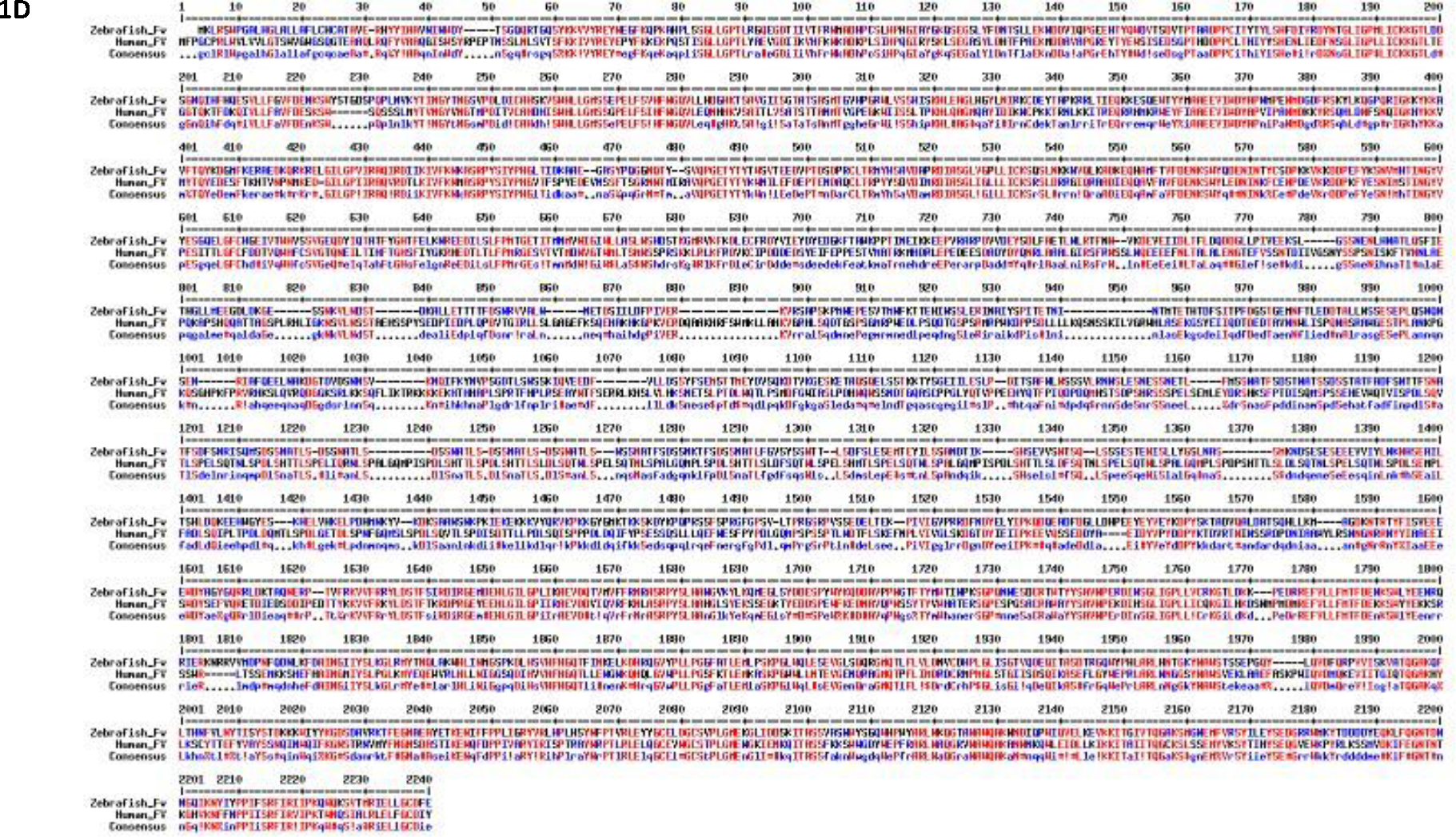
The aim of this study is to characterize zebrafish coagulation cofactors fviii and fv mutant fish and assess if they phenocopy classical hemophilia A and factor V deficiency in humans. The embryos from fviii and fv zebrafish heterozygote mutants generated by ENU mutagenesis were purchased from the ZIRC repository. They were reared to adulthood and genotyped. The heterozygote male and female were crossed to get homozygote, heterozygote, and wild-type fish. Functional kinetic coagulation assays and bleeding assays were performed on normal and mutant adult fish, and venous laser injury assays were performed on the larvae. The DNA from fviii and fv mutants were sequenced to confirm if they have a premature stop codon in exon 19, and in exon 2, respectively, and in both mutants, the amino acid glutamine is replaced with a stop codon. Homozygous and heterozygous 5 days post fertilization (dpf) larvae for fviii and fv deficient mutants exhibited prolonged time to occlusion after venous laser injury compared to wild-type controls. The homozygous and heterozygous fviii adult mutants showed modest bleeding and delayed fibrin formation in the kinetic partial thromboplastin time (kPTT) assay with their plasma. fv homozygous larvae had poor survival beyond 12 dpf. However, heterozygous fv mutants exhibited heavy bleeding and prolonged fibrin formation in the kPTT and kPT assay compared with wild-type siblings. Our characterization showed fviii and fv mutants from ZIRC phenocopied to a considerable extent classical hemophilia A and factor V deficiency in humans, respectively. These models should be useful in studying and developing novel drugs that reverse the phenotype and in generating suppressor mutations to identify novel factors that compensate for these deficiencies.


Conflict of interest statement

Declaration of interest

The authors report no conflict of interest.

Figures

Figure 1:
Figure 1:
Image showing domain structures and MultAlin analysis of FVIII and FV. (1A) The domain structure of active and inactive human FVIII gene and (1B) FV gene. The darker shades of purple and orange represent the domains of the heavy chain, and the lighter shades represent the light chain of FVIII and FV genes respectively. (1C) MultAlin sequence of the zebrafish and Fviii protein compared to human FVIII and (1D) zebrafish Fv protein sequence compared with the human FV protein sequence. The amino acids highlighted as red indicate high consensus between humans and zebrafish sequence, whereas black indicate neutral and blue indicate low consensus.
Figure 1:
Figure 1:
Image showing domain structures and MultAlin analysis of FVIII and FV. (1A) The domain structure of active and inactive human FVIII gene and (1B) FV gene. The darker shades of purple and orange represent the domains of the heavy chain, and the lighter shades represent the light chain of FVIII and FV genes respectively. (1C) MultAlin sequence of the zebrafish and Fviii protein compared to human FVIII and (1D) zebrafish Fv protein sequence compared with the human FV protein sequence. The amino acids highlighted as red indicate high consensus between humans and zebrafish sequence, whereas black indicate neutral and blue indicate low consensus.
Figure 2:
Figure 2:
Sequence analysis of fviii and fv mutants. Founder mutants obtained from ZIRC contained wild-type and heterozygote fviii and fv mutant embryos separately these embryos were reared to adulthood and DNA was extracted from their tails clips and amplified using PCR and resolved on 1.2% agarose gel electrophoresis (Supplementary Figures S1 and S2). The DNA bands obtained here were eluted and sent for sequencing. The progeny of the F1 generation, obtained by heterozygote in-crosses were genotyped similarly (2A, 2B and 2C) chromatograms of wild-type, heterozygote, and homozygote fviii mutants. Chromatograms of wild-type, heterozygote, and homozygote fv mutants (2D, 2E and 2F). The red arrow indicates the change of amino acids. For both the genes, in their cDNA transcript, C is replaced by T. So, for both genes, Glutamine codon CAG is replaced by a stop codon TAG. The wild-types show the original sequence, the heterozygotes show two peaks, indicating of the two alleles one with the stop codon, and the other like the wild-type sequence. The change of nucleotide is shown by a red arrow and the change of codon is highlighted with a red box. The homozygotes for both genes show both alleles with the stop codon.
Figure 3:
Figure 3:
Venous laser injury of 5 dpf fviii and fv mutant larvae. (3A) Progeny of wild-type x wild-type, homozygote x wild-type and homozygote x homozygote fviii fish crosses were subjected to venous laser injury in the caudal vein. Heterozygote (green) and homozygote (red) larvae both showed prolonged TTO in seconds compared to wild-type (blue) by one-way ANOVA. (3B) Progeny of wild-type x wild-type, heterozygote x heterozygote fv fish were subjected to venous laser injury in the caudal vein. The heterozygote in crosses larvae (red) show prolonged TTO in seconds compared to wild-type by student t test (black). The analysis for this study was done by one-way ANOVA and student t-test for fviii and fv mutant larvae respectively. Error bars represent ± SD; **** indicates a p < 0.0001, ns indicates not significant, N = number of larvae.
Figure 4:
Figure 4:
Survival curves for F-II generation fv mutant fish resulting from heterozygote x heterozygote crosses vs wild-type/wild-type crosses. Starting at 3dp, the offspring were observed for a period of 39 days. On day 12, 83% wild-type larvae survived but only 55% of the heterozygote survived past day 12 indicating that most of these progenies started to die by the 12th day post fertilization. By day 15, 80% wild-type survived whereas only 36.644% of the heterozygote in-cross progeny survived. This was analyzed by Log-rank test where p<0.0001.
Figure 5:
Figure 5:
Representative phenotypic observations in fv heterozygote incross embryos. (5A) Wild-type larva 5B and 5C shows bleeding pointed out by red arrows in the thoracic area of a fv deficient heterozygote in-cross progeny.
Figure 6:
Figure 6:
Representative images of bleeding of wild-type and heterozygote, homozygote, fviii mutant fish and of wild-type and heterozygote fv mutant fish. (6A) The fish were allowed to bleed for 1 minute and photographs were taken of the bleeding pattern. (6B) These images were analyzed using ImageJ software, where the total red pixel intensities were measured by multiplying the red pixel intensities to the area of the bleeding. The intensities of wild-type fish were compared to fviii heterozygote and homozygote fish by one-way ANOVA. (N=3). (6C, 6D) Similarly, the bleeding patterns of fv heterozygote fish were compared to its wild-type sibling by student t-test (N=4). * and ** indicates p values < 0.05 and < 0.01, respectively. Error bars represent standard deviation.
Figure 7:
Figure 7:
Representative kinetic coagulation activity for fviii and fv mutant fish. The kinetic curves were plotted against wild-type sibling in presence of Dade ACTIN in kPTT assay or zebrafish muscle thromboplastin in kPT assay. The curves were plotted to detect the time taken for fibrin formation in presence of either of the agonist, at absorbance 405 nm. (7A) Representative kPTT assay curves of fibrin formation for fviii mutants. The blue color represents wild-type fviii fish, green indicates heterozygote fish, and red indicates homozygote fish (N=3). Both the heterozygote and homozygote curves have shifted to the right indicating delayed time for fibrin formation. (7B) Representative kPTT assay and (7C) kPT assay curves of fibrin formation for fv mutants. The green color represents wild-type fish and the red curve shifted to right represents the delayed plasma activity of heterozygote fv mutant fish.

Similar articles

Cited by

References

    1. Arsiccio A, Beavis J, Raut S, & Coxon CH (2021). FVIII inhibitors display FV-neutralizing activity in the prothrombin time assay. J Thromb Haemost, 19(8), 1907–1913. 10.1111/jth.15355 - DOI - PMC - PubMed
    1. Bernal S, Pelaez I, Alias L, Baena M, De Pablo-Moreno JA, Serrano LJ, Camero MD, Tizzano EF, Berrueco R, & Liras A (2021). High Mutational Heterogeneity, and New Mutations in the Human Coagulation Factor V Gene. Future Perspectives for Factor V Deficiency Using Recombinant and Advanced Therapies. Int J Mol Sci, 22(18). 10.3390/ijms22189705 - DOI - PMC - PubMed
    1. Blanchette VS, Key NS, Ljung LR, Manco-Johnson MJ, van den Berg HM, Srivastava A, Subcommittee on Factor Viii, F. I. X., Rare Coagulation Disorders of the, S., Standardization Committee of the International Society on, T., & Hemostasis. (2014). Definitions in hemophilia: communication from the SSC of the ISTH. J Thromb Haemost, 12(11), 1935–1939. 10.1111/jth.12672 - DOI - PubMed
    1. Cui J, O’Shea KS, Purkayastha A, Saunders TL, & Ginsburg D (1996). Fatal haemorrhage and incomplete block to embryogenesis in mice lacking coagulation factor V. Nature, 384(6604), 66–68. 10.1038/384066a0 - DOI - PubMed
    1. Dahlback B, & Tran S (2022). A hydrophobic patch (PLVIVG; 1481–1486) in the B-domain of factor V-short is crucial for its synergistic TFPIalpha-cofactor activity with protein S and for the formation of the FXa-inhibitory complex comprising FV-short, TFPIalpha, and protein S. J Thromb Haemost, 20(5), 1146–1157. 10.1111/jth.15690 - DOI - PMC - PubMed

LinkOut - more resources